15 artigo 414
ORIGINAL ARTICLE
MAGNETIC RESONANCE IMAGING FOR DIAGNOSING THE PRE-SLIP
STAGE OF THE CONTRALATERAL PROXIMAL FEMORAL EPIPHYSIS
IN PATIENTS WITH UNILATERAL EPIPHYSIOLYSIS
Nei Botter Montenegro1, Victor Fruges Junior2, Riccardo Grinfeld2,
Marcelo Bordalo Rodrigues3,Edgard dos Santos Pereira4,
Carlos Gorios5
1 – PhD. Physician and Professor in the Discipline of Pediatric Orthopedics, Hospital das Clínicas, School of Medicine, University of São Paulo, São Paulo, Brazil. 2 – Resident Physician in Orthopedics and Traumatology, Orthopedics and Traumatology Service, University of Santo Amaro (UNISA), São Paulo, Brazil.
3 – Radiologist and Director of the Radiology Service, Institute of Orthopedics and Traumatology, Hospital das Clínicas, School of Medicine, University of São Paulo, São Paulo, Brazil.
4 – Physician and Titular Professor of the Discipline of Orthopedics and Traumatology, University of Santo Amaro (UNISA), São Paulo, Brazil. 5 – Physician and Coordinator of Residence in Orthopedics and Traumatology, University of Santo Amaro (UNISA), São Paulo, Brazil.
Work performed in the Medical Investigation Laboratory for the Musculoskeletal System (LIM 41), Department of Orthopedics and Traumatology, School of Medicine, University of São Paulo.
Correspondence: Rua Dr. Ovídio Pires de Campos, 333, Térreo – 05403-010 – São Paulo, SP – E-mail: [email protected]
Work received for publication: September 20, 2010; accepted for publication: September 29, 2010.
ABSTRACT
Objective: To assess the importance of using conventional magnetic resonance imaging and T2 mapping to determine the pre-slip stage of the contralateral epiphysis in patients with a clinical and radiographic diagnosis of unilateral proximal femoral epiphysiolysis who were initially tre-ated with in-situ fixation. Methods: This prospective cli-nical study on 11 patients with unilateral epiphysiolysis was conducted between February 2009 and August 2010,
INTRODUCTION
Proximal femoral epiphysiolysis is characterized by anterosuperior displacement of the femoral neck in relation to the capital epiphysis, in the hypertrophic layer of the growth plate. It is also known as adoles-cent coxa vara(1)
.
Its prevalence varies depending on the ethnic region studied. On average, it occurs in two adolescents per 100,000 individuals(2)
. It is most common between the
ages of 10 and 16 years, in males (male-female ratio of 1.43:1), predominantly on the left side (2:1) and among blacks. Patients with epiphysiolysis can be grouped into two biotypes: Frolich (adipose genital type) and, in a smaller proportion, Mikulicz (tall and thin type). Symptomatic bilateral involvement may occur at rates
between 9 and 25%, according to the literature(3-5).
Ho-wever, after months of follow-up, this incidence may increase(3,4), with bilateral involvement appearing in
60 to 80% within 18 months after the first slippage(1) .
The objective of this prospective study was to assess the importance of using conventional magne-tic resonance imaging and T2 mapping to determine the diagnosis of the pre-slip stage in the contralateral epiphysis, in patients with a clinical and radiographic diagnosis of unilateral proximal femoral epiphysio-lysis who were initially treated with in-situ fixation.
MATERIAL
This sample for this prospective clinical study con-ducted between February 2009 and August 2010 was
using magnetic resonance imaging on the contralateral hip. Results: We observed abnormalities in the proximal femoral capital physis of the contralateral unaffected hip, with edema under the growth plate in 27% of the patients assessed. Conclusion: Magnetic resonance imaging is an early and sensitive method for detecting the pre-slip stage of the proximal femoral epiphysis.
Keywords - Epiphysis, Slipped; Magnetic Resonance
Ima-ging; Diagnosis
The authors declare that there was no conflict of interest in conducting this work
composed of 11 patients with signs and symptoms suggestive of unilateral epiphysiolysis. The diagno-sis was confirmed using radiographic examinations in anteroposterior and Lauenstein views. There were six male patients and five female patients, of ages between nine and fifteen years, who were operated with in-situ fixation. Four other individuals without clinical or radiographic signs or symptoms of the disease formed a control group (eight hips). Patients with radiographic signs of bilateral epiphysiolysis (seen previously on magnetic resonance imaging), endocrine disease or renal osteodystrophy were ex-cluded from this study. The patients were treated at the Orthopedics and Traumatology Service of the Uni-versity of Santo Amaro (Tables 1 and 2).
All the magnetic resonance examinations were per-formed in a magnet of 1.5 Tesla (HDXT, General Elec-tric Medical Systems, Milwaukee, Wisconsin, United States) using a surface spool (Medrad, Indianola, PA, United States). All the hips were maintained in the neutral position, with the patient in dorsal decubitus. Images were produced in the axial, sagittal and coro-nal planes. The sequences used were: axial T2 with fat saturation (TR/TE: 3,500-4,400/55-65), axial T1 (400-600/10-16), sagittal T2 with fat saturation (3,500-4,600/55-65), sagittal T1 (400-650/10-16) and coronal T2 with fat saturation (3,500-4,200/52-65). Multi-echo sequences for producing T2 maps of the growth plate were also performed. Coronal T2 maps were obtai-ned from multi-spin echo sequences (TR, 1,500; eight echoes spaced between 10 and 90 milliseconds).
RESULTS
Clinically, 10 patients presented the Frolich bio-type (adipose genital) and one had the Mikulicz bio-type, without associated endocrine diseases.
The imaging findings from the magnetic resonance examinations on the hips contralateral to the treated hips are represented in Table 3. Eight hips from eight asymptomatic individuals were also analyzed, without findings of abnormalities on conventional magnetic resonance imaging (Table 4). These examinations were taken to be the standard for assessing the contralateral hips of the patients with epiphysiolysis (Figure 1).
We found abnormalities in the proximal femoral
Table 1 – Patients treated for unilateral proximal femoral
epiphysiolysis, according to sex, age, side affected, grade of slippage (Bianco method), date of surgery and date of magnetic resonance imaging.
Patient Sex Age Side Grade Date of Surgery
1 M 9 E I 22.02.2009
2 F 11 E I 27.08.2009
3 M 15 E I 15.10.2009
4 M 11 E II 15.11.2009
5 F 12 D I 16.11.2009
6 M 13 E I 18.11.2009
7 M 12 E II 14.12.2009
8 M 14 E I 18.02.2010
9 F 13 D I 10.03.2010
10 F 11 E I 02.07.2010
11 F 12 E II 19.08.2010
L – left side; R – right side.
Table 2 – Control group patients.
Patient Sex Age
12 M 13
13 M 12
14 M 11
15 M 11
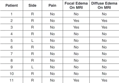
Table 3 – Patients studied and respective imaging findings from
magnetic resonance examinations on the hips contralateral to the hips treated for epiphysiolysis.
Patient Side Pain Focal Edema On MRI
Diffuse Edema On MRI
1 R No No No 2 R No Yes Yes
3 R No Yes Yes
4 R No No No
5 L No No No
6 R No No No
7 R No No No
8 R No No No
9 L No No No
10 R No No No 11 R No Yes Yes
MRI – magnetic resonance imaging; L – left side; R – right side.
METHODS
441
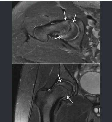
capital growth plate region on the side contralateral to the disease, with edema under the growth plate in 27% of the patients analyzed (Figures 2 and 3).
After the magnetic resonance examinations, during the outpatient follow-up, two patients presented pain in the contralateral hip on walking, but no radiogra-phic signs of pre-slip or epiphysiolysis were found in these patients.
DISCUSSION
The most serious early complications from epiphy-siolysis are chondrolysis and avascular necrosis of the femoral head. The first of these may occur during the natural evolution of the disease, or as a result of the treatment used. The second of these is closely related to certain procedures during the treatment, such as untimely in-situ reduction or fixation maneuvers in the posterior superolateral quadrant of the epiphysis(6)
.
The most feared late complication is early osteoar-throsis of the hip.
The rates of bilaterality reported in the literature have ranged from 9 to 25% according to the metho-dology used, with occurrences in 60 to 80% within 18 months after the first slippage. These numbers are a cause for concern, since the great majority of con-tralateral slippages occur without symptoms and the diagnosis is made in adulthood, in the form of dege-nerative disease(7)
.
In a study on in-situ fixation of the diseased hip, Simbalista Neto et al(8) observed that in cases of
pre--slip and grades I and II slippage, 100% of the results were good or excellent. On the other hand, in cases of excessive epiphyseal slippage, poor results could be
Table 4 – Study on control group individuals: magnetic resonance
examinations on right and left hips.
Patient Side Pain Focal edema on MRI Diffuse edema on MRI
12 R/L No No No
13 R/L No No No
14 R/L No No No
15 R/L No No No
L – left side; R – right side.
Figure 1 – Normal hip. T2-weighted coronal image with fat
saturation on the right hip of an asymptomatic 11-year-old boy.
Figure 3 – T2-weighted axial (A) and coronal (B) images with
fat saturation on the right hip showing diffuse edema around the growth plate, which also presents slight edema (arrows).
FEMORAL EPIPHYSIS IN PATIENTS WITH UNILATERAL EPIPHYSIOLYSIS
Figure 2 – T2-weighted axial image with fat saturation on the
right hip showing edema in the metaphysis and growth plate, with slight epiphyseal focal misalignment (arrow).
↓
↓
↓
↓
attributed to poor positioning of the implant, which could lead to necrosis, chondrolysis and non-closure of the growth plate. Along this line of thinking, pro-phylactic fixation of the contralateral hip would keep this better positioned, thereby preventing the possibi-lity of arthrosis.
Crawford(2) and Ferreira(9) concluded in their
stu-dies that the possible complications from prophylactic fixation (chondrolysis and avascular necrosis) outwei-ghed the benefits. Hence, they reserved this procedure for cases with hormonal disease and patients who were not in a socioeconomic condition conducive to good follow-up.
Laredo Filho et al(4) indicated contralateral in-situ
fixation for at-risk hips (endocrine disease, female sex and black race), and also for patients of low so-cioeconomic condition who did not maintain outpa-tient follow-up.
Jerre et al(10) recommended prophylactic fixation
when there were radiographic signs of pre-slip, endo-crine disease or recognized metabolic abnormalities, and typical biotypes with a propensity and high po-tential for residual growth.
Nelson Elias et al(7) performed prophylactic
fixa-tion of the contralateral hip because they believed that this was a bilateral disease and that the risks of slippage, whether diagnosed or not, outweighed the risks of a procedure on the non-slipped side.
Based on a large number of successful cases of in-situ prophylactic fixation, with low morbidity, Santili(6) recommended this procedure for girls
un-der the age of 12 years and boys unun-der the age of 14 years, as well as for individuals with endocrine diseases or recognized metabolic abnormalities, at any age.
Emery et al(11)indicated prophylactic fixation in
all cases, in view of the high rates of contralateral slippage and late osteoarthrosis.
Umans et al(12)studied 13 patients, among whom
15 hips were symptomatic, with the aim of detec-ting early lesions through evaluations using radiogra-phy, computed tomography and magnetic resonance imaging. They reached the conclusion that the last of these methods was the most sensitive for early diagnosis, since it showed focal and diffuse edema in the epiphyseal region. The authors did not report using these methods for prophylactic detection of the
disease in the contralateral hips, except in two cases that were already symptomatic.
In the literature, conventional magnetic resonance imaging has been used for early detection of epiphy-siolysis. The so-called T2 mapping technique is a new and recently developed method for analyzing cartila-ge, and this is being introduced into medical practice for joint cartilage analyses. With the aim of achie-ving early detection of the disease in the contralateral hips of the patients in our sample, we performed both conventional and T2 mapping magnetic resonance imaging. All the examinations were evaluated by the same radiologist, a specialist in musculoskeletal analysis. The following parameters were taken into consideration: focal edema around the growth plate, diffuse edema under the growth plate and misalig-nment/epiphyseal slippage in relation to the meta-physis. We found signs of pre-slip (focal and diffuse edema) in three of the eleven patients that had not been seen on simple radiographs. After performing this last imaging examination, we performed clinical analysis on the patients and observed that two of the three patients with abnormalities on magnetic reso-nance imaging presented complaints of pain in the contralateral hip. This showed the capacity of this method for early diagnosis in relation to symptoms. The T2 mapping study was assessed through calcula-ting the T2 time, but in all cases the thinness of the growth plate cartilage meant that no adequate calcula-tion for analyzing the alteracalcula-tions in this tissue through T2 mapping was possible.
We agree with the authors who indicated pro-phylactic fixation of the non-slipped hip for patients with gigantism, hypogonadism, hypothyroidism and hyperparathyroidism (endocrine diseases), renal osteodystrophy and high growth potential(6). We
di-sagree with Crawford(2) and Ferreira(9) regarding the
lack of benefit from contralateral fixation for patients without endocrine disease, given that patients who were not in the high-risk groups listed also presented the possibility that bilateral disease could occur.
443
done to show the possible growth plate abnormalities in the contralateral hip, in patients who have not been operated prophylactically.
Although this examination is not available in many medical centers and has a high cost, we believe that it would be prudent to perform it in order to prevent future deformities in the contralateral hip.
Prospective clinical studies with a larger case
se-ries might help to define the best strategy for when and how often to request magnetic resonance imaging for the contralateral hip.
CONCLUSION
Magnetic resonance imaging was shown to be an early and sensitive method for detecting the pre-slip stage of the proximal femoral epiphysis.
REFERENCES
1. Kehl DK. Deslizamento da epífise femoral capital. In: Morrissy RT, Weinstein SL. Lovell and Winter’s pediatric orthopaedics. 5a. ed. Tradução de Lambrinides CD, Nascimento FG, Strong S. São Paulo: Manole; 2001. p.1087-124. 2. Crawford AH. Current concepts review slipped capital femoral epiphysis. J Bone
Joint Surg Am. 1998;70(9):1422-7.
3. Hägglund G, Hansson LI, Ordeberg G, Sandström S. Bilaterality in slipped upper femoral epiphysis. J Bone Joint Surg Br. 1988;70(2):179-81.
4. Laredo Filho J, Braga Júnior MB, Ishida A, Botoletto A. Estudo crítico da indi-cação da pinagem preventiva do lado sadio na epifisiólise proximal do fêmur unilateral. Rev Bras Ortop. 1987;22(6):173-6.
5. Neto AK. Epifisiólise proximal do fêmur. In: Hebert S, Xavier R, Pardini Júnior AG, Barros Filho TE. Ortopedia e traumatologia: princípios e prática. 3a. ed. Porto Alegre: Artmed; 2003. p. 321-33.
6. Santili C. Epifisiólise. Rev Bras Ortop. 2001;36(3): 49-56.
7. Elias N, Simbalista Neto L, Jorge FVF, Tamanini A, Cerqueira F, Syllos A, Abreu AV. Epifisiólise proximal do fêmur. Rev Bras Ortop. 1999;34(5): 333-8. 8. Simbalista Neto L, Elias N, Cerqueira F, Vassimon F, Tamanini A, Syllos A.
Epifisiólise proximal do fêmur: estudo da fixação “in situ” com um parafuso esponjosa AO 6,5mm. Rev Bras Ortop. 1998;33(10): 815-21.
9. Ferreira JCA. Considerações sobre o escorregamento epifisário do fêmur. Rev Bras Ortop. 1996; 31(10): 809-14.
10. Jerre R, Billing L, Hansson G, Wallin J. The contralateral hip in patients primarily treated for unilateral slipped upper femoral epiphysis. Long-term follow-up of 61 hips. J Bone Joint Surg Br. 1994;76(4):563-7.
11. Emery RJ, Todd RC, Dunn DM. Prophylactic pinning in slipped upper femoral epiphysis. Prevention of complications. J Bone Joint Surg Br. 1990;72(2):217-9. 12. Umans H, Liebling MS, Moy L, Haramati N, Macy NJ, Pritzker HA. Slipped
capital femoral epiphysis: a physeal lesion diagnosed by MRI, with radiographic and CT correlation. Skeletal Radiol. 1998; 27(3):139-44.