The association between the outcomes of extraperitoneal
laparoscopic radical prostatectomy and the anthropometric
measurements of the prostate by magnetic resonance
imaging
_______________________________________________
Sompol Permpongkosol
1, Supanun Aramay
2, Thawanrat Vattanakul
2, Sith Phongkitkarun
21 Division of Urology, Department of Surgery, Faculty of Medicine, Ramathibodi Hospital, Mahidol
University, Bangkok 10400, Thailand; 2 Department of Diagnostic and Therapeutic Radiology, Faculty of
Medicine, Ramathibodi Hospital, Mahidol University, Bangkok, Thailand
ABSTRACT
ARTICLE
INFO
______________________________________________________________ ______________________
Introduction and objective: To determine the association between the anthropometric measurements by magnetic resonance imaging (MRI) and perioperative outcomes of extraperitoneal laparoscopic radical prostatectomy (ELRP).
Materials and Methods: From 2008 to June 2016, 86 patients underwent preoperative MRI prior to undergoing ELRP for localized prostate cancer. We analyzed the associa-tions between anthropometric measurements of MRI and the perioperative outcomes of patients who underwent ELRP.
Results: The mean patient age was 69.61±8.30 years. The medians of operating time and blood loss were 2.30 hours and 725.30ml, respectively. The total post-surgical complication rate was 1.16%. The median hospital stay was 6.50 days. The pathologi-cal stages for T2 and T3 were 45.74% and 34.04%, respectively. The rate as positive surgical margins (PSMs) was 18.09% (pT2 and pT3; 6.38% and 9.57%). The angles between pubic bone and prostate gland (angle 1&2), were significantly associated with operative time and hospital stay, respectively (p<0.05). There was no correlation be-tween the pelvimetry and positive surgical margin.
Conclusions: The findings of the present study suggest that anthropometric measure-ments of the MRI are related to operative difficulties in ELRP. This study confirmed that MRI planning is the key to preventing complications in ELRP.
INTRODUCTION
Prostate cancer (PCa) can be treated by radical prostatectomy (RP) which may provoke a troublesome side effect: urinary incontinence (UI). In addition, Lee CH (1) also suggest the likelihood of postoperative UI in patients undergoing LRP is markedly higher in those with larger intravesical prostatic protrusion. The keys to preventing
com-plications of laparoscopic radical prostatectomy (LP) are meticulous preoperative evaluation of pa-tients, magnetic resonance imaging (MRI) planning, and early diagnosis and management of complica-tions (2). The extraperitoneal laparoscopic radical prostatectomy (ELRP) technique proved to be a safe and effective procedure in the treatment of prostate cancer when compared with the transperitoneal (TLRP) approach, with low morbidity (3).
Keywords:
Prostatectomy; Prostatic Neoplasms; Magnetic Resonance Imaging, Laparoscopy
Int Braz J Urol. 2017; 43: 238-47
_____________________ Submitted for publication: April 24, 2017
_____________________ Accepted after revision: July 29, 2017
There are few studies that have evaluated the influence of anthropometric measurements by MRI on perioperative outcomes in patients who underwent ELRP. In addition, there is contro-versy regarding the association between body habitus and perioperative outcomes of surgery, including bleeding, operative time (OT), and re-section margins. Weimin (4) demonstrated that the poor view of the prostatic apex (VPA), pro-trusion of the prostate into the bladder, and high body mass index (BMI) were related to operative difficulties in ELRP. Also, Rue E (5) concluded that MRI before surgery did not provide a defi-nite benefit to help the surgeon tailor LRP more accurately, according to the location and extent of the tumor, and thereby reduce the rate of positive surgical margins (PSMs). In addition, to our knowledge, no association has been reported between the curve distance, periprostatic plexus diameter and the outcomes of ELRP.
Thus, the aim of this study was to de-termine the association between anthropometric measurements of the MRI and perioperative out-comes on the OT, estimated blood loss (EBL), PSA, Gleason grade, pathological stage and PSMs in patients who underwent ELRP.
MATERIALS AND METHODS
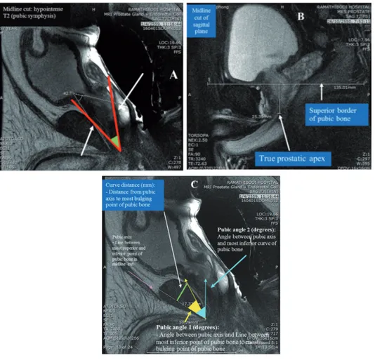
From 2008 to 2014, 94 patients underwent ELRP for localized prostate cancer by the same ex-perienced urologist (SP). In 86 patients, pelvic MR images were obtained at the time of prostate MRI before ELRP. For each patient, two clinically ex-perienced radiologists (SA and TV), independently performed all the anthropometric measurements of MRI twice in each patient, in order to determine the mean value. The anthropometric measure-ments of MRI included prostatic size in volume by the ellipsoid formula [AP (cm) x Transverse (cm) x Vertical (cm) x 0.52], the angle between pubic bone and prostate (degree) (Figure-1A), depth of prostatic apex (mm) (Figure-1B), curve of pubic bone (Figure-1C) including curve distance (mm), pubic angles 1 (degrees) and 2 (degrees), abdomi-nal wall thickness (mm), work space in AP (mm) and work space in transverse during surgery (mm)
bladder (mm) (Figure-2B), and retropubic fat and peri-prostatic plexus diameter.
The degree of the angle between the pubic bone and prostate gland was measured by draw-ing a line along the plane of the prostatic ure-thra and the line between the lowest points of the prostatic urethra to the most bulging point of the posterior cortex of the pubic bone. Curve distance (mm) was the perpendicular distance from pubic axis to the most bulging point of the posterior cortex of the pubic bone. Pubic angle 1 (degrees) was the angle between the pubic axis and the line between the most inferior point of the pubic bone to the most bulging point of the posterior cortex. Pubic angle 2 (degrees) was the angle between pubic axis and the most inferior curve of the pu-bic bone. The pupu-bic axis was the line between the most superior and inferior points of the pu-bic bone in a midline cut. Workspace transverse width (mm) in AP was from the anterior perito-neum to the anterior border of the coccyx (inner border) and Transverse was the distance between the medial borders of the acetabulum. Protrusion to the bladder base (mm) was from the most su-perior point of the prostate in the bladder to the outer border of the bladder wall.
The institutional review board for research involving human subjects approved the retro-spective analysis. We analyzed the associations between anthropometric measurements and pa-tient demographics, including age, body mass in-dex (BMI), preoperative prostate-specific antigen (PSA) level, pathologic stage, pathologic Gleason score, OT, EBL, surgical margin status and ‘30-day surgical-related complications’ defined as any complication rate.
MRI Technique
ex-Figure 1 - Anthropometric measurements by magnetic resonance imaging. (A) angle between the pubic bone and the prostate gland in midline cut: hypointense T2 (pubic symphysis); B) depth of prostatic apex; C) curve of public bone.
All patients were imaged in the supine position. After the acquisition of localizing images, sagittal, coronal, axial thin-slice T2-weighted fast spin-echo (FSE) images through the prostate gland and seminal vesicles were obtained using the following parameters: TR range, 3,000-6,000 milliseconds (msec); TE, 104 milliseconds; echo-train length, 18; field of view (FOV), 16x16cm; section thickness, 3mm; interslice gap, 0mm; matrix 512x256; and num-ber of excitations (NEX), 4. The transverse axial T1-weighted fast spine echo (FSE) images with a TR/TE of 400-600/10-15; matrix, 320x224; and all other parameters matched to the axial thin-slice T2W FSE sequence were obtained. The axial thin-slice T2-weighted images were used to calculate prostatic volume by Functool package post processing with the GE advantage workstation (GE Medical Systems).
Axial free-breathing DWI was performed using a single-shot echo-planar imaging tech-nique with a TR of 3,000-6,000msec and a TE of 60-120msec; FOV, 18x18cm; section thickness, 5mm; interslice gap, 1mm; matrix 128x128; and NEX, 6. ADC values were obtained from the DWI sequences, which were performed with b values of 0.50 or 100, 800 or 1000, and 1500s/mm2. The ADC maps were generated by auto-calculation of the ADC value in each pixel of each slice.
Dynamic contrast enhanced MR imaging was performed by injecting a 0.1mmol/kg bolus of gadolinium-based contrast agent at a rate of 3ml/sec, followed by a 30ml saline flush at the same rate and serial T1W 3D images were ob-tained every 12 seconds through the entire pros-tate, using an MR-compatible automated injector (MedRad, USA). To allow acquisition of non-en-hanced baseline images, the sequence and injec-tion of the contrast agent were initiated simulta-neously. A fast saturation-recovery TurboFLASH (fast low angle shot) sequence (TR 4.1msec, TE 1.9msec, flip angle 12º, matrix 256x192, FOV 200x240mm, slice thickness 5mm) was acquired. Total scan time was 5 minutes.
Statistical analysis
Analysis of variance and
compari-Simple linear and logistic regression analyses were used to identify associative factors for OT, EBL, and PSMs. All tests were two-sided, with p≤0.05 considered statistically significant. Statistical analyses were conducted with use of Stata version 14.0 (Stata Corp, College Station, Texas, USA).
RESULTS
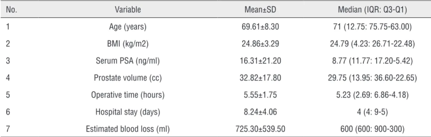
The mean patient age was 69.61±8.30 years, and the patient’s mean BMI was 24.86±3.29kg/m2. The mean preoperative se-rum PSA level was 16.31±21.20ng/ml. The me-dian of OT and EBL was 5.23 (2.69; 6.86-4.18) hours and 600 (600; 900-300) ml, respectively. The only post-surgical complication was a sin-gle case (1.16%) of wound infection. The me-dian hospital stay was 6.50 days (4.00; 9.00-5.00). The pathological stages, T2 and T3, were 45.74% and 34.04%, respectively. The rate of PSMs was 18.09% (17/94) (pT2 and pT3; 6.38% and 9.57%). Tables-1 and 2 demonstrates pa-tient’s characteristics and perioperative out-comes.
For the anthropometric measurements of MRI (Table-3), the mean prostatic volume was 32.72±17.41cc, the mean angle between the pu-bic bone and the prostate gland was 53.24±8.68 degrees; the mean depth of the prostatic apex was 29.00±6.10mm; the mean of curve distance was 14.28±2.70mm; the means of the pubic an-gles 1 and 2 were 23.10±3.81 and 48.70±10.11 degrees, respectively; and the mean abdomi-nal wall thickness was 20.00±6.36mm. Dur-ing surgery, the mean of workspace AP was 139.51±10.83mm and the mean of workspace transverse was 103.89±6.07mm; the mean of protrusion of the prostate into the bladder was 2.80±4.56mm and the means of retropu-bic fat and peri-prostatic plexus diameter were 3.20±2.02mm, and 3.30±0.79mm, respectively.
operative time and hospital stay, respectively (p<0.05). Interestingly, pubic angle 2, curve distance and bladder protrusion were signifi-cantly associated with PSA (p<0.05). For multi-variate analysis using simple linear regression analysis, pubic angle 2 was only significantly associated with PSA (p<0.05). There was no correlation between the pelvimetry and posi-tive surgical margin. For the correlation among PSA, Gleason grade, pathological stage and the perioperative outcomes, PSA level was signifi-cantly associated with hospital stay (p<0.05) (Table-4).
DISCUSSION
ELRP allows direct access to the retropu-bic space, avoiding potential bowel injury, and it represents the technique that best replicates stan-dard RP (6). There was no statistical difference from the transperitoneal techniques in OT, com-plication rates, or PSMs (7). Patients with a low-grade impact of intravesical prostatic protrusion (IPP≤5mm) have significantly higher chances of recovering full continence (8). Multiparametric MRI positivity can independently predict bio-chemical recurrence after RP (9).
Table 1 - Patient characteristics and perioperative outcomes.
No. Variable Mean±SD Median (IQR: Q3-Q1)
1 Age (years) 69.61±8.30 71 (12.75: 75.75-63.00)
2 BMI (kg/m2) 24.86±3.29 24.79 (4.23: 26.71-22.48)
3 Serum PSA (ng/ml) 16.31±21.20 8.77 (11.77: 17.20-5.42)
4 Prostate volume (cc) 32.82±17.80 29.75 (13.95: 36.60-22.65)
5 Operative time (hours) 5.55±1.75 5.23 (2.69: 6.86-4.18)
6 Hospital stay (days) 8.24±4.06 4 (4: 9-5)
7 Estimated blood loss (ml) 725.30±539.50 600 (600: 900-300)
SD: standard deviation; BMI: body mass index; PSA: prostate-specific antigen
Table - 2 - Patient pathological reports and positive surgical margins.
No. Variable N Percentage (%)
1 Pathologic Gleason score
≤6 29 30.85
7 48 51.06
≥8 14 14.89
2 Pathologic stage
T1 7 7.45
T2 42 44.68
T3 31 32.98
T4 2 2.13
However, few studies have evaluated the influence of the anthropometric measure-ments of the prostate MRI on perioperative outcomes in patients who underwent ELRP. In 2010, Deok-Hyun et al. (6) determined the effect of pelvic arch interference and the depth of the pelvic cavity, as shown on preoperative MRI, on the performance of ELRP. The authors suggest-ed that the depth of the pelvic cavity and pros-tate volume might increase surgical difficulty in patients undergoing ELRP by prolonging op-erative time. They measured the true conjugate diameter, the obstetric conjugate diameter, the difference between the true and obstetric diam-eters and the pelvic depth (the distance between the true conjugate and the prostate apex). Al-though the study was done in the same Asian population, all factors were different from the present study.
Our study demonstrated that the pubic angle 2 might increase surgical difficulty in patients undergoing ELRP by prolonging op-erative time. Prostate volume did not correlate with any anthropometric measurements of MRI. In our routine MRI of the prostate, the sagittal plane scanning technique does not include the sacral promontory due to the small field of view (FOV) in order to focus on the prostate gland. This prevented measurement of the true conju-gate diameter. In this study, therefore, the pel-vimetry was measured at the level of acetabu-lum on the axial image (both AP and transverse dimensions) to represent the working space for the urologists. However, this measurement was not correlated with operative time.
In the year 2010, Matikainen et al. re-ported the depth of prostatic apex is an inde-pendent predictor of positive apical margins at radical prostatectomy and confirmed MRI pel-vimetry might allow for preoperative planning of open retropubic prostatectomy (RRP) or LRP (10) Interestingly, the pelvimetric measurements of the study were different from Deok-Hyun et al. method (6); the interspinous distance (ISD), the body width of the pelvis at the mid-femoral head level (BFW), the soft tissue width (SW) the apical depth (AD) and symphysis angle, In
addi-al. (11) as the pelvic dimension index (PDI; de-fined as ISD/AD), the bony width (BWI; dede-fined as BFW/AD) and the SW index (SWI defined as SW/AD), The authors included each of PDI, AD, BWI and SWI as a measure of a “hostile” pelvis which is deep and narrow. The symphysis angle was defined as the angle axis of the symphy-sis pubis and the horizontal on the mid-sagittal T2-weighted sequence image.
Although the studies by Hong et al. (11) and Matikainen (10) were done in an Asian and the USA populations, respectively, these retro-spective studies are limited by the small num-ber of the population. The measurement of the pelvimetry in the present study was different from two previous reports, but very similar to the recently report by Weimin (4) that includ-ed the angle between the prostate and pubic bone and also the depth of prostate apex, in which, both parameters showed negative cor-relation with operative time. The surgery will be more difficult when the prostatic apex is lo-cated deep. Our study did not specifically men-tion a good and poor view of the prostatic apex (VPA), however, we developed two parameters i.e., curve distance and pubic angles that might influence laparoscopic techniques to approach the prostatic apex. The results showed that greater curve distances result in prolonged op-erative times. Since the present MRI technique did not demonstrate the perpendicular line from the promontory of the pelvis due to the narrow field of view (FOV), this study evaluated 4 different factors; first, curve distance, second, abdominal wall thickness, third, peri-prostatic plexus diameter and, fourth, the working space.
|
EXTRAPERIT
ONEAL LAP
AROSCOPIC RADICAL PROST
A
TECT
OMY AND PROST
A
TE MEASUREMENTS B
Y MRI
244
MRI Mean±SD
c.c. P c.c. P c.c. P c.c. P c.c. P c.c. P c.c. P c.c. P
Prostate volume 32.717±17.41 0.02 0.409 0.01 0.942 0.00 0.996 -0.00 0.932 -0.02 0.551 -0.00 0.801 0.01 0.471 0.00 0.415
Angle between pubic bone and prostate (degree)
53.24±8.68 0.01 0.897 0.19 0.471 0.03 0.163 0.01 0.296 0.03 0.592 -0.01 0.165 -0.02 0.454 -0.00 0.702
Depth of prosatic apex (mm)
29.00±6.10 -0.01 0.821 0.30 0.425 0.00 0.982 0.02 0.124 -0.09 0.237 0.00 0.96 0.02 0.487 0.01 0.276
Curve distance
(mm) 14.28±2.70 -0.14 0.272 -1.64 0.043 -0.03 0.663 -0.01 0.770 -0.30 0.061 0.01 0.707 0.01 0.921 0.04 0.171
Pubic angle 1
(degree) 23.10±3.81 -0.11 0.251 -0.90 0.123 -0.04 0.434 0.00 0.999 -0.29 0.015 0.00 0.847 0.05 0.253 0.02 0.329
Pubic angle 2
(degree) 48.70±10.11 0.01 0.804 -0.58 0.008 -0.04 0.025 0.01 0.203 -0.03 0.542 -0.00 0.875 -0.01 0.596 -0.01 0.480
Abd wall
thickness (mm) 20.00±6.36 0.09 0.117 0.40 0.252 0.01 0.670 -0.00 0.901 0.12 0.087 0.01 0.408 0.00 0.971 0.01 0.264
Work space AP
(mm) 139.51±10.83 0.03 0.403 -0.37 0.076 0.00 0.927 0.01 0.399 0.04 0.372 0.01 0.055 -0.01 0.770 -0.00 0.866
Work space
transverse (mm) 103.89±6.07 0.02 0.780 -0.09 0.800 0.03 0.451 0.01 0.665 -0.07 0.361 -0.01 0.068 -0.01 0.769 0.00 0.980
Bladder
protrusion (mm) 2.80±4.56 -0.01 0.856 0.97 0.049 0.02 0.606 0.02 0.177 -0.05 0.612 0.00 0.894 -0.06 0.114 0.02 0.360
Retropubic fat 3.20±2.02 -0.03 0.854 -2.12 0.057 0.00 0.979 -0.02 0.663 0.30 0.187 -0.01 0.540 -0.10 0.245 -0.05 0.177
Periprostatic plexus diameter (mm)
3.30 ±0.79 0.15 0.724 -0.18 0.951 0.37 0.119 0.07 0.516 -0.04 0.943 0.04 0.385 -0.16 0.475 0.02 0.840
ing. In addition, pubic angle 1 was significantly associated with hospital stay (p<0.05).
Weimin study (4) also reported that pro-trusion of the prostate into the bladder was significantly associated with positive resection margins (p=04) in multiple logistic regression analysis. Their positive surgical margin was very high (37%). The series of Matikainen (10) showed PSM rates of 10.4%, which were con-sistent with our literature. Our rate of PSMs of pT2 and pT3 was 6.38% and 9.57%, respective-ly. Moreover, the present study did not demon-strate any correlation between the pelvimetry and positive surgical margin, similar to Rud et al. results (5). High serum PSA, biopsy Gleason score of 7, low prostate volume, and interfas-cial NVB dissection were independently asso-ciated with side-specific PSMs after LRP, and should be considered during planning of the LRP surgical strategy (12). Moreover, our re-sults confirmed PSA level was associated with prolonged LOS and confirmed the conclusion of Pearce report (13).
Although the study demonstrated that radiologists can work in a team with urologists, trying to obtain better results, there are several limitations to this study that should be noted. Firstly, this study is limited in that the data were collected retrospectively and the patients were not randomized for study. A high proportion of enrolled patients with more aggressive, inter-mediate and high risk tumors might have been
(14) indicated the effectiveness of 3.0T MRI and 1.5T endorectal MRI were similar in assessing diagnostic performance of cancer localization, extraprostatic extension, and seminal vesicle involvement; they demonstrated prostate an-terior-posterior diameter measured was signifi-cantly shorter with 1.5T endorectal MRI than with 3.0T MRI. In addition, Albert et al. (15) also reported staging endorectal MRI should not be routinely used for treatment planning because it produces anatomic distortion. There-fore, further study is needed to clarify the dif-ferences between the two MRI systems in the association between the outcomes of ELRP and the anthropometric measurements of the pros-tate and illuminate whether MRI positivity can independently predict biochemical recurrence after LRP.
Finally, the most challenging laparo-scopic surgery in Urology is ELRP (16). Birk-meyer et al. reported a variation in surgeon’s technical skill based on peer-rated video-re-cording (17). Moreover, many studies reflect that during the learning curve a significant reduction in the average time to perform the urethral-bladder anastomosis, the estimated blood loss and the removal time of the urinary catheter have not caused any important com-plication while performing ELRP (3, 18). Our re-search design is from a Cross-Sectional analytic study with a 7-years series (from 2008-2014) and may have discrepancies when compared to Table 4 - Association among PSA, Gleason grade and pathological stage and perioperative outcomes and using simple linear regression analysis.
Variable of MRI Operative Time (hr) Blood loss (ml) Hospital stay (days)
c.c. P c.c. P c.c. P
BMI 0.06 0.281 0.04 0.159 -0.05 0.734
PSA 0.02 0.035 0.01 0.100 0.01 0.535
Positive margin 0.42 0.390 0.13 0.563 1.82 0.116
Pathological Gleason score -0.19 0.092 -0.06 0.258 -0.18 0.489
Pathological stage -0.31 0.304 -0.09 0.545 -0.98 0.150
experience and surgical procedures, similar to Ploussard et al. report (19).
CONCLUSIONS
We believe there are benefits of perform-ing MRI before ELRP to prevent complications of ELRP and suggest that anthropometric mea-surements of the MRI are related to operative difficulties. However, positive surgical margin was not influenced by the pelvimetry.
ACKNOWLEDGEMENTS
We wish to thank Mr. Terry King for English proof reading. Miss Kornkanok Sombo-onpun for help with data search and Pattawia Choikrua for data analysis
CONFLICT OF INTEREST
None declared.
REFERENCES
1. Lee CH, Ha HK. Intravesical prostatic protrusion as a predictor of early urinary continence recovery after laparoscopic radical prostatectomy. Int J Urol. 2014;21:653-6.
2. Ou YC, Yang CK, Chang KS, Wang J, Hung SW, Tung MC, et al. Prevention and Management of Complications During Robotic-assisted Laparoscopic Radical Prostatectomy Following Comprehensive Planning: A Large Series Involving a Single Surgeon. Anticancer Res. 2016;36:1991-8.
3. Barbosa Hdo N Jr, Siqueira TM Jr, Barreto F, Menezes LG, Luna MJ, Calado AA. 4-Ports endoscopic extraperitoneal radical prostatectomy: preliminary and learning curve results. Int Braz J Urol. 2016;42:438-48.
4. Weimin Y, Haga N, Yanagida T, Kurita N, Akihata H, Kojima Y. Impact of Body Habitus on Operative Difficulties during Extraperitoneal Laparoscopic Radical Prostatectomy. Urol J. 2016;13:2519-26.
5. Rud E, Baco E, Klotz D, Rennesund K, Svindland A, Berge V, et al. Does preoperative magnetic resonance imaging reduce the rate of positive surgical margins at radical prostatectomy in a randomised clinical trial? Eur Urol. 2015;68:487-96.
6. Nam DH, Hwang EC, Im CM, Kim SO, Jung SI, Kwon DD, et al. Factors affecting the outcome of extraperitoneal laparoscopic radical prostatectomy: pelvic arch interference and depth of the pelvic cavity. Korean J Urol. 2011;52:39-43.
7. Erdogru T, Teber D, Frede T, Marrero R, Hammady A, Seemann O, et al. Comparison of transperitoneal and extraperitoneal laparoscopic radical prostatectomy using match-pair analysis. Eur Urol. 2004;46:312-9.
8. Jo JK, Hong SK, Byun SS, Zargar H, Autorino R, Lee SE. Urinary Continence after Robot-Assisted Laparoscopic Radical Prostatectomy: The Impact of Intravesical Prostatic Protrusion. Yonsei Med J. 2016;57:1145-51. 9. Hattori S, Kosaka T, Mizuno R, Kanao K, Miyajima A,
Yasumizu Y, et al. Prognostic value of preoperative multiparametric magnetic resonance imaging (MRI) for predicting biochemical recurrence after radical prostatectomy. BJU Int. 2014;113:741-7.
10. Matikainen MP, von Bodman CJ, Secin FP, Yunis LH, Vora K, Guillonneau B, et al. The depth of the prostatic apex is an independent predictor of positive apical margins at radical prostatectomy. BJU Int. 2010;106:622-6.
11. Hong SK, Chang IH, Han BK, Yu JH, Han JH, Jeong SJ, et al. Impact of variations in bony pelvic dimensions on performing radical retropubic prostatectomy. Urology. 2007;69:907-11.
12. Secin FP, Serio A, Bianco FJ Jr, Karanikolas NT, Kuroiwa K, Vickers A, et al. Preoperative and intraoperative risk factors for side-specific positive surgical margins in laparoscopic radical prostatectomy for prostate cancer. Eur Urol. 2007;51:764-71.
13. Pearce SM, Richards KA, Patel SG, Pariser JJ, Eggener SE. Population-based analysis of salvage radical prostatectomy with examination of factors associated with adverse perioperative outcomes. Urol Oncol. 2015;33:163.e1-6.
14. Shah ZK, Elias SN, Abaza R, Zynger DL, DeRenne LA, Knopp MV, et al. Performance comparison of 1.5-T endorectal coil MRI with 3.0-T nonendorectal coil MRI in patients with prostate cancer. Acad Radiol. 2015;22:467-74.
15. Albert JM, Swanson DA, Pugh TJ, Zhang M, Bruno TL, Kudchadker RJ, et al. Magnetic resonance imaging-based treatment planning for prostate brachytherapy. Brachytherapy. 2013;12:30-7.
17. Paterson C, McLuckie S, Yew-Fung C, Tang B, Lang S, Nabi G. Videotaping of surgical procedures and outcomes following extraperitoneal laparoscopic radical prostatectomy for clinically localized prostate cancer. J Surg Oncol. 2016;114:1016-1023.
18. Peña González JA, González Sala JL, García Rojo D, Prera Vilaseca A, Hannaoui N, Vicente Palacio E, et al. Extra peritoneal laparoscopic radical prostatectomy. Preliminary results. Arch Esp Urol. 2005;58:937-46.
19. Ploussard G, de la Taille A, Xylinas E, Allory Y, Vordos D, Hoznek A, et al. Prospective evaluation of combined oncological and functional outcomes after laparoscopic radical prostatectomy: trifecta rate of achieving continence, potency and cancer control at 2 years. BJU Int. 2011;107:274-9.
_______________________ Correspondence address: