Effects of certain drugs on the in vitro
proliferation of fibroblasts in primary pterygium
Efeitos de algumas drogas sobre a proliferação
de fibroblastos de pterígio primário in-vitro
Juliana Almodin¹, Flavia Almodin
2, Edna Almodin
3, Vânia Cibele Minguetti-Câmara
4, João Paulo Neves
5, Ana
Karina Teixeira Bezzon
6,Carla Emília Diniz Maciel Safar
7, Helaine Belato Bertanha Amadeu
8¹ Glaucoma Department, Próvisão Eye Hospital – Maringá/PR, Brazil. 2 Tadeu Cvintal Ophthalmology Centre – São Paulo/SP, Brazil. 3 Próvisão Eye Hospital – Maringá/PR, Brazil.
4 Biochemistry Department, Maringá University Hospital – Maringá/PR, Brazil. 5 Retina and Vitreous Department, Hospital de Base – São José do Rio Preto/SP, Brazil. 6 Visclin Ophthalmology Clinic – São Paulo/SP, Brasil.
7 Oncoclínica – Maringá/PR, Brasil.
8 Post-graduation Programme (Master’s Degree), Maringá State University (UEM) – Maringá/PR, Brazil. Redentora Eye Hospital – São José do Rio Preto/SP, Brazil.
A
BSTRACTObjective: This study aims at observing the inhibition of cell proliferation in primary pterygium by the use in vitro of mitomicyn C, cyclophosphamide, methotrexate. Methods: Pterigyum was removed from seven pacients between 30 and 60 years and were submitted to culture of epithelial cells. Later the effect of drugs was tested on the cells: cyclophsphamide, methotrexate, mitomicyn. The cells were observed for five days under the microscope to assess cellular proliferation, and the experiments were repeated five times. Results:
When mitomicyn was used, a marked inhibition of cellular proliferation was observed. When cyclophosphamide was used there also was inhibition of cellular proliferation, 50% within 24 hs of the culture exposition to the drug increasing in the following days. Conclusion:
The inhibition effects in the cellular proliferation by the use of mitomicyn C was already expected, but the use of cyclophosphamide was also very effective. The cyclophsphamide inhibitory action on fibroblastic proliferation in vitro lead us to believe that it may be used to prevent pterygium recurrence after incision. However, tests in animals and later in humans are necessary.
Keywords: Cell proliferation/drug effects; Mitomycin/therapeutic use; Cyclophosphamide/; Pterygium/drug therapy; Methotrexate/ therapeutic use; Recurrence/prevention & control
R
ESUMOObjetivo: Este estudo tem por objetivo observar a inibição da proliferação celular in vitro em pterígios primários utilizando mitomicina C, ciclofosfamida e metotrexato. Métodos: Os pterígios foram retirados de 7 pacientes com idade entre 30 e 60 anos e foram submetidos à cultura de suas células epiteliais. Foi então verificado o efeito de drogas sobre as células: ciclofosfamida, metotrexato e mitomicina. As células foram observadas por 5 dias ao microscópio para avaliar a proliferação celular e os experimentos foram repetidos 5 vezes. Resultados: Quando a mitomicina foi utilizada observou-se importante inibição da proliferação celular. Quando a ciclofosfamida foi utilizada houve também inibição do crescimento, 50% após 24 horas de cultura após a exposição da droga aumen-tando nos dias subsequentes. Nenhum efeito foi observado quando o metotrexato foi utilizado. Conclusão: Os efeitos de inibição da proliferação celular pela mitomicina C já eram esperados, porém a ciclofosfamida também apresentou-se bastante eficaz. A ação inibitória da ciclofosfamida sobre a proliferação fibroblástica in vitro nos leva a acreditar que ela possa ser usada para prevenir a recorrência do pterígio depois da excisão. Entretanto, testes em animais e posteriormente em humanos se fazem necessários para se chegar a essa conclusão.
Descritores: Proliferação celular/efeito de drogas; Mitomicina/uso terapêutico; Ciclofosfamida; Pterígio/quimioterapia; Metotrexato/uso terapêutico; Recidiva/prevenção & controle
O
RIGINALA
RTICLEThe authors declare no conflicts of interest
Received for publication: 20/10/2011 - Accepted for publication: 14/10/2012
109
I
NTRODUCTIONP
terygium, from the Greek pterygos, meaning “arm”, is a fibrovascular, degenerative, elastotic, basophilic subepithelial tissue of triangular shape which grows from the bulbar conjunctiva toward the cornea. It can be primary or recurrent; the latter is defined as the proliferation of fibrovascular tissue similar to a previously removed primary pterygium and secondary to an inflammatory reaction. Recurrence is associated with conjunctival inflammation and pronounced corneal involvement. Multiple surgical interventions in the limbic area lead to severe local dysfunction and growth of fibrous tissue, which can result in the formation of symblepharon.Pterygium affects a large number of people worldwide, especially in areas closer to the equator, tropical and subtropical regions where the climate is warmer and ultra-violet rays are more intense. This, together with the higher prevalence in individuals who work outdoors, points to a strong relationship with exposure to actinic radiation — specifically, A and UV-B rays(1). In fact, UV radiation induces damage to limbal stem
cells thus increasing the risk of pterygium, which is one of the most frequent reasons for visits to ophthalmologists. Pterygium is a multifactorial disease which is also associated with chronic inflammation, repeated microtrauma, heredity, age, exposure to wind, sand, dust, and immune disorders(2).
Its course is slow and progressive, often associated with irritative symptoms (burning, foreign body sensation, redness) and decreased visual acuity (VA), which can occur due to invasion of the visual axis (pupillary area), changes in the tear film, and induced irregular astigmatism (which can reach 3 diopters)(1).
Currently, the only available treatment for complete resolution is surgical removal, which is indicated in cases with impaired VA, chronic inflammation, cosmetic changes, restriction of ocular motility, and persistent irritative symptoms(2). The high
rates of recurrence after excision have led to the development of various surgical techniques and adjuvant therapies(3). Among
the techniques employed for removal are conjunctival flap, autologous conjunctival graft(4), oral mucosa graft(5), lamellar
keratoplasty, penetrating keratoplasty(4), scleral keratoplasty, and
amniotic membrane graft(4). Excision with the bare sclera
technique, first described by D’Obraim (1948), is the most common procedure for treating pterygium. However, this technique is associated with a recurrence rate of 5-89%. Different adjuvant therapies have been shown to decrease the rate of recurrence, but various complications have been reported with their use(3).
Our work aims to find new drugs that can help reduce the recurrence of pterygium. To this end, we examined the in vitro inhibition of pterygium fibroblast growth in the presence of certain drugs.
M
ETHODSPatients
Pterygia were removed from 7 patients (3 men and 4 women) aged 30-60 years, after informed consent. All pterygia were primary, with no associated eye disease.
Culture of pterygium epithelial cells
The protocol for cell culture was based on modifications to the technique developed by Kria et al.(6), where immediately
after removal of the pterygium, it was washed in Earle’s balanced salt solution (Materbaby, Maringá, PR, Brazil) containing penicillin (200 ìg/ml), streptomycin (100 ìg/ml) (Sigma, St. Louis,
MO, USA), amphotericin B (100 ìg/ml), and Fungizone (5 ìg/ml) (GIBCO, Grand Island, NY, USA). Pterygia were cut into pieces of approximately 2x2 or 2x3 mm and placed in 60-mm plastic plates containing “Minimal Essential Medium” (MEM) (Funda-ção Ezequiel Dias, Belo Horizonte/MG, Brazil) supplemented with 10% foetal bovine serum (Nutricell, Campinas/SP, Brazil) and antibiotics at the concentrations described above. The plates were then incubated at 37ºC in a humidified incubator containing 5% CO2. The medium was changed twice a week, with 70% of the medium removed and replaced with fresh medium.
Testing the effect of drugs on cells
After the first layer of cells was formed, these were detached using a trypsin solution (0.25% EDTA without Ca++
and Mg++ [GIBCO, Grand Island, NY, USA]) at room
temperature for 1 minute. After trypsinisation, trypsin was inactivated by adding MEM with 10% foetal bovine serum and the cells were washed with the same medium, centrifuged and resuspended for primary counting. Cells were then added to four-hole plates at a density of 2 x 10⁴ cells/ml to be subcultured.
After 2 days of growth, drugs were added to the cells at the following concentrations: 1000 ìg/ml or 20 mg/ml: cyclophosphamide (Baxter Oncology GmbH, Frankfurt, Germany), 500 ìg/ml or 10 mg/ml; methotrexate (Galena Quími-ca e FarmacêutiQuími-ca Ltda, Campinas/SP, Brazil), 1000 ìg/ml or 20 mg/ml; and mitomycin (Bristol-Myers Squibb Brasil S.A, Santo Amaro/SP, Brazil), 20 ìg/ml or 0.4 mg/ml. Drugs were diluted in MEM medium with 10% serum placed in an incubator for 12 hours before use to stabilise its pH. Immediately after dilution the drugs were added to the cells and the plates were returned to the incubator. The cells were observed daily for 5 days on a phase contrast microscope to assess proliferation. Negative control was performed by adding no drugs. The experiments were repeated 5 times.
Results
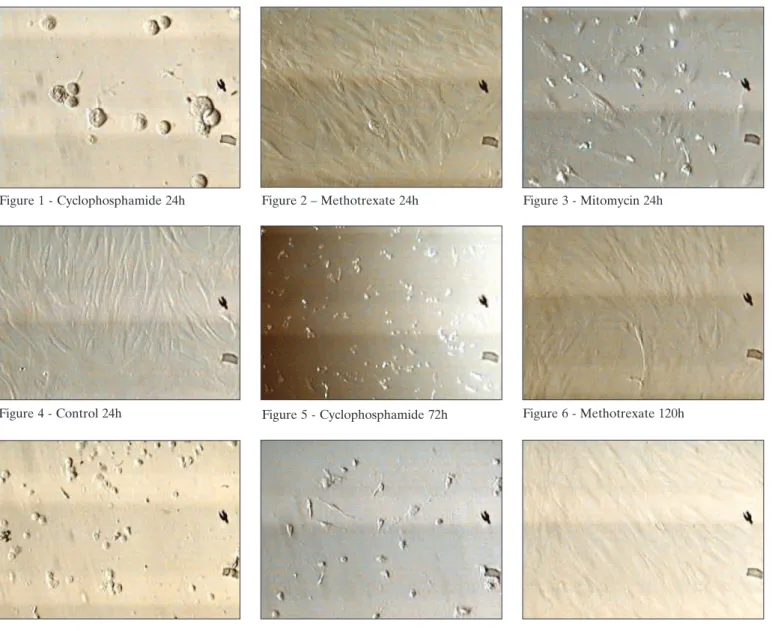
With mitomycin, a significant inhibition of cell proliferation was observed. In the first 24 hours the cells were almost completely detached from the plate. With cyclophosphamide, growth inhibition was also observed. Cells were approximately 50% detached from the plate 24 hours after exposure to the drug and detachment increased in the following days, with cells being almost completely detached after 72 hours. However, the cells showed no apparent degeneration even after 120 hours of culture in the presence of the drug. No effect was observed with methotrexate, even after 120 hours of culture; the cells remained attached to the plates and growth was observed. The same was observed in the control plate, with cell growth and cells attached to the plates. The pH of the plates was measured after 24 hours; plates with mitomycin, methotrexate and cyclophosphamide had a pH around 7.0 (Figures 1-9).
Discussion
Mitomycin C has been widely used either intraoperatively or postoperatively as an adjuvant therapy of pterygium excision to prevent recurrence. Singh et al. reported a recurrence rate of 2.2% after pterygium resection with the bare sclera technique and mitomycin C eye drops, compared with 88.9% in placebo-treated controls(7). Thereafter this antimitotic agent became
more popular and is currently indicated to reduce the postoperative recurrence of pterygium. However, serious visual complications following this approach cause concern among many surgeons. Effects such as conjunctival irritation and complications such as keratitis, necrosis of the sclera with or without
110
Figure 1 - Cyclophosphamide 24h Figure 2 – Methotrexate 24h Figure 3 - Mitomycin 24h
Figure 4 - Control 24h Figure 5 - Cyclophosphamide 72h Figure 6 - Methotrexate 120h
Figure 7 - Cyclophosphamide 72h Figure 8 - Mitomycin 72h Figure 9 - Control 72h
Rev Bras Oftalmol. 2013; 72 (2): 108-11
Almodin J, Almodin F, Almodin E, Minguetti-Câmara VC, Neves JP, Bezzon AKT, Safar CEDM, Amadeu HBB
inflammation (scleromalacia), secondary glaucoma, cataract, corneal oedema and perforation, and calcification of the sclera have been reported(8). Mitomycin C is a radiomimetic agent that
can cause avascular necrosis similar to beta irradiation. In 1992, Rubinfeld et al. reported 10 cases of serious complications related to topical mitomycin C after pterygium surgery using the bare sclera technique. This technique predisposes the eye to the avascular effects of mitomycin, which can lead to corrosion of the sclera, but it is still the most used technique, since it is the easiest to perform(9).
The search for greater safety in the use of mitomycin C has led to increasingly lower concentrations of the drug, which are still effective in reducing recurrence. However, a case was recently reported where a low dose (0.02%) of mitomycin C, used for only 3 min, led to perforation of the cornea and sclera(10).
Mitomycin has been widely used, but some precautions are needed. The drug should not come into contact with de-epithelised areas, and the sclera should not be left exposed after its use. When mitomycin comes into contact with areas with corneal epithelial defects, such as those produced during pterygium removal, regeneration is delayed(11). Excessive cauterisation of
the sclera should be avoided, and the resected area should not be left exposed at the end of the surgical procedure. Scleral damage due to delamination and excessive cauterisation, as well as the vessel occlusion effect of mitomycin C and tear film instability, may predispose to thinning and necrosis of the sclera in these areas(11-13). Also, mitomycin C should be avoided in the
elderly and in patients with atrophic pterygium, where the chance of recurrence is low. The drug should not to be used in dry eyes or in eyes with surface changes(9).
Beta irradiation also reduces recurrence rates to 5-33%, but it is also associated with serious complications(14). Because
of the unsatisfactory results with mitomycin and other adjuvant therapies, we decided to investigate other drugs that could help reduce the recurrence of pterygium. Since recurrence does not seem to be associated with exposure to ultraviolet light but to rapid fibroblast proliferation produced by the surgical trauma(15),
we decided to conduct in vitro experiments to assess pterygium fibroblast growth inhibition with certain drugs. These drugs, which are used in the treatment of cancer, were chosen because they are not vesicant(16), as we believe that the unwanted ocular effects
111
Corresponding author:
Juliana Almodin
Rua Silva Jardim, 359 Maringá (PR), Brazil
Cep 87013-000
Tel: +5544 3262 2061
e-mail: [email protected]
Rev Bras Oftalmol. 2013; 72 (2): 108-11 Effects of certain drugs on the in vitro proliferation of fibroblasts in primary pterygium
are those that cause severe irritation with blistering and tissue destruction when infiltrated outside blood vessels, and they may cause necrosis.
The inhibition of pterygium fibroblast proliferation with mitomycin in our experiments was already expected, but cyclophosphamide also showed to be very effective. Cyclophosphamide belongs to the group of chloroethylamines and is a bifunctional alkylating agent with no specificity for any phase of the cell cycle(17). On the other hand, mitomycin is an
alkylating antimetabolite agent which binds to the DNA causing anomalous bonds and breaks in its structure, inhibiting mitosis and protein synthesis, and leading to cell death(18). The inhibitory
action of cyclophosphamide on the in vitro proliferation of fibroblasts suggests that it could be used to prevent the recurrence of pterygium after its removal. In 1983 mitomycin C was first described as a potent fibroblast inhibitor in filtration surgery. Since then, several authors have reported excellent results with its intraoperative use, especially in refractory glaucoma(19). This
suggests that cyclophosphamide could also be used in filtration surgery as a substitute to mitomycin. Cyclophosphamide is a cyclic phosphamide ester of mechlorethamine which prevents cell division primarily by crosslinking DNA strands. It was used at larger doses than mitomycin C, since the concentrations used in this study were the same as those indicated for cancer, where the dose of mitomycin is always much lower than cyclophosphamide.
The observed effect of cyclophosphamide on cell proliferation was not seen with methotrexate, showing that not every antimitotic drug affects pterygium growth. It is still unclear whether cyclophosphamide will produce any adverse effects that contraindicate its use for the prevention of pterygium recurrence or even for filtration surgery. To clarify this issue, tests in animals and later in humans are needed.
R
EFERENCES1. Rossi EE. Broetto D, Grumann Júnior A. Análise dos pterígios operados no Hospital Regional de São José. Rev Bras Oftalmol. 2003;62(1):44-9.
2. Ferraz FHS, Schellini SA, Hoyama E, Bernardes SR, Padovani CR. Pterígio e alterações da curvatura corneana. Arq Bras Oftalmol. 2002;65(5):533-6.
3. Alves RA, Potério MB, Potério CB, Cardillo JA, José NK. Pterígio: terapêutica adjuvante. www.hospvirt.org.br
4. Samahá JT, Schellini SA, Sakamoto RH, Padovani CR. Tratamento do pterígio recidivado por transplante autólogo da conjuntiva. Arq Bras Oftalmol. 2002;65(4):415-8.
5. Fairbanks D, Vieira LA, Santos WD, Attie GCG, Gomes JAP, Freitas D. Membrana amniótica no tratamento dos afinamentos corneais e esclerais. Arq Bras Oftalmol. 2003;66(1):71-6.
6. Kria L, Ohira A, Amemiya T. TNP-470 (a fungus-derived inhibitor ofangiogenesis) reduces proliferation of cultured fibroblasts isolated from primarypterygia: a possible drug therapy for pterygia. Curr Eye Res. 1998;17(10):986-93.
7. Singh G, Wilson MR, Foster CS. Mitomycin eye drops as treatment for pterygium. Ophthalmology. 1998;95(6):813-21.
8. Chen S, Noonan C. Scleral dellen complicating primary pterygium excision. Eye (Lond). 2000;14(Pt 1):100-1.
9. Rubinfeld RS, Pfister RR, Stein RM, Foster CS, Martin NF, Stoleru S, et al. Serious complications of topical mitomycin-C after pterygium sur-gery. Ophthalmology. 1992;99(11):1647-54. Comment in Ophthalmol-ogy. 1993;100(7):976; author reply 977-8. OphthalmolOphthalmol-ogy. 1992;99(11):1645-6. Ophthalmology. 1993;100(3):292-3. Ophthalmol-ogy. 1993;100(7):976-7; author reply 977-8.
10. Dadeya S, Fatima S. Comeoscleral perforation after pterygium exci-sion and intraoperative mitomycin C. Ophthalmic Surg Lasers Imag-ing. 2003;34(2):146-8.
11. Alves MR, Potério MB, Cardillo JA. Nova técnica cirúrgica para ressecção de pterígio em associação com o uso intra-operatório de mitomicina C. Rev Bras Oftalmol. 1997;56(6):441-3.
12. Alves MR, Saldiva PHN, Lemos M, José NK. Efeitos do uso tópico da mitomicina C no epitélio corneano de coelhas: análise histopatológica pela morfometria. Arq Bras Oftalmol. 1996;59(5):431-7.
13. Potério MB, Alves MR, Cardillo JA, José NK. An improved surgical technique for pterygium excision with intraoperative application of mitomycin C. Ophthalmic Surg Lasers. 1998;29(8):685-7.
14. Haik GM, Ellis GS, Nowell JF. The management of pterygia, with special reference to surgery combined with beta irradiation. Trans Am Acad Ophthalmol Otolaryngol. 1962;66:776-84.
15. Cameron ME. Histology of pterygium: an electron microscopic study. Br J Ophthalmol. 1983;67(9):604-8.
16. Goodman LS. Goodman & Gilman as bases farmacológicas da terapêutica. 9a ed. Rio de Janeiro: McGraw-Hill; c1996.
17. Arencibia DF, Vidal A, Rosario LA, Suárez YE, Delgado L. Biomodelos para la inducción de micronúcleos en células de la medula ósea por ciclofosfamida y bleomicina. Vaccimonitor. 2011;20(1):28-33. 18. Cronemberger S, Santos DV, Ramos LF, Oliveira ACM, Maestrini HA,
Calixto N. Trabeculectomia com mitomocina C em pacientes com glaucoma congênito refratário. Arq Bra. Oftalmol. 2004;67(3):475-9. 19. Costa VP, Vasconcellos JP, Comegno PE, José NK. O uso da mitomicina