Bruno Felipe Gaia(a) Marcelo Augusto Oliveira de Sales(b)
Andréia Perrella(c) Marlene Fenyo-Pereira(d) Marcelo Gusmão Paraíso Cavalcanti(d)
(a)Stomatology Department, Dental School,
University of São Paulo, São Paulo, SP, Brazil.
(b)Department of Radiology, Dental School,
University of Paraíba, João Pessoa, PB, Brazil.
(c)Private practice, São Paulo, SP, Brazil. (d)Department of Radiology, Dental School,
University of São Paulo, São Paulo, SP, Brazil.
Corresponding author:
Marcelo Gusmão Paraíso Cavalcanti E-mail: [email protected]
Received for publication on Apr 19, 2011 Accepted for publication on Jun 09, 2011
Comparison between cone-beam and
multislice computed tomography for
identification of simulated bone lesions
Abstract: There are many studies that compare the accuracy of mul-tislice (MSCT) and cone beam (CBCT) computed tomography for evalu-ations in the maxillofacial region. However, further studies comparing both acquisition techniques for the evaluation of simulated mandibular bone lesions are needed. The aim of this study was to compare the ac-curacy of MSCT and CBCT in the diagnosis of simulated mandibular bone lesions by means of cross sectional images and axial/MPR slices. Lesions with different dimensions, shape and locularity were produced in 15 dry mandibles. The images were obtained following the cross sec-tional and axial/MPR (Multiplanar Reconstruction) imaging protocols and were interpreted independently. CBCT and MSCT showed similar results in depicting the percentage of cortical bone involvement, with great sensitivity and speciicity (p < 0.005). There were no signiicant in-tra- or inter-examiner differences between axial/MPR images and cross sectional images with regard to sensitivity and speciicity. CBCT showed results similar to those of MSCT for the identiication of the number of simulated bone lesions. Cross sectional slices and axial/MPR images pre-sented high accuracy, proving useful for bone lesion diagnosis.
Descriptors: Cone-Beam Computed Tomography; Tomography, Spiral Computed; Mandible.
Introduction
Rapid development of computer technology, improved performance, accessibility and lower cost have pathed the way for digital and advanced imaging modalities. Computed tomography (CT) images provide impor-tant information regarding the individual characteristics of pathological lesions, information which is particularly useful for diagnoses and treat-ments.1 The introduction of multislice computed tomography (MSCT)
represented a fundamental evolutionary step in the development and on-going reinement of CT imaging techniques.2 A single MSCT scan can
yield multiple, thin, overlapping slices that can be rapidly reconstructed, resulting in higher-quality reconstructed images and precluding the need for further patient radiation exposure.3
Cone-beam computed tomography (CBCT) stands out as an alter-native to MSCT. This recently-designed technology became a relevant tool for oral and maxillofacial diagnostic osseous imaging, providing to professionals access to excellent image quality and greater diagnostic ac-Declaration of Interests: The authors
curacy and sensitivity.4,5 In addition, CBCT allows
images to be acquired using a low dose of radiation, shorter patient examination time, and lower costs than MSCT, which makes its routine use feasible for oral and maxillofacial procedures.6,7 It has been
demonstrated that MSCT and CBCT are useful for evaluations in the oral and maxillofacial regions,8-11
although further studies are necessary comparing both acquisition techniques for the evaluation of bone lesions.
Several studies have reported that the interpre-tation allowed by different CT imaging modalities supply more information than axial images, aiding in achieving more reliable diagnosis and effective therapeutics. Furthermore, multiplanar reconstruc-tion (MPR) and cross secreconstruc-tional images provide improvements in the diagnosis, rehabilitation and evaluation of bone pathological processes in the maxillofacial complex.12-14
The purpose of this research was to compare the accuracy of MSCT and CBCT in the diagnosis of simulated mandibular bone lesions by means of cross sectional images and axial/MPR slices.
Methodology
The present study was submitted to and ap-proved by the Committee of Ethics and Research of our Institution, under protocol # 151/2003.
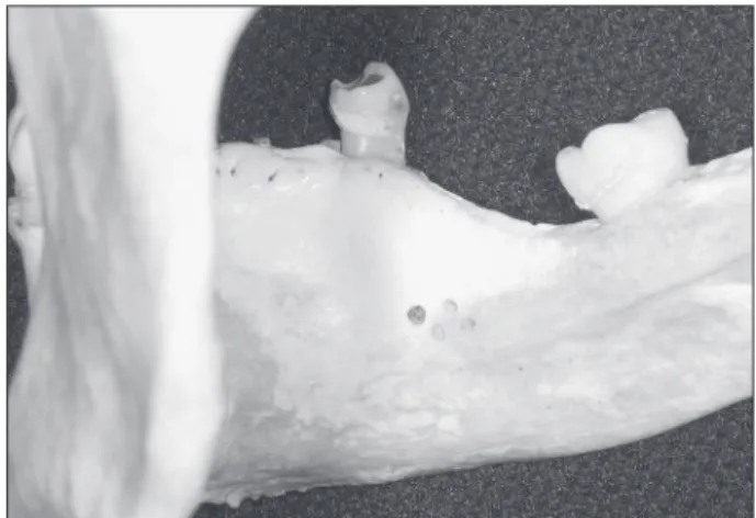
A total of 15 dry mandibles were examined. Le-sions involving cortical bone were produced using a round bur (H1, number 8, Komet Brasil, Santo André, Brazil) and a high-speed handpiece (Silent MS 350PB, Dabi Atlante, Ribeirão Preto, Brazil). The lesions, which differed in dimension and shape, were produced either in the buccal cortical bone or in the lingual cortical bone of the mandibular body. In some cases, the bur just touched the cortical bone, whereas in others, it perforated the medullary bone. A total of 52 perforations were made, ranging in diameter from 1 mm to 3 mm and in depth from 0.5 mm to 3.0 mm. In 7 mandibles, unilocular le-sions were produced and, in 8 mandibles, multilocu-lar lesions (having 3 to 9 loculi each) were produced on the lingual surface of the body of the mandible. In 2 of these mandibles, the buccal cortical bone of the mandibular body was perforated (Figure 1).
The mandibles were submitted to a CBCT scan-ner (i-CAT Cone Beam 3-D Dental Imaging System; Imaging Sciences International, Hatield, USA) with the following parameters: voxel size of 0.25 mm; raw data acquisition of 40 s; exposure settings of 90 kVp and 7 mA; and a display ield of view of 15 cm. Subsequently, MSCT was performed (Aquil-ion 64; Toshiba Medical Systems, Tustin, USA) with the following parameters: slice thickness of 0.5 mm; reconstruction interval of 0.3 mm; exposure time of 0.4 s (120 kVp, 300 mA and 512 × 512 pixel ma-trix); bone tissue ilter; and a ield of view of 18 cm.
For image acquisition, the specimens were placed in a plastic bucket, completely covered with water (in order to simulate soft tissue) and maintained in the same position as that used in in vivo studies (us-ing cotton sheets for support). Axial slices were ac-quired, the specimens being scanned from the man-dibular base to the condyle. The scanning plane was parallel to the mandibular base. Gantry angles var-ied according to mandibular base angles.
The original data were sent to a workstation. The association of MPR and axial images (with simulta-neous analysis of coronal and sagittal views) were displayed and analyzed using Vitrea software, ver-sion 3.4.5 (Vital Images Inc., Plymouth, USA), and the cross sectional images were analyzed using Im-aging Studio software, version 2.556 (Anne Solu-tions, São Paulo, Brazil). Both protocols were inter-preted independently by two experienced examiners (oral and maxillofacial radiologists). The analyses of
the images were performed in a random order of the protocols, in different sessions (with an interval of at least two weeks between sessions). The examiners had no contact with the specimens and were blinded to the acquisition technique used and to the charac-teristics of the lesions in each mandible.
The examiners were asked to identify the differ-ent characteristics of the lesions (whether the cor-tical bone had been perforated and the number of lesions in each mandible) in each protocol. The per-forations in the dry mandibles were considered the gold standard.
During the analysis of the images, only the pro-tocol images were displayed on the computer moni-tor in order not to inluence the interpretation (Fig-ures 2 and 3).
Intra- and inter-examiner reliability was cal-culated using the kappa statistic, the validity test (sensitivity and speciicity) and the chi-square test. The validity test is represented by the Youden index, which attempts to represent test accuracy by a single numerical value (sensitivity + speciicity - 1). The program Statistical Package for the Social Scienc-es, version 12.0 for Windows (SPSS Inc., Chicago, USA) was used. A 95% (p < 0.05) conidence
inter-val was used.
Results
Results regarding the detection of the number of lesions showed high agreement between examiners, as shown in Table 1.
The number of bone lesions was related to the in-terpretation criteria used for the detection of simu-lated bone lesions (absence, unilocular or multilocu-lar lesion), according to the protocol used and the gold standard.
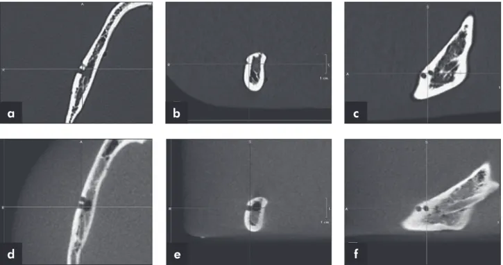
Figure 2 - MSCT (a, b, c) and CBCT (d, e, f) axial/MPR images.
Figure 3 - MSCT (a) and CBCT (b) cross sectional images.
a
a
d
b
b
e
c
The data collected showed that both examiners were able to identify simulated mandibular lesions using MSCT and CBCT following both protocols, with no statistical difference between them (over 95% of the lesions were identiied) (Table 1). The analyses involving the protocols (axial/MPR and cross sectional slices) were highly effective in aiding both examiners in identifying the number of lesions.
The kappa statistic was used to assess agree-ment between examiners. In the sample as a whole, a kappa value of 0.869 was found between MSCT and CBCT in the overall evaluation. In addition, the Kappa statistic was used for an individual evalua-tion of the protocols, comparing the results obtained by examiner 1, examiner 2, and the gold standard (Table 2).
The protocols were statistically signiicant for the intra- and inter-examiner analyses regarding the number of lesions, which proved the validity of the methods.
As regards the sensitivity and speciicity in de-tecting the number of mandibular lesions, the asso-ciation between axial slices and MPR showed better results than the cross sectional images, with no sig-niicant intra- or inter-examiner differences (Table 3).
MSCT and CBCT showed excellent results for both protocols, with no signiicant intra- or inter-examiner differences.
Discussion
Imaging of the maxillofacial region is limited with conventional radiography due to the overlap of anatomical structures, making visualization of the area of interest very dificult.8,15 MSCT and CBCT
represent an important advance because, using these methods, observers can reconstruct and manipulate high resolution images, thus improving the diagno-sis.15,16
Both modalities of image acquisition have advan-tages and disadvanadvan-tages regarding radiation dose,
Table 1 - Frequency of detection of simulated bone lesions.
Examiner 1 Examiner 2 Gold Standard Absence 348 351 380 Unilocular 317 309 290 Multilocular 235 240 230 Total 900 900 900
Number of cavities
Examiner 1 X Examiner 2 Examiner 1 X Gold Standard Examiner 2 X Gold Standard Kappa p Kappa p Kappa p MSCT
MPR/axial 0.935 < 0.005 0.951 < 0.005 0.886 < 0.005 MSCT
parasagittal 0.838 < 0.005 0.855 < 0.005 0.886 < 0.005 CBCT
MPR/axial 0.952 < 0.005 0.935 < 0.005 0.951 < 0.005 CBCT
parasagittal 0.707 < 0.005 0.803 < 0.005 0.886 < 0.005
Table 2 - Kappa and p values for the comparison between examiner 1, examiner 2, and gold standard for the identification of the number of simulated bone lesions.
Examiner 1 MSCT MPR/axial MSCT parasagittal CBCT MPR/axial CBCT parasagittal Sensitivity 92.6% 86.3% 92% 86.6% Specificity 94.5% 94.5% 90.4% 97.7% Examiner 2 MSCT MPR/axial MSCT parasagittal CBCT MPR/axial CBCT parasagittal
Sensitivity 95% 88% 98% 86.3% Specificity 96.2% 100% 97.4% 98.1%
acquisition time, cost, scattered radiation and arti-facts.6,7 Drawbacks should be taken into
consider-ation, since they can inluence image quality and interpretation accuracy. The applicability of CT im-ages has been broadly shown for several purposes such as craniometric measurements and anatomical identiication, forensic identiication, diagnosis and surgical planning of fractures, and maxillofacial im-plants.7,17 However, few studies have compared the
accuracy of MSCT and CBCT for the identiication of mandibular lesions, indicating the need for con-ducting new studies.
Simulated bone lesions have been widely used to compare radiological techniques for bone observa-tion.16 Pinsky et al.18 used simulated lesions to test
linear measurements in CBCT, but they made 4-8-mm defects, which are larger than those prepared in the present work. In these experiments, water was added to produce an environment closer to bone in vivo. This methodological procedure was the same used in the present experiment to attenuate the x-ray. This study used mechanical pseudo-lesions that are not radiographically identical to those developed naturally; simulated lesions were chosen in order to have a deined pattern to allow a comparison of the image acquisition modalities and the observation protocols, in order to approximate our indings to a true pathological bone lesion condition. The valid-ity of the methods was conirmed when the results obtained by examiners 1 and 2 were compared with the gold standard (Kappa > 0.869) for identiication of the number of simulated bone lesions.
Perrella et al.13 showed that the techniques based
on helical acquisition were recognizably capable of providing high sensitivity and speciicity in the diag-nosis of simulated mandibular bone lesions with and without metallic artifacts. Mischkowski et al.19 and
Dreiseidler et al.20 reported that the diagnostic value
was the same between CBCT and MSCT images, both methods providing good visualization of vari-ous mandibular anatomical structures. Our study corroborated to the results of these authors in that both modalities were equally capable of detecting the presence of simulated mandibular bone lesions, showing high speciicity and sensitivity values.
The importance of acquisition parameters, such
as slice thickness, in bone lesion evaluation were discussed previously by many authors.21-23 Shaha23
stated that for detailed evaluation of the mandible it is essential to obtain CT scans with bone windows and narrow cuts, and the accuracy found in his work was 68%. According to Baxter and Sorenson21,
identiication of the number of lesions was inaccu-rate when the diameter was comparable to or less than the CT slice thickness, not allowing a correct identiication of bone lesions. Furthermore, Caval-canti et al.22 demonstrated high false positive and
false negative rates when determining mandibular bone invasion using 3-mm-thick axial slices. In the present work, using a 0.5 mm slice thickness with a thinner interval of reconstruction (0.3 mm) for MSCT and 0.25 mm voxel size for CBCT, 90.4% and 90.7% of sensitivity and 96.3% and 95.9% of speciicity (median values) were found regarding the number of simulated lesions respectively.
CT technology used for image manipulation and visualization of structures in multiple planes provides better identiication of anatomical struc-tures in the axial, coronal and sagittal planes, thus increasing image interpretation. The evaluation of complex diseases and conditions in the maxillofa-cial region requires more accurate image exams as-sociated with multiplanar analysis for a complete delimitation and identiication of the pathological process and a better understanding of the disease behavior.24 Currently, MPR and cross sectional
images associated with tridimensional (3D) recon-struction represent the most useful imaging modali-ties for diagnoses and surgical planning in the oral and maxillofacial regions.13,14,16,24 Cross sectional
images, extensively used for the planning of dental implants, were obtained from original CT images which were directed perpendicularly to a plane pre-viously outlined, and depicted the anatomy better than straight sagittal planes, justifying the choice of that protocol in our research.
Utumi et al.14 Perrella et al.16 found differences
of accuracy regarding mandibular lesions when they used, respectively, different post-processing images and different slice thicknesses from helical CT. Warnke et al.25 evaluated the applicability of a
and quantitative evaluation of the temporomandibu-lar joint in the sagittal and coronal planes. Those authors observed that the combination of different image visualization planes provided more accu-rate images than conventional CT. In their study, 100% of the pathological bone alterations were ob-served. Additionally, the use of reduced thicknesses in MSCT and smaller voxels in CBCT, as our work did, allows greater sensitivity and speciicity, which corroborates previous studies.
Cara et al.15 compared the validity of different
single- and multislice CT for analyses of simulated lesions in the head of the mandible. Sensitivity re-sults were: axial single slice (62.7%), axial multislice (66.2%), axial/MPR single slice (72.7%), axial/ MPR multislice (93.1%). The association of axial images with MPR, using MSCT scans, demonstrat-ed a higher accuracy than the single slice method. The present study determined the validity of images acquired by using MSCT and CBCT with different protocols (axial/MPR images and cross sectional slices). Our results showed that both observation protocols had signiicant sensitivity and speciicity results in the detection of simulated bone lesions for both examiners compared to the gold standard, with no signiicant statistical difference (Table 3).
The association of CT protocols for visualization of simulated bone lesions was established with the aim of improving the visualization of the presence of mandibular bone alterations. Despite the high values of speciicity and sensitivity found, cross sec-tional slices should be used together with the
visu-alization of axial, and MPR images. These simul-taneous image analyses improve interpretation and prevent a misleading visualization due to laws in the process of image reconstruction. It is our opin-ion that MSCT and CBCT may improve the results of early detection of bone lesions in vivo, since good sensitivity and speciicity rates were obtained, even with tiny simulated lesions.
Conclusion
CBCT may be considered a valuable imaging tool for the identiication of simulated bone lesions, showing results similar to those of MSCT. The cross sectional slices and axial/MPR images were highly accurate in the identiication of simulated mandibu-lar bone lesions (cortical bone destruction and num-ber of the lesions in each mandible) proving to be useful for bone lesion diagnosis.
Acknowledgements
The authors would like to thank the São Paulo Research Foundation (FAPESP) for the inancial support provided through grant no. 2005/02157-8 and PhD grant no. 2006/05251-8, and the Coordi-nation for the Advancement of Higher Education Personnel (CAPES) for the inancial support pro-vided through a PhD grant. The authors are also grateful to Odonto X (Dr. Reinaldo Rosa), Rio de Janeiro, Brazil, where the CBCT images were pro-duced, and to the Department of Anatomy of the Gama Filho University School of Medicine, Rio de Janeiro, Brazil, for providing the dry mandibles.
References
1. Ozen T, Kamburog˘lu K, Cebeci AR, Yüksel SP, Paksoy CS. Interpretation of chemically created periapical lesions us-ing 2 different dental cone-beam computerized tomography units, an intraoral digital sensor, and conventional film. Oral Surg Oral Med Oral Pathol Oral Radiol Endod. 2009 Mar;107(3):426-32.
2. Mahesh M. Search for isotropic resolution in CT from con-ventional through multiple-row detector. Radiographics. 2002 Jul-Aug;22(4):949-62.
3. Cavalcanti MG, Ruprecht A, Vannier MW. 3D volume render-ing usrender-ing multislice CT for dental implants. Dentomaxillofac Radiol. 2002 Jul;31(4):218-23.
4. Guerrero ME, Jacobs R, Loubele M, Schutyser F, Suetens P, van Steenberghe D. State-of-the-art on cone beam CT imag-ing for preoperative plannimag-ing of implant placement. Clin Oral Investig. 2006 Mar;10(1):1-7.
5. Scarfe WC, Farman AG, Sukovic P. Clinical applications of cone-beam computed tomography in dental practice. J Can Dent Assoc. 2006 Feb;72(1):75-80.
7. Hassan B, van der Stelt P, Sanderink G. Accuracy of three-dimensional measurements obtained from cone beam com-puted tomography surface-rendered images for cephalometric analysis: influence of patient scanning position. Eur J Orthod. 2009 Apr;31(2):129-34.
8. Bernaerts A, Vanhoenacker FM, Hintjens J, Chapelle K, De Schepper AM. Imaging approach for differential diagnosis of jaw lesions: a quick reference guide. JBR-BTR. 2006 Jan-Feb;89(1):43-6.
9. Ludlow JB, Davies-Ludlow LE, Brooks SL, Howerton WB. Dosimetry of 3 CBCT devices for oral and maxillofacial radi-ology: CB Mercuray, New Tom 3G and i-CAT. Dentomaxil-lofac Radiol. 2006 Jul;35(4):219-26.
10. Cavalcanti MG, Haller JW, Vannier MW. Three-dimensional computed tomography landmark measurement in craniofacial surgical planning: experimental validation in vitro. J Oral Maxillofac Surg. 1999 Jun;57(6):690-4.
11. Schulze D, Blessmann M, Pohlenz P, Wagner KW, Heiland M. Diagnostic criteria for the detection of mandibular osteomyeli-tis using cone beam computed tomography. Dentomaxillofac Radiol. 2006 Jul;35(4):232-5.
12. Suomalainen A, Vehmas T, Kortesniemi M, Robinson S, Pel-tola J. Accuracy of linear measurements using dental cone beam and conventional multislice computed tomography. Dentomaxillofac Radiol. 2008 Jan;37(1):10-7.
13. Perrella A, Lopes PM, Rocha RG, Fenyo-Pereira M, Cavalcanti MG. Influence of dental metallic artifact from multislice CT in the assessment of simulated mandibular lesions. J Appl Oral Sci. 2010 Mar-Apr;18(2):149-54.
14. Utumi ER, Perrella A, Albuquerque MA, Adde CA, Rocha RG, Cavalcanti MG. Evaluation of simulated bone lesion in the head of the mandible by using multislice computed tomog-raphy. J Appl Oral Sci. 2009 Sep-Oct;17(5):521-6.
15. Cara AC, Gaia BF, Perrella A, Oliveira JX, Lopes PM, Ca-valcanti MG. Validity of single- and multislice CT for assess-ment of mandibular condyle lesions. Dentomaxillofac Radiol. 2007 Jan;36(1):24-7.
16. Perrella A, Borsatti MA, Tortamano IP, Rocha RG, Caval-canti MG. Validation of computed tomography protocols for simulated mandibular lesions: a comparison study. Braz Oral Res. 2007 Apr-Jun;21(2):165-9.
17. Naitoh M, Nakahara K, Suenaga Y, Gotoh K, Kondo S, Ariji E. Comparison between cone-beam and multislice computed tomography depicting mandibular neurovascular canal struc-tures. Oral Surg Oral Med Oral Pathol Oral Radiol Endod. 2010 Jan;109(1):e25-31.
18. Pinsky HM, Dyda S, Pinsky RW, Misch KA, Sarment DP. Ac-curacy of three-dimensional measurements using cone-beam CT. Dentomaxillofac Radiol. 2006 Nov;35(6):410-6. 19. Mischkowski RA, Scherer P, Ritter L, Neugebauer J, Keeve
E, Zöeller JE. Diagnostic quality of multiplanar reformations obtained with a newly developed cone beam device for maxil-lofacial imaging. Dentomaxillofac Radiol. 2008 Jan;37(1):1-9. 20. Dreiseidler T, Mischkowski RA, Neugebauer J, Ritter L, Zöel-ler JE. Comparison of cone-beam imaging with orthopanto-mography and computerized toorthopanto-mography for assessment in presurgical implant dentistry. Int J Oral Maxillofac Implants. 2009 Mar-Apr;24(2):216-25.
21. Baxter BS, Sorenson JA. Factors affecting the measurement of size and CT number in computed tomography. Invest Radiol. 1981 Jul-Aug;16(4):337-41.
22. Cavalcanti MGP, Santos DT, Perrella A, Vannier MW. CT- based analysis of malign tumor volume and localization. A preliminary study. Braz Oral Res. 2004 Oct-Dec;18(4):338-44. 23. Shaha AR. Preoperative evaluation of the mandible in patients
with carcinoma of the floor of the mouth. Head Neck. 1991 Sep-Oct;13(5):398-402.
24. Ahmad M, Freymiller E. Cone Beam Computed Tomography: Evaluation of Maxillofacial Pathology. J Calif Dent Assoc. 2010 Jan;38(1):41-7.