Effects of chronic heart disease on
skeletal muscle fiber size
1Departamento de Biomecânica, Medicina e Reabilitação do Aparelho Locomotor, Faculdade de Medicina de Ribeirão Preto, Universidade de São Paulo,
Ribeirão Preto, SP, Brasil
2Departamento de Patologia, Faculdade de Medicina,
Universidade Federal do Rio de Janeiro, Rio de Janeiro, RJ, Brasil 3Departamento de Estatística, Universidade Federal de São Carlos, São Carlos, SP, Brasil
A.C. Mattiello-Sverzut1, L. Chimelli2, S. Teixeira1, M. Pierre3 and L. Oliveira3
Abstract
Size changes in muscle fibers of subjects with chronic heart disease (CHD) have been reported, although a consensus has not been achieved. The aims of the present study were to investigate a possible association between CHD and fiber size changes in the brachial biceps compared to subjects without heart disease. Forty-six muscle samples were obtained in autopsies of individuals (13 to 84 years) without neuro-muscular disorders, 19 (10 males and 9 females) with, and 27 (14 males and 13 females) without CHD. In all cases muscle sections were stained with hematoxylin and eosin and processed for the visualiza-tion of myofibrillar ATPase activity. The lesser diameter of type 1 and type 2 fibers was obtained tracing their outlines (at least 150 fibers of each type per sample) onto an image analyzer connected to a com-puter. The results were analyzed statistically comparing males and females with and without CHD. Type 1 fiber mean lesser diameters were 51.51 and 54.52 µm in males (normal range 34-71 µm) and 45.65 and 55.42 µm in females (normal range 34-65 µm) without and with CHD, respectively; type 2 fibers measured 54.31, 58.23, 41.15, and 49.57 µm, respectively (normal range 36-79 µm for males and 32-59 µm for females). No significant difference in fiber size was detected in 24 males with and without CHD, while in 22 females there was a significant increase in size in those with cardiomyopathy. We con-cluded that CHD does not determine significant changes in fiber size. However, in females, there is some hypertrophy which, despite within normal range, may reflect morphologic heterogeneity of the sample, or the daily life activities in the upper limbs as a compensatory mechan-ism to fatigability that affect predominantly the lower limbs in sub-jects with CHD.
Correspondence
L. Chimelli
Serviço de Anatomia Patológica Hospital Universitário, UFRJ 21941-590 Rio de Janeiro, RJ Brasil
Fax: +55-21-526-2450 E-mail: chimelli@hucff.ufrj.br
Publication supported by FAPESP.
Received August 1, 2003 Accepted August 31, 2004
Key words
•Morphometry •Brachial biceps •Chronic heart disease •Post mortem muscle •Type 1 fiber hypertrophy •Type 2 fiber hypertrophy
Introduction
In 1972, Shafiq et al. (1) reported a histo-pathological study of skeletal muscle in pa-tients with heart disease. After this first
is type 1 and/or 2 fiber atrophy (9-13) or whether there exists any degree of fiber atro-phy (14). The aim of the present study was to compare the size of skeletal muscle fibers obtained at autopsy from subjects with CHD to the size of fibers obtained from patients without heart disease.
Subjects and Methods
Muscle samples were obtained at au-topsy, 3 to 9 h after death, from the belly of the brachial biceps of 46 subjects (22 fe-males and 24 fe-males) ranging in age from 13 to 84 years (mean female age: 45.6 ± 19 years; mean male age: 51.8 ± 18.1 years). Nineteen samples (10 males, mean age 51.8 ± 18.8, and 9 females, mean age 64.2 ± 10) were from subjects with chronic cardiomy-opathy confirmed at autopsy. Twenty-seven samples (14 males, mean age 52.8 ± 16.4, and 13 females, mean age 32.5 ± 11) were from subjects who had suffered sudden death or who had died after a short acute illness, without involvement of the neuromuscular system, as confirmed morphologically. In all cases, the muscle samples were immediately immersed in liquid nitrogen and oriented in order to obtain transverse sections, which were cut into 10-µm thick sections with a cryostat at -20ºC. Each section was placed on a glass coverslip, stained with hematoxy-lin and eosin and processed for the visualiza-tion of myofibrillar ATPase activity at pH 9.6, 4.6 and 4.3. The fiber outlines were traced onto an Image Analyzer Kontron, KS300, Carl Zeiss (Oberkochen, Germany) connected to an IBM-PC computer. In each muscle sample, at least 150 fibers of each type were measured to obtain the lesser di-ameter (type 1 fiber lesser didi-ameter, and type 2 fiber lesser diameter). To facilitate the presentation of the results, the morphomet-ric data were divided into four subgroups according to sex and presence or absence of CHD.
The Ethics Committee of the University
Hospital, School of Medicine of Ribeirão Preto, University of São Paulo, approved this study.
Statistical analysis
The results were analyzed statistically by the Levene test to identify variance equality and by the Student t-test to compare classes. The Mann-Whitney test was used to com-pare the male and female subgroups in the groups with and without cardiomyopathy. The SPSS software (version 10.0) was used for statistical analysis.
Results and Discussion
Morphological examinations of hema-toxylin and eosin-stained sections showed no structural changes in muscle fibers, ex-cept for a variation in fiber size, which did not include angulated fibers or atrophy of fascicles. ATPase-stained sections did not show type grouping.
The most frequent cardiac disorder in the group with CHD was hypertensive cardiopa-thy, but there was also cardiac failure associ-ated with Chagas’ disease, cor pulmonale and chronic ischemic myocardiopathy. In most of the patients of the group without CHD the death was related to hypovolemic or septic shock, due to digestive hemorrhage or acute infections (meningitis, pneumonia, intestinal perfuration) respectively; dissect-ing aneurism of the aorta, acute myocardial infarct and pulmonary thrombo-embolism were also causes of death in this group.
significant difference between males and females (P < 0.05).
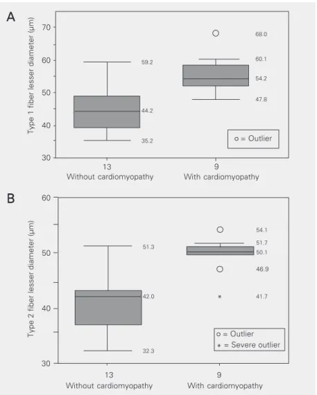
Regarding subjects with CHD, class com-parison (male, autopsy - without cardiomy-opathy, type 1 and type 2 fibers, N = 14, and male, autopsy - chronic cardiomyopathy, type 1 and type 2 fibers, N = 10) showed no significant difference. The results obtained for females showed a statistically significant difference in mean lesser diameter of type 1 and type 2 fibers between subjects with car-diomyopathy (N = 9) and subjects without heart disease (N = 13; P < 0.05; Figure 1A,B), with higher mean values for females with chronic cardiomyopathy.
The mean lesser diameter obtained for all subjects of both sexes was within the normal values established by Brooke and Engel (15), 40-80 µm for males and 30-70 µm for fe-males, using 103 samples, and by Mattiello-Sverzut et al. (16), 34-71 and 34-65 µm for type 1 fibers, and 36-79 and 32-59 µm for type 2 fibers, in males and females, respec-tively, using 72 samples. In males with or without chronic cardiomyopathy, type 2 fi-bers were larger than type 1 fifi-bers, while in females, type 1 fibers were larger than type 2 fibers, as observed by Brooke and Engel (15) in normal muscles. However, among females, subjects with heart disease had a significant-ly larger mean lesser diameter of type 1 and type 2 fibers than subjects without cardio-myopathy. No literature reports are available about the effects of chronic cardiomyopathy on skeletal muscle in females. For males, the data indicate the presence of mild atrophy, predominantly in type 2 fibers of the vastus lateralis, quadriceps and gastrocnemius muscles of patients with chronic cardiomy-opathy (12,15,17). Buller et al. (17) evalu-ated morphologically and functionally the adductor muscle of the thumb and did not find any size abnormality, not even in strength performance, suggesting only an effect of chronic cardiomyopathy on large muscle groups involved in locomotion. In the bra-chial biceps muscle, as studied in our cases,
Table 1. Muscle fiber diameters of males (M) and females (F) with and without chronic cardiomyopathy.
Sex Without Chronic cardiomyopathy cardiomyopathy
Type 1 fiber mean M 51.51 ± 1.62 54.52 ± 3.07 lesser diameter (µm) F 45.65 ± 2.08 55.42 ± 2.04* Type 2 fiber mean M 54.31 ± 3.42 58.23 ± 3.37
lesser diameter (µm) F 41.15 ± 1.46 49.57 ± 1.17*
Data are reported as means ± SD for 19 (10 males and 9 females) with, and 27 (14 males and 13 females) without cardiomyopathy.
*P < 0.05 compared to subjects without cardiomyopathy (Student t-test).
Figure 1. Box plot showing muscle fiber diameters of female autopsy subjects with (N = 9) and without (N = 13) cardiomyopathy. A, Type 1 fibers; B, type 2 fibers. *P < 0.05 compared to subjects without cardiomyopathy (Student t-test and when necessary, Mann-Whitney test). (Fifty percent of the subjects are contained inside the boxes and the central line represents the median. The other 50% are distributed between the box and the horizontal lines that are above and below the box; the numbers inside the panel represent the values obtained during statistical analysis).
Type 1 fiber lesser diameter (µm)
59.2
68.0
60.1
54.2
47.8
44.2
35.2
70
60
50
40
30
Without cardiomyopathy With cardiomyopathy
A
A
A
A
A
42.0
32.3
46.9
54.1
51.7 50.1
With cardiomyopathy
Type 2 fiber lesser diameter (µm)
60
50
40
30
51.3
B
B
B
B
B
Without cardiomyopathy
* 41.7
13 9
13 9
= Outlier
= Outlier
Caforio et al. (10) observed a reduction of type 1 fibers in one third of the muscle samples from patients with chronic dilated or hypertrophic cardiomyopathy, while the remaining ones presented normal values of type 1 and 2 fibers. In experimental models, both muscle fiber types of the sural triceps of dogs (9) and of the plantaris and sural triceps of rats (18) presented a reduced diameter.
The present results do not confirm the existence of morphological or morphomet-ric changes in upper limb muscles of patients with CHD, as previously reported (1-5). Our findings are intriguing and cannot be fully explained. The increase in size of both fiber types in subjects with chronic cardiomyopa-thy, more marked in females, may reflect morphologic heterogeneity of the sample, as suggested by Johnson et al. (19), and tro-phism, as suggested by Shorey and Cleland (20) of the muscles in the human body. This increase may also be determined by effects of daily life on the upper limbs as a
compen-satory mechanism for the fatigability and intolerance to exercise predominantly af-fecting the lower limbs of subjects with chronic cardiomyopathy.
CHD was not associated with significant morphological changes in the brachial bi-ceps muscle of these subjects, although the fibers of females with heart disease were significantly larger than in controls. How-ever, the values obtained were within the normal range established in the literature and in our own observations.
Acknowledgments
We are grateful to CAPES for providing a Doctoral fellowship for A.C. Mattiello-Sverzut, Department of Pathology, School of Medicine of Ribeirão Preto, USP. The authors also thank M.P.M. Scandar, Depart-ment of Pathology, School of Medicine of Ribeirão Preto, USP,for technical assistance.
References
1. Shafiq SA, Sande MA, Carruthers RR, Killip T & Milborat AT (1972). Skeletal muscle in idiopathic cardiomyopathy. Journal of Neurologi-cal Sciences, 15: 303-320.
2. Isaacs H & Muncke G (1975). Idiopathic cardiomyopathy and skel-etal muscle abnormalities. American Heart Journal, 90: 767-773. 3. Dunnigan A, Pierpont ME, Smith SA, Breningstall G, Benditt DG &
Benson DW (1984). Cardiac and skeletal myopathy associated with cardiac dysrhythmias. American Journal of Cardiology, 53: 731-737. 4. Dunnigan A, Staley NA, Smith SA, Pierpont ME, Judd D, Benditt DG & Benson Jr DW (1987). Cardiac and skeletal muscle abnormalities in cardiomyopathy: comparison of patients with ventricular tachy-cardia or congestive heart failure. Journal of the American College of Cardiology, 10: 608-618.
5. Smith ER, Heffernan LP, Sangaland VE, Vaughan LM & Flamington CS (1976). Voluntary muscle involvement in hypertrophic cardiomy-opathy: a study of 11 patients. Annals of Internal Medicine, 85: 566-572.
6. Hootsmans WJM & Meerschwam IS (1971). Electromyography in patients with hypertrophic obstructive cardiomyopathy. Neurology, 21: 810-816.
7. Przybojewski JZ, Hoffman H, de Graaf AS, van der Walt JJ, Tiedt FA, O’Kennedy A, Torrington M, Lochner A & Hewlett R (1981). A study of a family with inherited disease of cardiac and skeletal muscle. Part 1: Clinical electrophysiological and electron micro-scopic studies. South African Medical Journal, 59: 363-373. 8. Lochner A, Hewlett RH, O’Kennedy A, van der Walt JJ, Tiedt FA,
Hoffman H, de Graaf AS, Przybojewski JZ & Torrington M (1981). A
study of a family with inherited disease of cardiac and skeletal muscle. Part 2: Skeletal muscle morphology and mitochondrial oxi-dative phosphorylation. South African Medical Journal, 59: 453-461. 9. Sabbah HN, Hansen-Smith F, Sharov VG, Kono T, Lesch M, Gengo PJ, Steffen RP, Levine TB & Goldstein S (1993). Decreased propor-tion of type I myofibers in skeletal muscle of dogs with chronic heart failure. Circulation, 87: 1729-1737.
10. Caforio ALP, Rossi B, Risaliti R, Siciliano G, Marchetti A, Angelini C, Crea F, Mariani M & Muratorio A (1989). Type 1 fiber abnormalities in skeletal muscle of patients with hypertrophic and dilated cardio-myopathy: evidence of subclinical myogenic myopathy. Journal of the American College of Cardiology, 14: 1464-1473.
11. Howell S, Maarek JM, Founier M, Sullivan K, Zhan W & Sieck G (1995). Congestive heart failure: differential adaptation of the dia-phragm and latissimus dorsi. Journal of Applied Physiology, 79: 389-397.
12. Mancini DM, Coyle E, Coogan A, Beltz J, Ferraro N, Montain S & Wilson JR (1989). Contribution of intrinsic skeletal muscle changes to 31P NMR skeletal muscle metabolic abnormalities in patients
with chronic heart failure. Circulation, 80: 1338-1346.
13. Sullivan MJ, Green HJ & Cobb FR (1990). Skeletal muscle biochem-istry and histology in ambulatory patients with long-term heart failure. Circulation, 81: 518-527.
15. Brooke MH & Engel WK (1969). The histographic analysis of human muscle biopsies with regard to fiber types. 1. Adult male and female. Neurology, 19: 221-234.
16. Mattiello-Sverzut AC, Chimelli L, Moura MSA, Teixeira S & Mello de Oliveira JA (2003). The effects of aging on brachial biceps muscle fiber size. A morphometrical study in biopsies and autopsies. Arqui-vos de Neuropsiquiatria,61: 555-560.
17. Buller NP, Jones D & Poole-Wilson PA (1991). Direct measurement of skeletal muscle fatigue in patients with chronic heart failure.
British Heart Journal, 65: 20-24.
18. Delp MD, Duan C, Mattson JP & Musch TI (1997). Changes in skeletal muscle biochemistry and histology relative to fiber type in rats with heart failure. Journal of Applied Physiology, 83: 1291-1299.
19. Johnson MA, Polgar J, Weightman D & Appleton D (1973). Data on the distribution of fibre types in thirty-six human muscles. An au-topsy study. Journal of the Neurological Sciences, 18: 111-129. 20. Shorey CD & Cleland KW (1988). Morphometric analysis of frozen
transverse sections of human skeletal muscle taken post-mortem.