www.bjorl.org
Brazilian
Journal
of
OTORHINOLARYNGOLOGY
ORIGINAL
ARTICLE
The
influence
of
growth
factors
on
skin
wound
healing
in
rats
夽
Elen
Carolina
David
João
De
Masi
a,∗,
Antonio
Carlos
Ligocki
Campos
a,
Flavia
David
João
De
Masi
b,
Marco
Aurelio
Soatti
Ratti
a,
Isabela
Shin
Ike
a,
Roberta
David
João
De
Masi
caUniversidadeFederaldoParaná(UFPR),Curitiba,PR,Brazil
bPontifíciaUniversidadeCatólicadoParaná(PUCPR),Curitiba,PR,Brazil cUniversidadedaRegiãodeJoinville(Univille),Joinville,SC,Brazil
Received27August2015;accepted3September2015
Availableonline7January2016
KEYWORDS Woundsinrats; Growthfactor; Healing
Abstract
Introduction:Healingisaprocessthatrestoresthephysicalintegrityofbodystructures.Itisa dynamic,complex,multicellularprocessthatinvolvestheextracellularmatrix,cytokines,blood cells,andgrowthfactors.Growthfactorsareproteinsthatactivateandstimulatecell prolifer-ationthroughtheactivationofangiogenesis,mitogenesis,andgenetranscription,accelerating thehealingprocess.
Objective:Toassesstheinfluenceofgrowthfactorsonthehealingprocessofwoundsmadeon thebacksoffemaleratscomparedtothecontrolwound,throughmacroandmicroscopy.
Methods:Thisstudyused45femaleWistarrats,inwhichthreewoundsweremadeontheback. Thefirstwasthecontrolwound,thesecondreceivedepithelialgrowthfactorinjection,and thethirdreceivedacombination offactors. Macroscopicandmicroscopic assessmentswere performedon thethird, seventh,and 15thdays ofthe experiment.For microscopic analy-sis,hematoxylin---eosinstainingwasutilizedtoassesstheinflammatoryprocess;vimentin,for assessmentofbloodvesselsandfibroblasts,andSiriusRedforcollagenassessment.
Results:Inthemacroscopicassessment, theuseofgrowthfactorsresultedinfasterhealing anddecrease ofgranulationtissue ondays seven and15; (80.31%reduction inthecontrol woundvs.83.24%intheepithelialwoundvs.100%inthemixedwound).Utilizingmicroscopy, atthethreestagesoftheexperiment,therewerenosignificantdifferencesbetweenthethree wounds;however,whencomparingthedayofeuthanizationforeachtypeofwound,therewas afavorableoutcomeforepithelialandmixedwounds(betweenthethirdvs.15thday,p<0.001, andinthecomparisonoftheseventhvs.15thday;p=0.002andp=0.001forepithelialand
夽 Pleasecitethisarticleas:JoãoDeMasiEC,CamposAC,JoãoDeMasiFD,RattiMA,IkeIS,JoãoDeMasiRD.Theinfluenceofgrowth
factorsonskinwoundhealinginrats.BrazJOtorhinolaryngol.2016;82:512---21.
∗Correspondingauthor.
E-mails:[email protected],[email protected](E.C.JoãoDeMasi).
http://dx.doi.org/10.1016/j.bjorl.2015.09.011
mixedwounds,respectively)withahighernumberoffibroblasts,angiogenesis,andcollagen typeI.
Conclusion: Theuseofgrowthfactorsaccelerateshealing,stimulatesgreaterangiogenic activ-ity,andacceleratesfibroplasiaandcollagenmaturation.
© 2015 Associac¸˜ao Brasileira de Otorrinolaringologia e Cirurgia C´ervico-Facial. Published by Elsevier Editora Ltda. This is an open access article under the CC BY license (http:// creativecommons.org/licenses/by/4.0/).
PALAVRAS-CHAVE Feridasemratas; Fatordecrescimento; Cicatrizac¸ão
Ainfluênciadefatoresdecrescimentonacicatrizac¸ãodeferidascutâneasderatas
Resumo
Introduc¸ão: Acicatrizac¸ãoéumprocessoderestaurac¸ãodaintegridadefísicadasestruturas docorpo.Éumprocesso dinâmico,complexo,multicelular queenvolvematrizextracelular, citosinas,célulassanguíneasefatoresdecrescimento.Osfatoresdecrescimentosãoproteínas queestimulameativamaproliferac¸ãocelularmedianteaativac¸ãodaangiogênese,mitogênese, transcric¸ãogenética,acelerandooprocessodecicatrizac¸ão.
Objetivo: Avaliarainfluência dosfatores de crescimento noprocesso cicatricial deferidas realizadasnodorsoderatasemcomparac¸ãocomaferida,controleatravésdamacroe micro-scopia.
Método: Foramutilizadas45 ratasWistar,submetidasàcriac¸ãodetrês feridas nodorso. A primeira controleasegundacominjec¸ãodefatordecrescimentoepitelialeaterceira com fatormisto.Asavaliac¸õesmacroscópicasemicroscópicasforamrealizadasno3◦,no7◦eno15◦
diadoexperimento.Paraanálisemicroscópica,utilizou-secolorac¸ãodeHematoxilina-Eosina para avaliaroprocessoinflamatório;vimentina,paraaavaliac¸ãodosvasosefibroblastos,e
SiriusRed,paraavaliarocolágeno.
Resultados: Na avaliac¸ão macroscópica, o uso de fatores de crescimento proporcionou cicatrizac¸ão maisrápida ediminuic¸ão dotecidode granulac¸ãono7◦ e15◦ dia;(80,31%de
reduc¸ãonaferidacontrolevs.83,24%naferidaepitelialvs.100%naferidamista).Na micro-scopia, nostrêsmomentosdoexperimento,não foramencontradasdiferenc¸as significativas entreastrêsferidas;entretanto,quandocomparadososdiasdemorteemrelac¸ãoacadatipo deferida,observou-seresultadofavorávelparaasferidasepiteliaisemistas(entre3◦×15◦dia
apresentoup<0,001enacomparac¸ãoentre7◦×15◦ dias;p=0,002ep=0,001paraasferidas
epiteliaisemistas)commaiornúmerodefibroblasto,angiogêneseecolágenotipo1.
Conclusão:autilizac¸ãodefatoresdecrescimentoaceleraacicatrizac¸ão,estimulamaior ativi-dadeangiogênica,aceleraafibroplasiaematurac¸ãodocolágeno.
© 2015 Associac¸˜ao Brasileira de Otorrinolaringologia e Cirurgia C´ervico-Facial. Publicado por Elsevier Editora Ltda. Este ´e um artigo Open Access sob uma licenc¸a CC BY (http:// creativecommons.org/licenses/by/4.0/).
Introduction
Healing is a process that restores the internal and/or externalphysical integrityofbodystructuresandinvolves complexinteractionsbetweencellsandseveralother fac-tors.Itisadynamicandcomplexprocess,consistingofthree phases:tissueinflammation,proliferation,andremodeling.1 The healing process comprises the extracellular matrix, cytokines,blood cells,andgrowthfactors.Growthfactors areproteins that stimulateandactivate cell proliferation throughactivationofangiogenesis,myelogenesis,andgene transcription, amongother reactions, which activate and acceleratethehealingprocess.1,2
Among the growth factors, the most important ones forwound healinginclude:epithelial growthfactor(EGF), platelet-derivedgrowthfactor(PDGF),transforminggrowth factor(TGF-b),vascularendothelial growthfactor (VEGF),
fibroblast growth factor (FGF), and insulin growth factor (IGF);thelatterstimulatescellproliferation,tissue remod-eling, and collagen and elastin increase. VEGF acts on angiogenesis and tissue granulation at the early stage of healing.PDGFiscrucialfor inflammation,granulation, re-epithelialization, and remodeling in the three stages of woundhealing.3,4
A great number of growth factors and cytokines are presentatthewoundsite.Theirdynamicexpression mani-feststemporalandspatialcharacteristicsintheregulation andchangesinthepatternofexpressionofgrowthfactors thatareassociatedwithimpairedwoundhealing.Important alterationsinthelevelsofonefactoreventuallyaffectthe productionofothergrowthfactorsandcytokines.Thus,it hasbeenshownthatpro-inflammatorycytokinesandgrowth factors are released in serum during the early phase of woundhealing,andactaspotentstimulatorsofthe expres-sionof several other growth factors. One example is the regulationofFGF7,agrowthfactorproducedbyfibroblasts at the wound site. Another example is the regulation of VEGF,amajorregulatorofangiogenesis,whichisproduced by keratinocytes and macrophages at the wound site. It wasfoundthatpro-inflammatorycytokinescaninduceVEGF expressioninbothcelltypes.Theseexampleshighlightthe complexinteractionsthatoccurduringwoundhealing.Such interactions should be considered when interpreting the resultsobtainedby theoverexpressionorelimination ofa singlegrowthfactoratthewoundsite.6
A study performed to compare the effectiveness of platelet-rich plasma (PRP) in the healing of wounds in rabbitscomparedtwogroups: onethat received chondro-cytes+PRP and the other that received only PRP. These components were subcutaneously injected on the dorsal regionoftherabbits;ascontrol,only PRPwasinjectedin four rabbits.After two months they underwent magnetic resonance imaging (MRI) assessment, histological analy-sis, and quantification of glycosaminoglycans. The MRI showed formation of new cartilage, indicating that PRP regenerates the cartilage, and demonstrating the poten-tialuse ofthismethod forthe reconstructionofcartilage defects.5,7
Shi8 showed that the conjunctival growth factor acti-vation and scar formation in corneal wounds in rabbits markedlyimprovedthearchitectureofthecornealstroma and reduced scar formation. However, the authors con-cluded thatthere is nomeasurable in vivoimpactof the cornealwoundscaranditshouldbeconsideredasaspecific targetofdrugtherapyforcornealscar.
Feng etal.9 carriedout astudy in diabeticrats; todo so,theymadewoundsontheanimals’backsandinjected keratinocytegrowthfactorstoassesshealing.Twowounds measuring 2cm in diameter were made on each side of thespinalcolumn;growthfactorwasinjectedinoneside, whereassalinesolutionwasinjectedintheotherside.The studyperiodlastedfourweeksandphotographsweretaken dailyoveraperiodof28days.Theresultsshowedcell pro-liferationanda significanthealingstimulus inthewounds withgrowthfactor.
ThepresentstudyusedEGFalone,andVEGFgrowth fac-tor together with IGF and FGF --- which we called mixed factors(MF)---toquantifythecollagen,elastin,vessels,and cellproliferationintheskinhealinginrats,aswellasthe effectivenessofgrowthfactorsinthehealingprocessin rela-tiontothecontrolwound,thatdidnotreceivegrowthfactor orsalineinjection,tomimicnaturalhealing.
Theobjectiveofthisstudywastoassesstheinfluenceof growthfactorsonthehealingprocessthroughmacroscopic evolution,andmicroscopyofthewoundhealingprocesson thebacksoffemaleratsthathadbeeninjectedwithEGFor
0 h
6 h
3 h 9 h
Figure1 Schematicdrawingshowingthelocationofgrowth
factorinjectionintheepithelialandmixedwoundsinthefour
quadrants:three,six,nine,and12o’clock.
VEGFcombinedwithMF,andtocomparetheresultsto con-trolwoundsthatdidnotreceiveinjectionofgrowthfactors.
Methods
The animals werehandled according tothe Brazilian Col-lege of Animal Experimentation (Colégio Brasileiro de Experimentac¸ãoAnimal---COBEA)criteriaandthe require-ments established in Guide for the Care and Use of Experimental Animals(Canadian Council onAnimal Care). The study wasapproved bythe EthicsCommittee on Ani-mal Use (Comissão de Ética no Uso de Animais --- CEUA), Decree 787/03-BL of June 11, 2003, under Process No. 23075.013736/2012-11.ThestudywasconductedfromJuly 2012toJuly2015.
Forty-fivefemaleWistarratswereused(Rattus norvegi-cus albinus, Rodentia mammalia), agedbetween 115 and 130days,weighing200---253g.Aftertheywereweighed,the animalswererandomlydividedintothreegroupsof15 ani-mals.Healingwasassessedatdifferentstagesineachgroup. InGroup1,healingwasevaluatedonthethirdday;inGroup 2,ontheseventhday,andinGroup3,onthe15thday.
Threewoundsweremade:onemeasuring1cmin diame-ter andtwomeasuring0.6cm.The wounds weremade in different sizes in order to measure their tension. Due to non-availability of a tensiometer, wound tension was not performedandthestudywascarriedoutbyevaluatingthe macroscopicandthemicroscopic aspects.Ineachanimal, theexcisionextendedfromtheskintothemusclelayer.
The woundswere named 1---3 (F1,F2, F3). F1 wasthe controlwound(proximal),F2(centralormedial),received aninjectionofEGF,andF3(distal)receivedaninjectionof VEGF combinedwithFGFand IGF(whichwe calledmixed factorMF).
The growth factorswere injected only on the day the woundsweremade.Fourinjectionsof0.5mLweregivenfor eachwoundatfourpoints:three,six,nine,and12o’clock intothedermisandsubcutaneoustissue(Figs.1and2).The woundswereallowedtohealspontaneously.
F1 F2 F3
Figure 2 Denomination ofthe wounds: wound 1--- control
(1.0cmindiameter);wound2---epithelial(0.6cmindiameter),
andwound3---mixed(0.6cmindiameter).
Aftertheeuthanizationoftheanimalsonthethird, sev-enth,and15thdaysoftheexperiment,theskin-aponeurotic flaps containingthewounds wereremoved. Subsequently, thematerialwassampledandsubmittedtoanautomated histologicalprocessandembeddedinparaffin.Histological
slidescontainingthesectionswerestainedbyhematoxylin and eosin (HE) and Sirius Red. Additional sections were preparedfortheimmunohistochemistryanalysis,usingthe antivimentinantibody.
Visualizing the sections stained with hematoxylin and eosin, the number of polymorphonuclear neutrophils, chronicinflammatorycells(lymphocytesandplasmacells), newly-formed capillaries, fibroblast proliferation, and amountofdepositedfiberswereassessed.Epithelial regen-erationwasconsideredaspartialorcomplete.
Sections stained in Sirius Red were used to assess the presenceandtypeofcollagen(matureorimmature)through polarized light microscopy, quantifying both types of col-lagen as percentages (immature collagen=green; mature collagen=red).
Histological sections, submitted to the immunohisto-chemical study and marked with antivimentin antibody, were used to identify fibroblasts and newly formed capillaries.
Afterassessingthemacroscopicandmicroscopicaspects ofF1,F2,andF3,theywerecomparedtotheHEstainingfor
Day 01 Day 02 Day 03
Day 06 Day 05
Day 04
Day 07
Day 10 Day 11 Day 12
Day 15 Day 14
Day 13
Day 08 Day 09
100%
90%
80%
70%
60%
50%
40%
30%
20%
10%
0%
0 1 2 3 4 5 6 7 8 9 10
Day
Wound reduction
11 12 13 14 15 Wound 1 Wound 2 Wound 3
Figure4 Chartcomparingofthehealingofwounds1,2,and3
bymeasuringthewounds1,2,and3inpercentages,throughout
the15daysoftheexperiment.
allstudyvariables(neutrophils,lymphocytes,macrophages, fibroblasts,vessels)andaseparateanalysiswasperformed foreachmomentofassessment(three,seven,and15days) andsubmittedtostatisticalanalysis,considering the non-parametric Friedman test. Times of euthanization were compared using the nonparametric Kruskal---Wallis test. p-Values<0.05wereconsideredstatisticallysignificant.Data wereanalyzedusingStatisticav.8.0software.
Results
The macroscopic wound assessment was performed daily andrecordedinphotographs.TheF1,F2,andF3ofallthe ratsweremeasured usingaruler graduatedinmillimeters positionedatthelevel ofthelesion,andthemeansofall woundsweretakenoneachdayoftheexperiment(Fig.3). On the third day of the experiment, all wounds were open;in F1,therewaslittlewoundcontraction and gran-ulationtissue;inF2andF3,therewasaslightimprovement inrelationtoF1.
Ontheseventhdayoftheexperiment,thewoundswere stillopenwithaslightcontractionofallwounds,butF1had granulationtissue,showingdelayedwoundhealing.
Onthe15thdayoftheexperiment,thewoundscarswere asfollows:inF1therewasa reductionof 80%±0.311%of
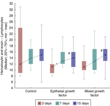
Note: * p < 0.05 for the 3-7 days difference in the wound with epithelial growth factor.
# p < 0.05 for the 3-5 days difference in the wound with epithelial factor and in the wound with mixed growth.
32
30
28
26
24
22
20
18
16
14
12
10
8
6
Control
*
# #
Mixed growth factor Epithelial growth
factor
Hemato
xylin and eosin – L
ymphocytes
(Median;
25
%–
75%;
min - max)
4
3 days 7 days 15 days
Figure5 Hematoxylinandeosinevolution---lymphocytesin
eachwound,assessment.
thewounddiameter;inF2,thereductionwas83%±0.201%, andinF3itwas100%(Fig.4).
Whencomparingthethreetypesofwoundoneach day oftheanimals’euthanization,therewerenosignificant dif-ferencesinthenumberofneutrophils(Table1).However, whenevaluatingtheprogress ofeach woundonthethree daysoftheanimals’euthanization,asignificantdifference wasfoundforF1,whichshowedthehighestnumberof neu-trophils. Forthiswound, there weredifferences between thethirdandseventhdays(p=0.029)andbetweenthe sev-enthand15thdays(p=0.007).Betweenthethirdand15th daystherewasnosignificant difference(p=0.557).ForF2 andF3, theresultswerelower,withatendency to signif-icanceamongthe threedaysof assessment (p=0.058 and p=0.076,respectively).
Regarding the lymphocytes, when comparing F1, F2, and F3 in each of the euthanization moments, it was
Table1 Neutrophilassessmentwithhematoxylinandeosinonthethird,seventh,and15thdaysineachwound;assessment betweenthegroups.
Euthanizationday Wound n Mean±SD pa
Thirdday Control 15 24.2±4.5 0.482
Epithelialwound 15 21.7±5.7
Mixedwound 15 22.9±5.2
Seventhday Control 15 31.6±8.4 0.155
Epithelialwound 15 24.8±5.5
Mixedwound 15 28.5±9.6
15thday Control 15 23.1±9.6 0.856
Epithelialwound 15 19.9±4.4
Mixedwound 15 20.7±6.2
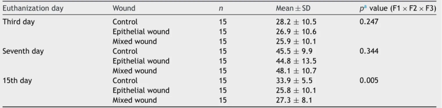
Table2 Macrophagecountperfieldandcomparisonofwounds1,2,and3ateachmomentofeuthanization.
Euthanizationday Wound n Mean±SD pavalue(F1×F2×F3)
Thirdday Control 15 28.2±10.5 0.247
Epithelialwound 15 26.9±10.6
Mixedwound 15 25.9±10.1
Seventhday Control 15 45.5±9.9 0.344
Epithelialwound 15 44.8±13.5
Mixedwound 15 48.1±10.7
15thday Control 15 33.9±5.5 0.005
Epithelialwound 15 25.8±10.1
Mixedwound 15 27.3±8.1
a NonparametricFriedmantest,p<0.05.
observed that there was no significant difference among them(p=0.270).Asfortheseventhday,therewasalsono significantdifferencebetweenthethreewounds(p=0.759). Thesamewasobservedforthe15thday(p=0.451).
In F1, the results were similar regarding lymphocytes in the three days of comparison (p=0.774). As for F2, an increase in the number of lymphocytes was observed betweenthethirdandseventhdays(p=0.007)andbetween thethirdandthe15thdays(p=0.007).Whencomparingthe assessment days for F3, an increasednumber of lympho-cyteswerefound onlywhencomparingthethirdand15th days(Fig.5).
Regardingmacrophages,theresultsweresimilaramong thethreewoundsfor boththethirdandtheseventhdays, (p=0.247andp=0.344,respectively).Asforthe15thday, theresultsshowedasignificantdifferenceamongthethree wounds, with a lower number of macrophages in F2 and F3 (p=0.005). In addition, for the 15th day, when com-paringthewoundstwoby two,adifferencewasobserved betweenF1andF2(p=0.001),withfewermacrophagesin
120
100
80
60
40
20
Control
* * *
* #
Hemato
xylin and eosin – Fibrob
lasts
(Median;
25
%–
75%;
min - max)
Epithelial growth factor
Mixed growth factor 0
3 days 7 days 15 days
Figure 6 Hematoxylin and eosin evolution --- fibroblasts in
eachwound,betweenthetimesofeuthanization,assessment
betweengroups.
F2;andbetweenF1andF3,therewerefewermacrophages inF3(p=0.004).F2andF3showednosignificantdifference (p=0.660;Table2).
When comparing the days of euthanization regarding fibroblasts, significant differences were obtained among them,withthehighest numberoffibroblasts inF2andF3 (p=0.018forF1;p=0.003forF2,andp<0.001forF3).When comparingthedaysofeuthanizationtwobytwo,therewere differencesbetweenthethird andseventhdays(p=0.004 forF1andp<0.001forF2andF3,whichshowedhigher num-beroffibroblastsondayseven).Whencomparingbetween thethirdand15thdays,asignificantdifferencewasverified onlyforF2 (p=0.044)andinthecomparisonbetweenthe seventhand15thdays,onlyforF3(p=0.003;Fig.6).
Whenassessingtheevolutionofeachwoundinthethree days of euthanization, significant differences were found regardingtheincreaseinthenumberofvessels(p<0.001).
Note: * p < 0.05 for the 3-7 days difference in the three wounds and 7-15 days difference in control wounds and with epithelial growth. # p < 0.05 for the 3-15 days difference in the three wounds. 50
45
40
35
30
25
20
15
10
5
0
Control
# # #
* *
* *
*
Hemato
xylin and eosin –V
essels
(Median;
25
%–
75%;
min - max)
Epithelial growth factor
Mixed growth factor
3 days 7 days 15 days
Figure7 Hematoxylinandeosinevolution---vesselsineach
wound,evaluationbetweenthetimesofeuthanization,
Wound 1 Wound 2 Wound 3
Figure8 Photomicrograph ofvimentin ---inwhich macrophages, fibroblasts, andvessels areevaluated, onthe third dayof
evolution,inthewounds1,2,and3(VIM×200).
Wound 1 Wound 2 Wound 3
Figure9 Photomicrograph of vimentin---where macrophages, fibroblasts,andvessels areevaluated, onthe seventh dayof
evolution,inwounds1,2and3(VIM×200).
Wound 1 Wound 2 Wound 3
Figure10 Photomicrographofvimentin---wheremacrophages,fibroblasts,andvesselsareevaluated,onthe15thdayofevolution,
inwounds1,2,and3(VIM×200).
When comparing the days of euthanization in pairs, sig-nificantdifferences wereobserved betweenthe thirdand seventh days (p<0.001), and there were also significant differences between the third and 15th days (p<0.001). Betweentheseventhand15thdays,therewasnosignificant differencefor F1 and F2 (p=0.044 and p=0.035, respec-tively)(Fig.7).
Figs.8---10showthehistologicaldetailsofthe
immunohis-tochemicalassessmentofscarsinallthreeassessedperiods oftime.
WhenassessingtypeIcollagenonthedaysof euthaniza-tion,therewasnostatisticalsignificanceforF1(p=0.240), whereas there was a greater amount of type I collagen forF2andF3(p≤0.001),demonstratingadecreaseinthe amount of inflammatory infiltrate, increased angiogenesis andfibroplasia,witha faster and moreorganized healing (Figs.11---13).
RegardingcollagentypeIII,significantdifferenceswere foundinF2andF3,withaloweramountoftypeIIIcollagen (p≤0.001).
Discussion
100% 90% 80% 70% 60% 50% 40% 30% 20% 10% 0%
72.1% 72.4% 70.4%
27.9% 27.6%
Wound 1
Collagen percentage
on the 3
th day
Wound 2 Wound 3 29.6%
Red Green
Figure11 DemonstrationofthepercentageoftypeIandtype
IIIcollagen onthe third dayof the experiment inthe three
wounds. 100% 90% 80% 70% 60% 50% 40% 30% 20% 10% 0% Collagen percentage
on the 7
th day
Wound 1 41.9% 58.1% 36.9% 63.1% 41.8% 58.2%
Wound 2 Wound 3
Red Green
Figure12 DemonstrationofthepercentageoftypeIandtype
IIIcollagenontheseventhdayoftheexperimentinthethree
wounds. Red Green 100% 90% 80% 70% 60% 50% 40% 30% 20% 10% 0% Collagen percentage
on the 15
th day
Wound 1 49.2% 50.8% 64.7% 35.3% 73.3% 26.7%
Wound 2 Wound 3
Figure13 DemonstrationofthepercentageoftypeIandtype
III collagenon the 15th day of the experiment in the three
wounds.
Wound healing is a complex series of reactions and interactions of inflammatory mediators and cell growth interactions.10---14Manyintrinsicandextrinsicfactorsaffect wound healing, and there is a great variety of commer-cialoptions that aim tocounteract negativeinterference or stimulate the healing process.14 The interrelationship between nutrition and the wound healing process acts together with the immune system and the immunomodu-latory function.15 The present study analyzed the healing process evaluating macroscopic and microscopic aspects, as well as the influence of growth factors on the heal-ing process. In the beginning of the healing process, thereisamigrationofneutrophilsstimulatedbyplatelets, and after that themacrophages, which contribute to the angiogenesisand fibroplasia.This occurred in thepresent
study, in which the wounds with EGF and MF infiltra-tion exhibited an increased number of neutrophils, with p=0.058andp=0.076;forthemacrophages,significant dif-ferences were found in the three wounds (p<0.01); but when comparing the wounds two by two, there was sig-nificant difference for the F2 and F3 (p<0.001). In the HE evaluation, regarding the vessels, there were signifi-cantdifferencesbetweenthethreewounds(p<0.001).The immunohistochemicalanalysisalsoshowedincreased fibro-blasts, macrophages, and vessels for wounds F2 and F3 (p<0.001). FortypeIcollagen, F2andF3 showedgreater depositionofthistypeofcollagen(p=0.001andp<0.001), demonstratinggreaterinflammatoryresponseandincreased angiogenesis,withfaster healing.The typeIcollagen per-centagesonthe 15thday of theexperiment were:for F1 (49.2%), for F2 (64.7%), and for F3 (73.3%). This demon-strates the improvement in wound healing where there wasinjectionof growth factors in relation tothe control wound.
Withregardtogrowthfactorsderivedfromplatelets,van den Dolder et al. carried out a study in rat bone matrix andperformedaninvitroculture,asampleofbonematrix coveredwith skin and PRP andanother sample withonly bone matrix with skin; they concluded that PRP stimu-lates the matrix and bone differentiation, as a positive correlationwasfoundbetween woundhealingandgrowth factor.16 Thepresent studyanalyzedtheuseofVEGF,EGF, FGF,andIGFtoacceleratewoundhealing;favorableresults were obtained when they were compared with the con-trolwound,withoutthepresenceofgrowthfactors.Wound areawasmacroscopicallyanalyzedandshowedthe follow-ing results: F1 showed an area of 0.031cm2 at the end
oftheexperiment andthe F2andF3 areas were, respec-tively, 0.008cm2 and 0, demonstrating a more effective
healing.
Thepercentageofcollageninthepresentstudy,observed throughthemicroscopyusingHE,washigherinwoundswith growthfactor,inadditiontoalowerinflammatoryresponse whencomparedwiththe control wound (p<0.05=0.016). Similarresults arefound inthe literature.Xieetal. used aratmodelandcomparedhealinginwoundstreatedwith growthfactorsandacontrolwound.Thewoundswere mon-itoreddailyandmeasuredwithacaliperondaysone,seven, 14,and28,wheneuthanasiaandexcisionalbiopsywere per-formed. The microscopic analysis wasperformed withHE andcollagenwasmeasuredinpercentages.3The methodol-ogywassimilartothat usedin thisstudyfor thecreation ofthewounds,measurements,biopsy,andmicroscopy.The resultswerealsosimilar,astheyshowedahigherpercentage ofcollageninwoundswithgrowthfactor.
showedthattopicaluseofgrowthfactorpromotedhealing improvement,asdemonstratedinthepresentstudy.17
Inanotherstudy,theauthorscomparedawoundhealing modelinWistarratsthroughtheexpressionofgrowth fac-tors---Group1:healthywithoutwound;Group2:excisional wound; Group 3: transcutaneous electrical nerve stimu-lation (TENS); Group 4: topical saline solution; Group 5: povidone iodine; Group 6: lavender oil. The experiment lastedfivedaysandthegrowthfactorsweremeasuredby immunoassayandimmunohistochemistryanalysis.The lev-elsofPDGFandEGFgrowthfactorsweresignificantlyhigher in theTENS group comparedtothe other groups andthe controlgroup (p<0.05).4 This study showed thedifferent treatments for wound healing, distinct from the present studyinwhichonlytheeffectofgrowthfactorsonwound healingwasassessed;therewasnoexternalstimulus,only growthfactorswereused.
Itisnoteworthythatalthoughthepresentstudyobtained favorableresultswiththeuseofEGF andMF(p<0.05)on thethree daysof assessment and a higherpercentage of maturecollagenforF2andF3,with64.7%and73.3%; respec-tively,whencomparedwithF1withonly49.2%,therewas astudythatevaluatedtheeffectsofPRPinhorsewounds withnegative results. Three wounds were created in the metacarpalregionof sixmares of differentbreeds, total-ing36wounds;of these36wounds,18weretreated with PRPandbandageand18werecontrolwounds,treatedonly withbandagewithoutmedication.Ananatomopathological studywasperformed one,two,andthreeweeksafterthe woundwasmade.Alargeamountofgranulationtissueand highconcentrationof transforminggrowth factorB1were observedinthewoundstreatedwithPRP, whencompared withwoundsthatwerenottreatedwithPRP.Histological, biochemicaldata,andgeneexpressiondidnotsignificantly differbetweenwoundstreatedwithPRPandtheuntreated. Theauthorsconcludedthatthetopicalapplicationof autol-ogousPRPdoesnotaccelerateorimprovehealingofwounds withgranulationinhorselimbs.Thiskindoftreatmentcan improvewoundswithextensivetissuelossoralternatively, chronicwoundsthatwouldbenefitmediatorstoaccelerate thehealingprocess.18
ThecareandobjectivesofthestudybyPradeepconsisted incomparingtheclinicaleffectivenessoftworegenerative techniques in the treatment of bone defects in humans. PRPandPRPassociatedwithinorganicmatrixderivedfrom bovinetissueandpeptide-15(ABM/P-15)wereused. Analyz-ingtheclinicalandradiologicalparametersin14patients, theycreatedbonedefectsandinjectedPRP,andinanother group,PRPassociatedwithAMB/P-15.Allanalyzedclinical parameters(plaqueindex,groovebleedingindex,levelof insertion,gingival margin level, probing depth) showed a statisticallysignificant difference(p<0.001) for the graft groupassociated withPRP.The computerized tomography (CT)scanshowed that bone growthwassignificantly bet-terwhenPRP+AMB/P-15werecombined(p<0.001).Itwas concludedthatthecombinationof PRPandAMB/P-15was more effective than PRP alone in treating intraosseous defects.19Similarresultswereobtainedinthepresentstudy, asthe wounds with MF showed slightly better healing in relation to wound with EGF, which leads to the conclu-sionthatacombinationofgrowthfactorsresultsinbetter healing.
Conclusion
The macroscopicresultsrevealed areduction oftheopen wound area, and the microscopic findings support the conclusionthattheuseofgrowthfactorsinjectedintothe bordersofskinwoundsinrats,bothepithelialgrowthanda combinationofvascularendothelialgrowthfactor,fibroblast growthfactorandinsulingrowthfactor,acceleratethe heal-ingprocesscomparedtothecontrolwound,astheyfoster greaterangiogenicactivity,andacceleratedfibroplasiaand thedepositionoftypeIcollagen,inadditiontoaccelerating thematurationofhealing.
Conflicts
of
interest
Theauthorsdeclarenoconflictsofinterest.
References
1.SenguptaM,BanerjeeP,PaulS,SenguptaJ,GhoshM.Healing effectofphenytoinonexcisionalwoundinexperimentalalbino rats.MullerJMedSciRes.2015;6:27---30.
2.HomDB,LinzieBM,HuangTC.Thehealingeffectsofautologous plateletgelonacutehumanskinwounds.ArchFacialPlastSurg. 2007;9:174---83.
3.Xie Z, Paras CB, Weng H, Punnakitikashem P, Su LC,Vu K, etal.Dualgrowthfactorreleasingmulti-functionalnanofibers forwoundhealing.ActaBiomater.2013;9:9351---9.
4.KutluAK,C¸ec¸enD,Gürgen SG,Sayin O,C¸etinF.A compari-sonstudyofgrowthfactorexpressionfollowingtreatmentwith transcutaneous electrical nerve stimulation, saline solution, povidone-iodine,andlavenderoilinwoundhealing.EvidBased ComplementAlternMed.2013;2013:1---9.
5.Cervelli V, Gentile P, Grimaldi M.Regenerative surgery: use offatgraftingcombinedwithplateletrichplasmaforchronic lower-extremityulcers.AesthetPlastSurg.2009;33:340---5. 6.WernerS,GroseR.Regulationofwoundhealingbygrowth
fac-torsandcytokines.PhysiolRev.2003;83:835---70.
7.Wu W, Chen F, Liu Y, Ma Q, Mao T. Autologous injectable tissue-engineered cartilage by using platelet-rich-plasma: experimentalstudyinarabbitmodel.JOralMaxillofacSurg. 2007;65:1951---7.
8.ShiL, Chang Y,YangY,ZhangY,Yu FSX,WuX. Activationof JNKsignalingmediatesconnectivetissuegrowthfactor expres-sionandscarformationincornealwoundhealing.PLoSONE. 2012;7:e32128.
9.Feng ZG, Pang SF, Guo DJ, YangYT, Liu B, Wang W, et al. Recombinantkeratinocytegrowth factor1 intobacco poten-tiallypromoteswoundhealingindiabeticsrats.BiomedResInt. 2014;2014:1---9.
10.WrotniakM,BieleckiT,GazdzikTS.Currentopinionaboutusing theplatelet richgelinorthopedicsa traumasurgery.Orthop TraumatolRehabil.2007;9:227---38.
11.YazawaM,OgataH,NakajimaT,MoriT,WatanabeN,HandaM. Basicstudiesontheclinicalapplicationsofplatelet-richplasma. CellTransplant.2003;12:509---18.
12.MarlovitsS,MoussaviM,GablerC,ErdusJ,VecseiV.Anew sim-plifiedtechnique for producingplatelet richplasma:a short technicalnote.EurSpineJ.2004;13:102---6.
13.Vick VL, Holds JB,Hartestein ME, Rich R, Davidson BR. Use ofautologousplatelet concentrateinblepharoplastysurgery. OphthalPlastReconstrSurg.2006;22:102---4.
15.CamposACL,BrancoAB,GrothAK.Assessmentandnutritional aspectsof wound healing. Curr Opin ClinNutr Metab Care. 2008;11:281---8.
16.VandendolderJ,MoorenR,VloonAPG,StoelingaPJW,Jansen JA.Plateletrichplasma:quantificationofgrowthfactorlevels andtheeffectongrowthanddifferentiationofratbonemarrow cells.TissueEng.2006;12:3067---73.
17.AsaiJ,TakenakaH,LiM,AsahiM,KishimotoS,KatohN,etal. Topicalapplicationofexvivoexpandedendothelialprogenitor
cells promotesvascularisation andwoundhealingin diabetic mice.IntWoundJ.2013;10:527---33.
18.MonteiroSO,LepageOM,TheoretCL.Effectsofplatelet-rich plasmaon therepair of woundson thedistal aspect ofthe forelimbinhorses.AmJVetRes.2009;70:277---82.