Tomographic and pathological findings in pulmonary
sarcoidosis
*
Aspectos tomográficos e anatomopatológicos da sarcoidose pulmonar
Alessandro Severo Alves de Melo1, Edson Marchiori2, Domenico Capone3
Objective: To analyze radiological findings observed at high-resolution computed tomography in patients with sarcoidosis, and establishing their correlation with pathological findings. Materials and Methods: High-resolution computed tomography findings in ten patients with sarcoidosis were reviewed and correlated with findings in specimens obtained by surgical biopsy or at necropsy of four of such patients. Results: The most frequently observed finding was presence of nodules with peri-lymphatic distribution, predominating along bronchovascular sheaths and pleural surface, with subpleural nodules and nodular scissurae. Other less frequent findings were ground-glass attenuation and interlobular septa thickening. Conclusion: In general, all the mentioned findings demonstrated anatomopathological correlation with development of granulomas in these regions.
Keywords: Computed tomography; Sarcoidosis; Pathology; Lung diseases.
Objetivo: Analisar os aspectos radiológicos observados nas tomografias computadorizadas de alta resolução de pacientes com sarcoidose e fazer a correlação com os achados anatomopatológicos. Materiais e Métodos: Foram revistos os aspec-tos radiológicos observados nas tomografias computadorizadas de alta resolução de dez pacientes com sarcoidose e feita correlação com material obtido de biópsias cirúrgicas ou necrópsias de quatro desses pacientes. Resultados: O aspecto mais frequentemente observado foi o de nódulos, com distribuição perilinfática, predominando ao longo das bainhas bron-covasculares e da superfície pleural, com nódulos subpleurais e cissuras nodulares. Outros achados menos comuns foram as opacidades em vidro fosco e o espessamento de septos interlobulares. Conclusão: Em geral, todos esses achados corresponderam, anatomopatologicamente, ao acúmulo de granulomas nessas regiões.
Unitermos: Tomografia computadorizada; Sarcoidose; Anatomopatologia; Doenças pulmonares.
Abstract
Resumo
* Study developed at Department of Radiology, Universidade Federal Fluminense (UFF), Niterói, RJ, and at Unit of Radiodiagno-sis, Hospital Universitário Clementino Fraga Filho (HUCFF) da Universidade Federal do Rio de Janeiro (UFRJ), Rio de Janeiro, RJ, Brazil.
1. Associate Professor, Department of Radiology, Universidade Federal Fluminense (UFF), Niterói, RJ, Brazil.
2. Titular Professor, Department of Radiology, Universidade Fe-deral Fluminense (UFF), Niterói, RJ, Adjunct Coordinator of Post-Graduation Course in Radiology, Universidade Federal do Rio de Janeiro (UFRJ), Rio de Janeiro, RJ, Brazil.
3. Assistant Professor of Pneumology, Universidade do Estado do Rio de Janeiro (UERJ), Rio de Janeiro, RJ, Brazil.
Mailing Address: Dr. Edson Marchiori. Rua Thomaz Cameron, 438, Valparaíso. Petrópolis, RJ, Brazil, 25685-120. E-mail: [email protected]
Received March 17, 2011. Accepted after revision May 20, 2011.
Melo ASA, Marchiori E, Capone D. Tomographic and pathological findings in pulmonary sarcoidosis. Radiol Bras. 2011 Jul/Ago;44(4):220– 224.
The present study discusses the main findings observed at HRCT in sarcoido-sis, correlating them with pathological findings.
MATERIALS AND METHODS
The present study reviewed HRCT findings in ten patients of hospitals in Rio de Janeiro, RJ, Brazil, with histologically confirmed diagnosis of sarcoidosis. Such radiological findings were correlated with those observed in specimens obtained from surgical biopsies or at necropsies of four of such patients. The age range was between 30 and 68 years (mean = 48 years), and the study sample included seven fe-male and three fe-male patients.
RESULTS
Presence of nodules with perilymphatic distribution was the most frequent find-use of immunosuppressant drugs or
anti-inflammatory agents, irreversible and ex-tensive pulmonary fibrosis may develop, leading to a progressive deterioration of the respiratory function(1).
An understanding of the normal anatomy as well as of the changes caused by the disease is required for an appro-priate analysis of any radiological image, and radiologic-pathologic correlation is critical to understand the anatomical foun-dations of images formation(2).
Many studies have described the find-ings of sarcoidosis at computed tomog-raphy; but only a small number of them have correlated high-resolution com-puted tomography (HRCT) findings with pathological findings(2–4). Many times,
such a correlation is hardly established, considering that the patients who die are generally those with extensive fibrotic lesions in the lungs, and open biopsy is rarely performed in cases o sarcoidosis(5).
INTRODUCTION
Sarcoidosis is a multisystem disease of unknown etiology, characterized by the presence of noncaseating granulomas, affecting many tissues and organs in the body, particularly the respiratory tract(1).
ing. The lesions were predominantly ob-served along bronchovascular sheaths and pleural surface, with subpleural nod-ules and nodular scissurae (Figure 1). In-terlobular septal thickening was a less
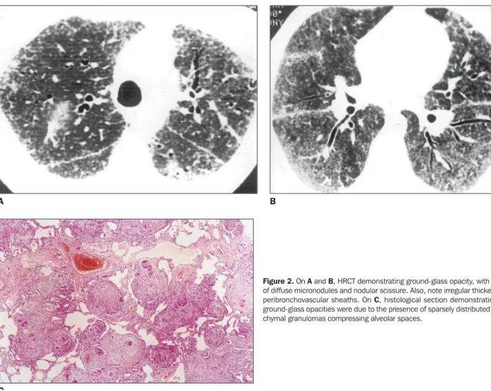
fre-quent finding. Ground-glass opacity was another less frequent finding, generally observed in association with the presence of small nodules (Figure 2). In general, all these findings correlated pathologically
with accumulation of granulomas in those regions. Air trapping was other interest-ing findinterest-ing determined by bronchial lu-men narrowing caused by the presence of peribronchial granulomas (Figure 3).
Figure 1. Three different patients. On A and B, presence of broncho-vascular sheaths thickening and clustered nodules in the subpleural re-gion. On B and C, nodular scissurae are also highlighted. On A, observe hilar lymph nodes enlargement, and on C, vessels with apparently increased caliber, particularly at right, determined by perivascular sheath thickening. On D and E, histological sections demonstrate clustered granulomas in the subpleural region (D) and along the interlobular septum (E), respon-sible for the nodular appearance of such structures at HRCT.
D E
A B
DISCUSSION
In sarcoidosis, granuloma is the basic histopathological finding. Noncaseating granulomas are confined to the interstitial compartment of the lungs, with a perilym-phatic distribution along bronchovascu-lar sheaths, interlobubronchovascu-lar septa and pleu-ral surface(4,6,7), including the scissural
pleura(7). Such a preference for
peribron-chovascular regions explains the high frequency of positive transbronchial bi-opsies(8). In general, predominant sites of
lesions are the upper, middle and poste-rior zoned os the lungs(8). In truth,
granu-lomas may determine three image pat-terns: nodules, irregular thickening of structures and ground-glass opacity(2).
Areas of consolidation may also be ob-served as a result from irregular conflu-ence of nodules(8).
Nodules represent the most frequent findings at HRCT(7). In general, they have
2 to 10 mm in diameter, irregular mar-gins(3,7), histologically representing
gra-nulomatous aggregates(3,6,7).
Less frequently, granulomatous aggre-gates may originate large nodules (> 1 cm) and large areas of opacity with ill-defined contours(2,3,5,7,8).
Small, individual granulomas sparsely distributed over the parenchyma produce ground-glass opacity(7). Such
granulo-mas are not sufficiently large to be indi-vidualized at HRCT, but produce a diffuse increase in lung attenuation originating this image pattern(6). Initially, some
au-thors suggested that such a pattern was related to alveolitis, but later studies have demonstrated that, as a rule in acute phases of sarcoidosis, this pattern was determined only by the presence of
gra-nulomas(2,3,7). Although ground-glass
opacity usually corresponds to the pres-ence of a potentially treatable or revers-ible disease, in some cases it results from fibrosis(1,7). The presence of fibrosis is
suggested by concomitance of traction bronchiectasis, bronchiolectasis and pa-renchymal distortion(1).
In spite of their perilymphatic distribu-tion, a higher number of sarcoid granulo-mas occur along bronchovascular sheaths and in lower number, in interlobu-lar septa and subpleural regions(3).
Inter-lobular septal thickening is a less frequent finding in sarcoidosis(7,9).
Linear opacities are less frequent and less profuse than nodules. Polygonal ar-cades such as those observed in carcino-matous lymphangitis, are extremely rare findings in sarcoidosis(7,9). In sarcoidosis,
nodules present irregular margins; in
lym-Figure 2. On A and B, HRCT demonstrating ground-glass opacity, with finding of diffuse micronodules and nodular scissure. Also, note irregular thickening of peribronchovascular sheaths. On C, histological section demonstrating that ground-glass opacities were due to the presence of sparsely distributed paren-chymal granulomas compressing alveolar spaces.
A B
phangitis, they are generally smoother(9).
Sometimes, the lymphatic distribution is histologically difficult to recognize, be-cause of the disproportionate involve-ment of an anatomical site as compared with another. In sarcoidosis, there is a much greater involvement of bronchovas-cular sheaths, and little involvement of septa and pleural surface, eventually sug-gesting a bronchiolocentric/angiocentric pattern rather than properly a lymphatic pattern(4).
Proliferation of granulomas along bronchovascular sheaths causes their irregular thickening(3), with arterial walls
and bronchi appearing like “rosary beads” because of their contour nodu-larity(7). Also, scissurae and interlobular
septa may present such “rosary beads” pattern(7).
At HRCT, the pleural surface may present a nodular pattern; pleural granu-lomas cannot be differentiated from those located in alveolar septa adjacent to the pleura(3).
It is important to recognize that the presence of cellular and granulomatous inflammatory elements indicates disease activity, reversibility and favorable re-sponse to treatment, while fibrotic in-volvement indicates disease irreversibil-ity and poor therapeutic response(1).
In general, alterations determined by granulomas (small nodules, large nodules, ground-glass opacity and nodular thick-ening – “rosary beads” pattern – of
in-terstitial structures) are reversible(3,5),
since they represent active inflammatory lesions(5). Either with or without
treat-ment, such lesions may persist for months or years, or otherwise be reabsorbed(1,8).
However some findings are irrevers-ible, representing manifestation of fibro-sis. A common sign observed in cases of fibrosis is architectural distortion that may cause posterior ballottement of the main bronchus or of the upper bronchi-oles(5,7,8). Other findings include traction
bronchiectasis, honeycombing(5,7,8), cysts
and bullae(7), and parenchymal bands,
septal and non-septal lines(5). Irreversible,
linear, irregular and elongated opacities may appear, particularly along broncho-vascular sheaths as an early
manifesta-Figure 3. On A, HRCT demonstrating inflated secondary pulmonary lobule in the right lower lobe, besides thickened septa with evidences of fibrosis. On B, section demonstrating granuloma compressing bronchiole and determining air trapping. On C, HRCT showing inflated pulmonary lobule at the bottom of the left lung, with a centrilobular arcade-like structure and nodular contour. On D, centrilobular region section showing multiple granulomas in the peribronchovascular sheath, deter-mining narrowing of the bronchiole lumen, explaining the valve emphysema and the arcade-like configuration with nodulations observed on C.
A B
tion of fibrosis(7). Irregular interfaces
simi-lar to those observed in idiopathic pulmo-nary fibrosis, have also been described, determined by focal septal fibrosis(8). The
fibrosis may be so extensive as to form perihilar masses resembling the progres-sive masprogres-sive fibrosis of silicosis(7). Other
possibility is the post-treatment persis-tence of ground-glass opacity, attributed to alveolar septal fibrosis(5).
As regards pulmonary function, stud-ies have demonstrated that the profusion of granulomas observed in biopsies is not correlated with functional deterioration(1).
Apparently, the functional alteration, rather than the quantity of lesions, is re-lated to the strategic location of some
granulomas (involving bronchioles and causing increased resistance to air flow)(1,6).
Airways narrowing determined by peri-bronchial granulomas results in air trap-ping at HRCT, particularly in studies per-formed during the expiratory phase, where the crazy-paving pattern can be seen(6).
REFERENCES
1. Remy-Jardin M, Giraud F, Remy J, et al. Pulmo-nary sarcoidosis: role of CT in the evaluation of dis-ease activity and functional impairment and in prog-nosis assessment. Radiology. 1994;191:675–80.
2. Müller NL, Miller RR. Ground-glass attenuation, nodules, alveolitis, and sarcoid granulomas. Radi-ology. 1993;189:31–2.
3. Nishimura K, Itoh H, Kitaichi M, et al. Pulmo-nary sarcoidosis: correlation of CT and histopatho-logic findings. Radiology. 1993;189:105–9.
4. Colby TV, Swensen SJ. Anatomic distribution and histopathologic patterns in diffuse lung disease: cor-relation with HRCT. J Thorac Imaging. 1996;11:1– 26.
5. Brauner MW, Lenoir S, Grenier P, et al. Pulmo-nary sarcoidosis: CT assessment of lesion reversi-bility. Radiology. 1992;182:349–54.
6. Gleeson FV, Traill ZC, Hansell DM. Evidence on expiratory CT scans of small-airway obstruction in sarcoidosis. AJR Am J Roentgenol. 1996;166: 1052–4.
7. Traill ZC, Maskell GF, Gleeson FV. High-resolu-tion CT findings of pulmonary sarcoidosis. AJR Am J Roentgenol. 1997;168:1557–60. 8. Brauner MW, Grenier P, Mompoint D, et al.
Pul-monary sarcoidosis: evaluation with high-resolu-tion CT. Radiology. 1989;172:467–71. 9. Müller NL, Kullnig P, Miller RR. The CT