244 Radiol Bras. 2011 Jul/Ago;44(4):244–248
Evaluation of medical X-ray machines in Paraíba state
radiology centers between 2008 and 2009
*
Avaliação das condições de funcionamento dos equipamentos de raios X médico em serviços de radiologia no Estado da Paraíba, durante os anos de 2008 e 2009
Adriana Carla Rodrigues Mendes1, Christiane Lucena Ramos2, Danilo Wanderley Matos de Abreu3
Objective: To evaluate the light and radiation fields congruence and radiation beam alignment in medical X-ray equipment in Paraíba state radiology centers by means of two quality control tests. Materials and Methods: A loaded cassette, a measuring tape and a bubble level were utilized in the field size and alignment testing. The evaluation of collimation systems accuracy and X-ray beam alignment was undertaken during health inspections performed in
radiology centers between 2008 and 2009. Results: In 2008, 121 X-ray machines were evaluated in the Paraíba
state. In 2009, 117 machines were tested. From this universe, 86 machines were selected for comparison, since they were evaluated both in 2008 and 2009, with 18.60% (n = 16) showing test results improvement from one year to
another. Conclusion: The percentage of problematic X-ray machines decreased between 2008 and 2009,
notwithstanding no quality assurance program has been observed in Paraíba state radiology centers. Keywords: Equipment; X-rays; Quality control.
Objetivo: Avaliar a coincidência entre o campo luminoso e o campo de radiação, e o alinhamento do feixe de radiação dos equipamentos de raios X médico no Estado da Paraíba, por meio de dois testes de controle de qualidade. Mate-riais e Métodos: Foram utilizados os dispositivos para os testes de tamanho de campo e de alinhamento, um chassi carregado, trena e nível de bolha. Os testes de exatidão do sistema de colimação e de alinhamento do raio central do feixe de raios X foram realizados durante as inspeções sanitárias em serviços de radiologia nos anos de 2008 e 2009. Resultados: No ano de 2008, 121 equipamentos de raios X foram testados no Estado da Paraíba. No ano de 2009, passaram pelos testes 117 equipamentos. Deste universo, 86 foram selecionados para a comparação por terem sido
avaliados tanto no ano de 2008 como em 2009, sendo observada uma melhoria de 18,60% (n = 16) nos resultados
dos testes realizados de um ano para outro. Conclusão: Pode-se concluir que o percentual de equipamentos apre-sentando problemas no seu desempenho sofreu uma diminuição entre 2008 e 2009, não sendo observado programa de garantia de qualidade em nenhum dos serviços de radiologia do Estado da Paraíba.
Unitermos: Equipamento; Raios X; Controle de qualidade. Abstract
Resumo
* Study developed at Agência Estadual de Vigilância Sanitá-ria da Paraíba (Agevisa-PB), João Pessoa, PB, Brazil.
1. Master of Preventive and Child Dentistry, Health Surveyor, Agência Estadual de Vigilância Sanitária da Paraíba (Agevisa-PB), João Pessoa, PB, Brazil.
2. Specialist in Pediatric Dentistry, Health Surveyor, Agência Estadual de Vigilância Sanitária da Paraíba (Agevisa-PB), João Pessoa, PB, Brazil.
3. Master of Urban Engineering, Health Surveyor, Agência Estadual de Vigilância Sanitária da Paraíba (Agevisa-PB), João Pessoa, PB, Brazil.
Mailing Address: Adriana Carla Rodrigues Mendes. Rua Lin-dolfo José Correia das Neves, 419, ap. 502, Jardim Oceania. João Pessoa, PB, Brazil, 58037-305. E-mail: mendesadriana1@ hotmail.com
Mendes ACR, Ramos CL, Abreu DWM. Evaluation of medical X-ray machines in Paraíba state radiology centers between 2008 and 2009. Radiol Bras. 2011 Jul/Ago;44(4):244–248.
radiology is defined as “an organized effort by the service management towards assur-ing that images are acquired with enough quality to allow an appropriate diagnosis with the lowest radiation dose to the pa-tient”(2).
In Brazil, the development of tech-niques and devices for quality control in ra-diology started at the Physics Department in the Ribeirão Preto Campus of Univer-sidade de São Paulo since the mid-1970s, by Professor Thomaz Guilardi Neto, as-sisted by Professor John Cameron. Profes-sionals were educated and prepared to per-form evaluation techniques, and have implemented the first quality programs of throughout São Paulo and other Brazilian states.
level of quality, so as to minimize errors of interpretation and identification of struc-tures, thus allowing an accurate diagnosis with low radiation levels. Otherwise a low quality image causes the repetition of im-aging studies and, consequently, the dupli-cation of radiation dose in the same patient, besides additional costs to the radiology service(1).
The benefits from an appropriate radio-logical image can be attained by means of the implementation of equipment quality assurance programs in radiodiagnosis cen-ters. According to the World Health Orga-nization, quality assurance in diagnostic
Received October 20, 2010. Accepted after revision June 29, 2011.
INTRODUCTION
With the publication of Resolution SS 625/94, of December 14, 1994, the imple-mentation of quality control programs be-came mandatory in the state of São Paulo(3).
Following the successful implementation of the programs in the state of São Paulo, the Ministry of Health issued the Order (Portaria) MS/SVS 453, on June 1st, 1998(1).
Order 453 establishes the need to imple-ment a quality assurance program (QAP) in radiology center(4,5). As a result, it
estab-lishes that every diagnostic X-ray equip-ment must be subject to periodical evalua-tion of their performance by means of sev-eral constancy tests, such as, for example: measurement of the representative dose value delivered to the patients submitted to radiography at the center; testing of the accuracy of X-ray tubes voltage indicator (kVp); evaluation of the accuracy of expo-sure time, as applicable; testing of the X-ray beam alignment; X-X-ray tube perfor-mance test; testing of the air kerma rate lin-earity as a function of the mAs, testing of automated exposure system reproducibil-ity; measurement of the focal point size; evaluation of collimation system accuracy; and grid alignment test(6).
For the present study, Agência Estadual de Vigilância Sanitária da Paraíba (Agevisa-PB) (Paraíba State Department of Health Surveillance) provided the research instru-ments required for the testing of collima-tion system accuracy and X-ray beam align-ment. Because of their simplicity and facil-ity, the tests allowed a critical evaluation of the apparatuses with respect to collimation of radiation in the patients’ areas of inter-est and the distortion of the radiographic image, allowing the minimization of risks resulting from radiation exposure, minimi-zation of the dose delivered both to patients and workers, costs optimization, reduction of films rejection and wear on the equip-ment, besides a better quality in radio-graphic image standards, thus contributing for accurate diagnoses(7).
The collimation system accuracy testing is aimed at evaluating the congruence be-tween the light and radiation fields limited by the equipment collimator, thus avoiding X-rays reaching unintended areas of the patient’s body during the radiological ex-amination. On the other hand, the testing of X-ray beam alignment is aimed at
evalu-ating the perpendicularity between the cen-ter of the light field and the plane of the image reception system, thus avoiding the radiographic image distortion(8).
The present study was aimed at evalu-ating the light and radiation fields congru-ence and radiation beam alignment in medical X-ray equipment in Paraíba state, private and philanthropic radiology centers by means of two quality control tests (QCT) as follows: testing of collimation system accuracy and X-ray beam alignment performed in the years of 2008 and 2009 by the technical team of the Agevisa-PB ionizing radiations sector.
MATERIALS AND METHODS
The present study is of the observa-tional, descriptive and comparative type, as it informs, in quantitative terms, on the dis-tribution of an event in the population, and compares the testing results in the same apparatuses in different years(9).
The data recording was performed by a single investigator and the visits were car-ried out during health surveillance inspec-tions following the normal operational rou-tine of the radiological centers. Because of ethical confidentiality considerations, the hospitals and radiological centers visited during the present study were not identi-fied. So, inspected institutions are only classified according to the service network they belong to, as public, private and phil-anthropic.
In 2008, 121 X-ray apparatuses belong-ing to 87 radiology services in the state of Paraiba underwent QCT. Among them, 42.14% (n = 51) belonged to the public health system, 55.37% (n = 67), to private clinics, and 2.47% (n = 3), to philanthropic centers.
In 2009, 117 apparatuses were submit-ted to the same tests in the Paraíba state, 38.46% (n = 45) of them in the public health network, 56.41% (n = 66), in the private network, and 5.12% (n = 6) in the philanthropic network.
Of this universe, 71.07 % (n = 86) of the apparatuses were selected for comparison, since they had been evaluated both in the years of 2008 and 2009.
The tests performed in the present study were the following: X-ray beam alignment
collimation system accuracy, according to the Brazilian protocols for QCTs(6,10) and a
medical radiodiagnosis handbook(11). The
following instruments were utilized for each evaluation: devices for field size test-ing (plate with radiopaque marktest-ings with two orthogonal axes in scales of 0.5 cm and two concentric circles) and, for the X-ray beam alignment (a plastic cylinder with 0.8 mm-diameter steel spheres placed at lower and upper bases at a distance of 15 cm), a (preferably 24 × 30) cassette loaded with film, a measuring tape and a bubble level(8,12).
For the evaluation of the collimation system accuracy, a 24 × 30 loaded cassette was placed on the table’s surface, and over such cassette the device for field size mea-surement, in such a way that such device was perpendicular to the X-ray beam. Af-ter adjustment of the focus-film distance to 1m, the adjustment of the edges of the light field was made so that it matched the rect-angular outline of the plate, through the X-ray equipment collimator. For the X-X-ray beam alignment, the beam alignment de-vice was placed at the center of the colli-mator test device. Subsequently, an expo-sure utilizing approximately 40 kVp and 3 mAs(11) was made.
After the evaluation of the tests results, in those cases in which the X-ray appara-tuses were not in compliance with the stan-dards, the institutions to which they be-longed were notified and deadlines were established for corrective maintenance, after which the inspection team would per-form new tests in the same apparatuses to check on the adjustments made.
In the present study, in order to evalu-ate the results, the X-ray apparatuses were classified according to their models, into fixed equipment and portable equipment. The portable apparatuses that were being utilized as fixed equipment were included in the present study as fixed equipment.
RESULTS
The data obtained by the present study developed in the years of 2008 and 2009 are categorical, with no margin for error. Among the apparatuses existing in the Paraíba state, a significant sample (71.07%;
The congruence between the light and radiation fields was evaluated for analysis and interpretation of the images of the col-limator test. The largest distance between the borders of the light field and those of the radiation field was measured. The greater the congruence between the light and the radiation fields, the better is the alignment. The difference between the borders of the radiation field and the borders of the light field must not be greater than 2% of the focus-film distance that is ±2 cm away from the line corresponding to the device’s rectangle(9,11). The images generated on this
test are represented on Figures 1 and 2. As regards the analysis and interpreta-tion of the X-ray beam alignment test, the
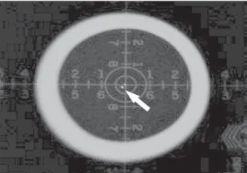
location of the image of the sphere on the top of the cylinder was verified. If the im-age of the upper sphere was within the first circle, the inclination corresponded to < 1.5°. In those cases where the image of the upper sphere was located between the first and the second circles, the inclination was < 3°(11). Whenever the image of the upper
sphere intercepted the second circle, the perpendicularity of the central ray must be corrected(8). Figures 3 and 4 represent the
images of the X-ray beam central axis alignment test.
In the present study, in the year of 2008, QCTs were performed in 92 fixed X-ray apparatuses and in 29 portable apparatuses, in different centers belonging to the
pub-lic health network, private network and philanthropic network.
Among the 121 tested apparatuses, 32.23% (n = 39) presented problems re-garding the collimation system accuracy and/or alignment of the X-ray beam central ray, most of those 39 apparatuses (n = 22) belonging to private centers. Among the tested apparatuses, 23.96% (n = 29) were classified as fixed X-ray apparatuses with problems, while 8.26% (n = 10) were por-table apparatuses that were not compliant with the quality assurance standards.
In 2009, 117 apparatuses (89 fixed and 28 portable) were tested in the Paraíba state. Among those apparatuses, 19.65% (n = 23) presented problems in the collimation
Figure 1. Collimation system accuracy test. Image demonstrating compliant fields congruence.
Figure 2. Collimation system accuracy test. Image demonstrating non com-pliant fields congruence.
Figure 4. Central ray perpendicularity test. Image demonstrating the non compliant incidence of the X-ray beam central ray.
system accuracy and/or X-ray beam align-ment, most of them (n = 13) belonging to private clinics. As regards the classification of the apparatuses, among all the appara-tuses monitored in 2009, 12.82% (n = 15) were classified as fixed apparatuses with problems, and 6.83% (n = 8), as portable, non compliant apparatuses.
Table 1 Classification of the fixed and portable X-ray apparatuses, according to the collimation system accuracy tests performed in 2008 and 2009.
Fixed and portable X-ray apparatuses
Compliant fields congruence Non compliant fields congruence Total number of X-ray apparatuses
n
67 19 86
%
77.90 22.10 100.00
n
78 8 86
%
90.69 9.31 100.00
2008 2009
n, number of X-ray apparatuses.
Table 2 Classification of fixed and portable X-ray apparatuses, according to X-ray beam central ray perpendicularity tests performed in 2008 and 2009.
Fixed and portable X-ray apparatuses
Compliant central ray incidence Non compliant central ray incidence Total number of X-ray apparatuses
n
76 10 86
%
88.37 11.63 100.00
n
81 5 86
%
94.18 5.82 100.00
2008 2009
n, number of X-ray apparatuses.
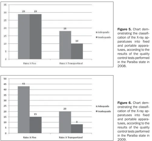
In the present study, 71.07% (n = 86) of the apparatuses tested in 2008 were tested again in 2009, with improvement in QCT results of 18.6% (n = 16) from one year to another (Figures 5 and 6). The other apparatuses tested in both years obtained the same satisfactory results on the colli-mation system accuracy and central ray
perpendicularity tests, with no change in results.
In the collimation system accuracy tests performed in 2008 and 2009, results im-provement was observed in 12.79% (n = 11) of the apparatuses with fields congru-ence in conformity with the standards (Table 1). Improvement was also observed in X-ray beam central ray perpendicularity test, in 5.82% (n = 5) of the apparatuses tested in both years (Table 2).
DISCUSSION
In the present study, it was observed that in all the visits, no institution had a QAP in radiodiagnosis, contravening the Order 453/98 of the Ministry of Health which recommends the periodical testing in all the diagnostic X-ray apparatuses with the pur-pose of maintaining such equipment in appropriate operational conditions. The collimation system accuracy test and X-ray beam alignment test recommended by the Order 453/98 must be performed every six months and annually, respectively. In the present study, the periodicity of both tests was annual, following the annual routine of health surveillance inspections at medi-cal X-ray centers. Additionally, the lack of such a program contributes to the image quality deterioration, with the consequen-tial increase in wasting of films and repeti-tion of imaging studies, thus increasing the radiation exposure for both patients and workers(4,5).
Currently, difficulties regarding the awareness of the regulating agencies on the importance of enforcing the implementa-tion of such programs in the instituimplementa-tions in the Paraíba state are still observed. Addi-tionally, there is a lack of qualified profes-sionals for such programs, and scarcity of qualified and experienced technicians for equipment maintenance once the problems are detected. Not only training in radiopro-tection for all individuals involved in the operation of radiation emitting equipment is necessary, but it also necessary to assure a perfect operation of such equipment.
The collimation system accuracy tests performed in 2008 demonstrated that 22.1% (n = 19) of the evaluated appara-tuses were non compliant with the light and radiation fields congruence tests (Table 1), Figure 5. Chart
dem-onstrating the classifi-cation of the X-ray ap-paratuses into fixed and portable appara-tuses, according to the results of the quality control tests performed in the Paraíba state in 2008.
with results similar to the ones reported by Medeiros & Alves(12) (17%) and lower than
those observed by Silva et al.(13) (30%) and
Carrizales & Cozman(14) (41%). As regards
the X-ray beam central ray alignment, the results observed in the present study (11.63%; n = 10) were very low as compared with the results observed by Carrizales & Cozman(14) (53%), in the cases where the
radiology centers did not follow a QAP. As regards the QCTs performed in 2009, the prevalence of non compliant ap-paratuses in the light and radiation fields congruence tests corresponded to 9.31% (n = 8) (Table 1), a percentage higher than the one reported by Bacelar et al.(15) (5%) and
lower than the one observed by Gori et al.(16) (13%). As regards the X-ray beam
central ray alignment, a difference was observed in the number of non compliant apparatuses in 2009 (5.82%; n = 5) in com-parison with the previous year, a percent-age considerably lower than the one re-ported by Silva et al.(13) (17,7%).
CONCLUSIONS
By testing the light and radiation fields congruence and the X-ray beam central ray perpendicularity in apparatuses at Paraíba state radiology centers, it was possible to observe that the non conformities observed were quite significant in the first year, dem-onstrating the importance of implementing quality assurance programs in radiology centers for prevention and maintenance of the X-ray apparatuses. Adjustments in non compliant apparatuses were only per-formed after inspection and notification on the non compliance by the inspecting agency (Agevisa-PB), as none of the radi-ology centers in the Paraíba state had a quality assurance program.
The QCTs performed in the first year of the present study (2008) revealed a high index of non conformities (45.34%), that was significantly reduced in the following year (26.74%), with 18.60% (n = 16) of the apparatuses tested in 2008 improving their performance in 2009. The remaining ap-paratuses tested in both years obtained the same satisfactory results.
A good X-ray equipment performance is not only a matter of complying with the regulations, but also, and more importantly, a matter of permanent interest in improv-ing the quality and efficiency at the radiol-ogy centers. Therefore the implementation of quality assurance programs at the insti-tutions operating diagnostic X-ray appara-tuses is proposed, with periodical evalua-tion and adjustment of the equipment by qualified professionals, with the purpose of producing high quality images to allow correct diagnoses, with a reduction of the radiation dose delivered to patients and involved professionals exposed to radia-tion, as well as reducing the costs for the centers, as a result of the reduction of im-aging studies repetitions.
Acknowledgements
The authors wish to thank the team of the Agevisa-PB Technical Board of Direc-tors of Medical Science and Technology and Correlates for the support in the devel-opment of the present study.
REFERENCES
1. Furquim TAC, Costa PR. Garantia de qualidade em radiologia diagnóstica. Rev Bras Fis Med. 2009;3:91–9.
2. Organización Panamericana de la Salud. Garan-tía de la calidad en radiodiagnóstico. México: OPS; 1984.
3. Secretaria de Estado da Saúde. Norma técnica que dispõe sobre o uso, posse e armazenamento de fontes de radiação ionizante, no âmbito do Estado
de São Paulo. Resolução SS 625/94. São Paulo, SP: Diário Oficial do Estado; 14/12/1994. 4. Moores BM, Watkinson AS, Henshaw ET, et al.
Quality control in diagnostic radiology. In: Oberhofer M, editor. Advances in radiation pro-tection. 1st ed. Brussels and Luxembourg: Kluwer Academic Publishers; 1991. p. 209–36. 5. Yacovenco A, Lira SH, Borges JC, et al. Programa de garantia de qualidade em radiologia diagnós-tica. RBE/CEB. 1994;10:7–19.
6. Brasil. Ministério da Saúde. Secretaria de Vigi-lância Sanitária. Diretrizes de proteção radioló-gica em radiodiagnóstico médico e odontológico. Portaria nº 453. Brasília, DF: Diário Oficial da União; 2/6/1998.
7. Ros RA. Metodologia de controle de qualidade de equipamentos de raios X (nível diagnóstico) uti-lizados em calibração de instrumentos [disserta-ção]. São Paulo, SP: IPEN/USP; 2000. 8. Netto TG. Garantia e controle de qualidade em
radiodiagnóstico. [acessado em 17 de fevereiro de 2010]. Disponível em: http://www.rxnet.com.br/ fique_informado/documentos/controle_%20 qualidade.pdf
9. Pereira MG. Métodos empregados em epidemio-logia. In: Pereira MG. Epidemiologia: teoria e prática. Rio de Janeiro, RJ: Guanabara Koogan; 1995. p. 269–88.
10. Protocolo de avaliação de proteção radiológica em radiodiagnóstico médico. Rio de Janeiro, RJ: RXD-URMCF, IRD/CNEN; 2000.
11. Brasil. Ministério da Saúde. Agência Nacional de Vigilância Sanitária. Radiodiagnóstico médico: desempenho de equipamentos e segurança. Bra-sília, DF: Editora Anvisa; 2005.
12. Medeiros RB, Alves FFR. Análise dos resultados do programa de gerenciamento da qualidade dos equipamentos radiológicos. Rev Imagem. 1997; 19:97–9.
13. Silva MO, Carvalho ACP, Azevedo ACP. Levan-tamento das condições de funcionamento dos serviços de radiologia de hospitais públicos e universitários do Rio de Janeiro. Radiol Bras. 2004;37:271–8.
14. Carrizales LI, Cozman A. Quality control in radiodiagnosis in Venezuela, 1989-1990. Phys Med. 1990;6:255–7.
15. Bacelar A, Oliveira SS, Streck EE, et al. Avalia-ção preliminar de parâmetros operacionais em equipamentos fixos de raios X diagnóstico. Ra-diol Bras. 1998;31:129–33.