425
Study of the ureterovesical jet by means of color Doppler
Radiol Bras 2006;39(6):425–428 Original Article
STUDY OF THE URETEROVESICAL JET BY MEANS OF COLOR DOPPLER
IN PATIENTS WITH AND WITHOUT VESICOURETERAL REFLUX*
Makoto Sakate1
, Altamir Santos Teixeira2
, Alzira Teruio Yida Sakate3
, Pedro Gabriel Silva4 , Ulisses Benedito Colombo5
, José Goldberg6
OBJECTIVE: The present study had as its objective to compare the findings of voiding cystourethrography with those of duplex color Doppler in patients with suspected vesicoureteral reflux. MATERIALS AND METH-ODS: The research was developed through the study of ureterovesical jet angles, both in axial and longitu-dinal planes. Also, the distance (in cm) between ureteral meatuses was analyzed. RESULTS: From a total sample of 32 patients (mean age of five years and two months), 18 presented with vesicoureteral reflux (10 with unilateral reflux — 4 right-sided and 6 left-sided —, and 8 with bilateral reflux) and 14 patients did not presented with reflux. The angles of ureterovesical jet and distances between the ureteral meatuses were measured in all patients, and mean values, standard deviation and coefficient of variance were calculated. CONCLUSION: Data showed that a trend towards meatus lateralization is a sign of predisposition to vesicoureteral reflux. Non-parametric Mann-Whitney statistical analysis did not show significant differences (p > 0.05) between groups (inclination angles of ureterovesical jet and distance between urinary meatuses). Keywords: Vesicoureteral reflux; Ureterovesical jet; Color Doppler ultrasound; Voiding cystourethrography.
Estudo do jato urinário intravesical com Doppler colorido em pacientes com e sem refluxo vesicoureteral. OBJETIVO: O presente estudo teve como objetivo comparar os achados da uretrocistografia miccional com o ultra-som Doppler duplex colorido, em pacientes com suspeita de refluxo vesicoureteral. MATERIAIS E MÉTODOS: A pesquisa foi realizada através do estudo dos ângulos dos jatos urinários intravesicais, nos planos axial e longitudinal. Foi analisada, também, a distância (em centímetros) entre os meatos ureterais. RESULTADOS: Do total de 32 pacientes estudados (com média de idade de 5 anos e 2 meses), 18 pacientes apresentaram refluxo vesicoureteral (10 com refluxo unilateral, sendo 4 no lado direito e 6 no lado esquerdo, e 8 com refluxo bilateral) e 14 pacientes não apresentaram refluxo. Os valores angulares dos jatos urinários intravesicais e as distâncias entre os meatos ureterais foram obtidos para todos os pacientes e foram calcu-lados a média, o desvio-padrão e o coeficiente de variação. CONCLUSÃO: Os dados evidenciaram tendência de que a lateralização do meato ureteral seja sinal de predisposição ao refluxo vesicoureteral. A análise es-tatística não-paramétrica de Mann-Whitney não evidenciou diferenças significativas (p > 0,05) entre os grupos (ângulos de inclinação dos jatos urinários intravesicais e distância entre os meatos ureterais).
Unitermos: Refluxo vesicoureteral; Jato urinário intravesical; Doppler colorido; Uretrocistografia miccional. Abstract
Resumo
* Study developed at Center of Diagnostic Imaging – Hospital das Clínicas da Faculdade de Medicina de Botucatu, Universidade Estadual Paulista, São Paulo, SP, Brazil.
1. Doctor, Assistant Professor of Radiodiagnosis. 2. Assistant Professor of Radiodiagnosis.
3. Doctor, Assistant Professor of Radiotherapy and Respon-sible for the Course of Improvement in Radiobiology and Photo-biology.
4. MD, Resident in Radiodiagnosis.
5. MD in Biomedical Sciences – Course of Improvement in Radiobiology and Photobiology.
6. Doctor, Assistant Professor of Urology.
Mailing address: Prof. Dr. Makoto Sakate. Rua Aleixo Varoli, 651, Jardim Paraíso. Botucatu, SP, Brazil 18610-295. E-mail: [email protected]
Received October 31, 2005. Accepted after revision April 18, 2006.
INTRODUCTION
The ureterovesical jet is the flow of the urine originating from kidneys through ureters and ureteral meatus into the blad-der. This flow is intermittent as a result of the ureteral peristalsis, and can be easily
observed with color Doppler ultrasound(1).
The ureters implantation and their localiza-tion typically present small variability and may change in the presence of some abnor-malities, causing vesicoureteral reflux(2),
which is the retrograde flow of vesical urine into the ureter because of a congeni-tal or secondary ureterovesical valve in-competence(3). Typically, the ureters
pen-etrate symmetrically, lateroposteriorly into the bladder floor, and ureteral meatus form the base of the trigone, with a 25–50 mm distance between each other(4).
Vesicoureteral reflux incidence in chil-dren at the first episode of urinary infection is 77% and 84% in children with pyelone-phritis(5). Considering this high correlation,
each and every child with urinary infection should be submitted to renal ultrasound and voiding cystourethrography for
investigat-ing the presence of vesicoureteral reflux and possible complications(6).
The voiding cystourethrography is a tra-ditional method for evaluating urethra and bladder, especially in cases of vesicoure-teral reflux. However, this technique uti-lizes ionizing radiation and is an invasive examination(7,8).
The renal ultrasound is indicated for de-tecting possible anatomic variations predis-posing to infections and eventual renal pelvis dilatation as a result of reflux. This is a safe and non-invasive method, allow-ing the evaluation of either normal or pathological structural anatomical de-tails(9,10). The arrival of the color Doppler
426
Sakate M et al.
Radiol Bras 2006;39(6):425–428 real visualization of the ureterovesical
jet(11,12).
The present study had the objective of analyzing the ureterovesical jet angle and the distance between the ureteral meatus, comparing the findings from voiding cystourethrography in patients with sus-pected vesicoureteral reflux.
MATERIALS AND METHODS
The sample of the present study con-sisted of 32 patients (13 male and 19 fe-male) with ages ranging between six months and 11 years (mean age = 5.2 years). All the patients with diagnostic hypothesis of vesicoureteral reflux were previously sub-mitted to voiding cystourethrography.
A Toshiba (Sonolayer Alfa SSH-140 A/ G) color Doppler ultrasound system equipped with a 3.5 MHz semi-convex type transducer was employed, and the record-ing was carried out with a Sony videocas-sette recorder, at the Ultrasound Division of Hospital das Clínicas da Faculdade de Medicina de Botucatu – Universidade Estadual Paulista (HC/FMB-Unesp).
The voiding cystourethrography was performed in compliance with the HC/ FMB-Unesp Radiodiagnosis Sector proto-col. Bladder catheterization was made with the patient in horizontal dorsal decubitus, after local asepsis. The vesical filling was made with iodine contrast media in a 20% saline solution, under a 70 to 100 cmH2O
positive pressure. X-ray anteroposterior, left and right oblique views were obtained during the vesical filling, and also during the voiding phase, in an attempt to detect the ureters enhancement. In the presence of reflux, x-ray films were recorded and clas-sified into grades, according to the Interna-tional Classification of Vesicoureteral Re-flux(13).
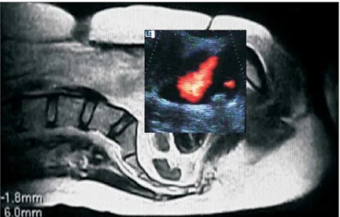
For the bladder ultrasound examination, the patients were orally hydrated with wa-ter (100 to 1,000 ml, according to the pa-tient age). With a partially filled bladder, ureteral orifices were localized by means of color Doppler ultrasound, with direct visu-alization of intermittent ureterovesical jets (left and right), which can be separately or simultaneously seen, projecting into the bladder, anteriorly to the pelvic cavity. Axially, the urinary jets project themselves
into the bladder forming acute angles on both sides of the vesical trigone (Figure 1). Sagittally, the urinary jets project them-selves from the upper portion of the trigone towards the craniocaudal and anteroposte-rior directions, forming an acute angle to the perineal region (Figure 2). By means of this approach, using the vesical floor as a reference point, the localization of ureteral orifices and urinary jet angulation were analyzed during a 10-minute period. Dur-ing this same period, urinary jets images were recorded in videocassette for angle calculations, with a protractor, and
calcu-lation of the distance between the ureteral orifices, with a pachymeter corrected for ul-trasound measurement scale.
The Mann-Whitney non-parametric test was employed for statistical analysis, with a p value of 0.05 being considered statisti-cally significant(14).
RESULTS
The previous voiding cystourethro-graphy in 32 patients had demonstrated the presence of grades I and II unilateral vesicoureteral reflux in 18 patients, and
Figure 1. Color Doppler ultrasound of bladder in the axial plane demonstrating bilateral ureterovesical jet.
427
Study of the ureterovesical jet by means of color Doppler
Radiol Bras 2006;39(6):425–428 bilateral in 8 patients, with predominance of grade 1 to the right, and grades II and III to the left (Table 1).
The ureterovesical jet mean angles in transverse and longitudinal planes, on both sides in all groups of patients with no re-flux, unilateral reflux or bilateral rere-flux, presented higher values on the right side than on its contralateral (Tables 2, 3 and 4). However, there was no statistically signifi-cant correlation between right and left sides.
In the present study, the distance be-tween ureteral meatus demonstrated a trend to be higher, although non-statistically sig-nificant (p = 0.41), in the group presenting bilateral reflux (Table 5).
DISCUSSION
With regard to the ureterovesical jet, it was bilaterally observed with color Dop-pler in all the patients, during the urinary bladder filling. For this, the bladder should not be completely filled and the patient should not be dehydrated(15,16). The
dura-tion, periodicity and size of the ureterovesi-cal jet varied on both sides(17,18).
The analysis of both right and left ure-terovesical jets angles, in transverse and longitudinal planes, presented high
vari-comparative mean between ureteral meatus in patients with and without reflux was sta-tistically significant, indicating that the ureteral meatus lateralization is a sign of predisposition to vesicoureteral reflux, since the ureters penetrate more into the vesical wall, reducing the distance from its intramural segment, and resulting in reflux. The present study has demonstrated a trend towards the existence of a correlation between the ureteral meatus lateralization and the reflux, in spite of not showing any significant difference between patients with and without bilateral reflux. This trend may present changes as the number of ana-lyzed patients increases, indicating that additional studies are necessary.
REFERENCES
1. Leung VYF, Metreweli C, Yeung CK. The ureteric jet Doppler as an indicator of vesicoureteric sphincter function in adults and children. An observational study. Ultrasound Med Biol 2002; 28:865–872.
2. Macedo CS, Riyuzo MC, Bastos HD. Vesicoure-teral grade I to III reflux disappearance frequency in pediatric patients. Rev Bras Saúde Mater In-fant 2004;4:299–307.
3. Gross GW, Lebowitz RL. Infection does not cause reflux. AJR Am J Roentgenol 1981;137:929–932. 4. Netter FH. Kidneys, ureters and urinary bladder. The Ciba collection of medical illustrations. Ro-chester, USA: Ciba, 1975;6:22–23.
5. Mcheik JN, Levard G. Vesicoureteral reflux: di-agnosis and management in children. Prog Urol 2002;12:646–650.
6. Souza AS. Uro-radiologia pediátrica. In: Prando A, Prando D, Caserta NMG, Bauab Jr T, editores. Urologia – diagnóstico por imagem. São Paulo. Sarvier, 1997;406–439.
7. Bjerklund Johansen TE. Diagnosis and imaging in urinary tract infections. Curr Opin Urol 2002; 12:39–43.
8. Dana A, Helenon O. Urinary tract imaging: con-ventional radiology and ultrasound. J Radiol 2004;85(2 Pt 2):159–168.
9. Radermacher J. Ultrasound of the kidney and re-nal vessels. I: Normal findings, congenital dis-eases, diseases of the kidney parenchyma. Inter-nist (Berl) 2003;44:1283–1297.
10. Rosi P, Del Zingaro M, Porena M. Ultrasound anatomy and normal ECD of the kidney. Arch Ital Urol Androl 2005;77:79–83.
11. Ramirez Lopera MC, Espla Garcia L. Pediatric serial urinary cystoureterography. Needs, nursing diagnosis, and care protocol. Rev Enferm 2002; 25:14–17.
12. Berrocal T, Rivas S, Jaureguizar E, et al. Contrast-enhanced sonourethrography versus conventional miction cystourethrography in the assessment of the urethra: preliminary study. Cir Pediatr 2004; 17:58–60.
13. Zar JH. Bioestatistical analysis. New Jersey: Pren-tice Hall, 1996;718.
14. Dubbins PA, Kurtz AB, Darby J, Goldberg BB. Ureteric jet effect: the echographic appearance of Table 1 Number of patients presenting
vesicouteral reflux at voiding cystourethrography and re-spective classification into grades.
Grades I II III Total RSU 2 2 0 4 LSU 3 3 0 6 RSB 5 1 2 8 LSB 1 4 3 8
RSU, right-sided unilateral; LSU, left-sided unilateral; RSB, right-sided bilateral; LSB, left-sided bilateral.
Table 2 Patients without vesicoureteral reflux (WO/ VUR). Urinary jet angle in transverse plane (right-sided – RUJAT; left-(right-sided – LUJAT) and longitudinal plane (right-sided – RUJAL; left-sided – LUJAL).
WO/VUR RUJAT LUJAT RUJAL LUJAL n 14 14 14 14 m 51° 42° 48° 37° sd 23° 22° 22° 17° vc 0.44 0.10 0.45 0.46
n, number of patients; m, mean angle; sd, standard deviation; vc, variation coefficient.
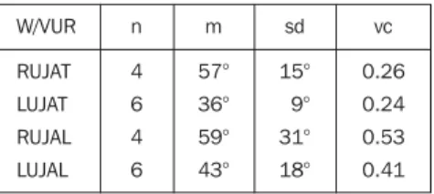
Table 3 Patients with vesicoureteral reflux (W/ VUR). Urinary jet angle in transverse plane (right-sided – RUJAT; left-(right-sided – LUJAT) and longitudinal plane (right-sided – RUJAL; left-sided – LUJAL).
W/VUR RUJAT LUJAT RUJAL LUJAL n 4 6 4 6 m 57° 36° 59° 43° sd 15° 9° 31° 18° vc 0.26 0.24 0.53 0.41
n, number of patients; m, mean angle; sd, standard deviation; vc, variation coefficient.
Table 4 Patients with bilateral vesicoureteral re-flux (W/BVUR). Urinary jet angle in transverse plane (right-sided – RUJAT; left-sided – LUJAT) and longi-tudinal plane (right-sided – RUJAL; left-sided – LUJAL). W/BVUR RUJAT LUJAT RUJAL LUJAL n 8 8 8 8 m 46° 35° 42° 27° sd 27° 21° 22° 10° vc 0.59 0.61 0.52 0.37
n, number of patients; m, mean angle; sd, standard deviation; vc, variation coefficient.
n, number of patients; m, mean angle; sd, standard deviation; vc, variation coefficient.
ability both in patients without reflux or with unilateral or bilateral reflux according to the previous voiding cystourethrography. For all the patients groups, right sided mean angles in axial and longitudinal planes pre-sented higher values than their contralat-eral, which is in disagreement with the lit-erature, where studies report that the higher the value of the angle of ureter entry into the bladder, the smaller the course of the intramural ureter, resulting in return of urine from the bladder back to the ure-ter(19,20). Also, in the present study, we have
observed a predisposition to predominance of left sided reflux in the group presenting unilateral reflux.
As regards the distance between ureteral meatus measured by means of color Dop-pler ultrasound, according Marshall(21), the
Table 5 Distances between ureteral meatus in patients without vesicoureteral reflux (WO/VUR), with vesicoureteral reflux (W/VUR), and with bilat-eral vesicouretbilat-eral reflux (W/BVUR).
428
Sakate M et al.
Radiol Bras 2006;39(6):425–428
urine entering the bladder. Radiology 1981;140: 513–515.
15. Jequier S, Paltiel H, Lafortune M. Ureterovesical jets in infants and children: duplex and color Dop-pler US studies. Radiology 1991;178:888–889. 16. Kusmic AC, Brkljacic B. Color Doppler ultra-sonography in the assessment of vesicoureteric reflux in children with bladder dysfunction. Pediatr Surg Int 2002;18:135–139.
17. Strehlau J, Winkler P, la Roche J. The uretero-vesi-cal jet as a functional diagnostic tool in childhood hydronephrosis. Pediatr Nephrol 1997;11:460– 467.
18. No authors listed. Medical versus surgical treat-ment of primary vesicoureteral reflux: report of the International Reflux Study Committee. Pedi-atrics 1981;67:392–400.
19. Hansson S, Jodal U. Urinary tract infection. In:
Avner ED, Harmon WE, Niaudet P, editors. Pe-diatric nephrology. 5th ed. Baltimore: Lippincott Williams & Wilkins, 2004;1008–1025. 20. Marshall JL, Johnson ND, De Campo MP.