Arq Neuropsiquiatr 2007;65(4-A):974-977
974
ULTRASTRUCTURAL STUDY OF THE LATERAL
VENTRICLE CHOROID PLEXUS IN EXPERIMENTAL
HYDROCEPHALUS IN WISTAR RATS
Daniela Pretti da Cunha Tirapelli
1, Luiza da Silva Lopes
2, João José Lachat
2,
Benedicto Oscar Colli
2, Luís Fernando Tirapelli
2ABSTRACT - Hydrocephalus is one of the most frequent and complex neurological diseases characterized by the abnormal buildup of cerebrospinal fluid (CSF) in the ventricles of the brain, due to an altered CSF dynamics. To detect possible ultrastructural alterations of the lateral ventricles choroid plexus (responsible for the CSF production), rats seven days after birth were submitted to an intracisternal injection of 20% ka-olim (hydrated aluminum silicate) for the hydrocephalus induction. Twenty-eight or 35 days after injection, injected animals and respective controls were processed for observation under a transmission electron mi-croscopy. Alterations found: presence of concentric cell membrane fragments, larger number of primary and secondary lysossomes, vacuoles, and cytoplasmic vesicles, and an enlargement of the intercellular space and between the basolateral interdigitation of the choroid epithelium. The alterations observed are prob-ably associated to an increase of the ventricular pressure, inducing morpho-functional effects on the chor-oid plexus integrity.
KEY WORDS: hydrocephalus, choroid plexus, ultrastructural study, rat.
Estudo ultraestrutural dos plexos corióides dos ventrículos laterais de ratos Wistar submetidos a hidroce-falia experimental
RESUMO - A hidrocefalia é uma das mais freqüentes e complexas doenças neurológicas caracterizada pelo acúmulo de líquido cefalorraquidiano (LCR) no interior dos ventrículos cerebrais e conseqüente alteração na dinâmica liquórica. Para detectar as possíveis alterações ultra-estruturais nos plexos corióides dos ven-trículos laterais (responsáveis pela produção do LCR), ratos sete dias após o nascimento, foram submeti-dos à indução de hidrocefalia pela injeção intracisternal de caulim a 20%.Após 28 e 35 dias da injeção, es-tes animais e seus respectivos controles foram processados para observação em um microscópio eletrôni-co de transmissão. Alterações observadas: presença de membranas eletrôni-concêntricas, maior número de lisosso-mos primários e secundários, vacúolos e vesículas citoplasmáticas, aumento do espaço intercelular e entre as interdigitações basolaterais das células do epitélio corióideo. As alterações observadas possivelmente es-tão associadas ao aumento da pressão nos ventrículos, induzindo efeitos morfo-funcionais na integridade dos plexos corióides.
PALAVRAS-CHAVE: hidrocefalia, plexos corióides, estudo ultra-estrutural, rato.
1Pos-graduate Student, Department of Surgery and Anatomy, Medicine School of Ribeirão Preto, University of São Paulo, Ribeirão
Preto SP, Brazil (FMRP/USP); 2Professor, Department of Surgery and Anatomy, FMRP/USP.
Received 11 April 2007, received in fi nal form 2 July 2007. Accepted 16 August 2007.
Dr. Luis Fernando Tirapelli - Departamento de Cirurgia e Anatomia / Campus Universitário / FMRP/USP - 14048-900 Ribeirão Preto SP - Brasil. E-mail: [email protected]
Hydrocephalus is one of the most frequent and complex neurological diseases characterized by the abnormal buildup of cerebrospinal fl uid (CSF) in the ventricles of the brain, due to an altered CSF dy-namics. The hydrocephalus is a result of multiple pathophysiological mechanisms, with many differ-ent causes including a birth defect, hemorrhage into the brain, infection, meningitis, tumor, or head inju-ry. In this way, the treatment possibilities as also the ideal time to introduce it has been widely studied1.
Kaolin has been widely used to induce
hydroceph-alus in several developmental stages of experimen-tal animals including neonates and adults. This mod-el of experimental induction was chosen due to it be-ing effi cient, inexpensive, and it does not need surgi-cal proceedings or cause anatomic modifi cations and does not even lead to other alterations unless those results that are totally of hydrocephalus2-7.
Arq Neuropsiquiatr 2007;65(4-A)
975
Hydrocephalus: choroid plexus ultrastructure Tirapelli et al.
are involved by hydrocephalus in its early stages8,9. Del
Bigio et al.10 and Kiefer et al.11 observed reduced
den-sity of ependymal microvilli, vacuolar formation and cellular inclusions on choroid cells, and intercellular space enlargement, after experimentally induced hy-drocephalus. Go et al.12 and Madhavi and Jacob13
ob-served the presence of Kolmer fagocytic cells on the ependymal cells surface and on the choroid cells in hydrocephalic animals. Also, by means of morphom-etry, these authors have found a signifi cant decrease of surface volume and area of the choroid epithelium in hydrocephalic animals, compared to controls. Alter-ations on the choroid plexus in hydrocephalic animals are not well established in the literature.
In order to contribute for a better understand-ing of the hydrocephalus pathologic alterations of brain structures, the aims of the present study were to detect, by means of transmission electron micros-copy (TEM), the alterations on lateral ventricles chor-oid plexus of Wistar rats, with experimentally induced hydrocephalus.
METHOD
Twenty-four litters of 7-day old Wistar rats were used, without sexual distinction. Each litter was constituted of the mother rat and 8 newborn rats. Two groups of animals were submitted to the hydrocephalus induction with
intracyster-nal injection of 20% kaolin (after 28 or 35 days of injection) and the third group was used as control14,15. Each pup rat was
held by an auxiliary, who held the head with one hand and the body with the other, bending the animal’s neck, leaving the posterior cervical area free. By palpation the space be-tween the posterior extremity of the foramen magnum, on the occipital bone, and the posterior arch of the fi rst cervical vertebra was identifi ed. With a Mise 0.3 odontologic needle, with a short bezel, the suboccipital percutaneous injection was done, and 0.4 mL of a kaolin suspension (Merck) diluted in distilled water of 20% was slowly injected. Following this the rats were put back into their cages with their mothers.
The animals of each group injected with kaolin, and their respective controls of the same age, were anesthetized with vapors of sulphuric ether and submitted to a transcardiac perfusion with PBS solution (about 1 mL/g of the animal’s weight), after the perfusion, with the same volume, with a 2% paraformaldehyde and 1% glutaraldehyde in a 0.1M, pH 7.4 phosphate buffer. The animals were decapitated, their brains removed in block by a craniectomy of the vertex and immersed in the same fi xative solution of formaldehyde and glutaraldehyde for more than 24 hours at 4oC.
After washing with a 0.1M phosphate buffer, the brains were sectioned at the coronal plane, and the choroid plex-uses identifi ed and removed. The samples from the choroid plexuses were fi xed in a 1% osmium tetroxide in a 0.2M phos-phate buffer for 2 hours, at 4oC, washed with a 0.1M
phos-phate buffer and then dehydrated in an increasing series of acetone and embedded in araldite. After the polymerization of the resin, the blocks were cut in sections of 0.5 µm
Arq Neuropsiquiatr 2007;65(4-A)
976
Hydrocephalus: choroid plexus ultrastructure Tirapelli et al.
fi nes) and after 500Å of thickness. The sections were stained with uranil and lead citrate and examined in a EM 208 Philips TEM. All experimental procedures were performed accord-ingly with the Ethical Principles in Animal Research adopted by Brazilian College of Animal Experimentation (COBEA) and was approved by the College of Medicine of Ribeirão Preto of the University of São Paulo – Ethical Commission of Ethics in Animal Research (CETEA), protocol n0 133/2006.
RESULTS
The choroid epithelium is ultrastructuraly de-scribed as simple columnar, presenting many microvilii on the ventricle surface and the occasional presence of cilia16 (Figs 1A and 1B). The cells cytoplasm presents
a large central nucleus, mitochondria, rough endo-plasmic reticulum, Golgi complex and occasional lys-ossomes. A joint complex is found on the apical junc-tion of the cells (Fig 1B) as a basolateral membrane
full of interdigitation (Fig 1C). Choroid epithelial cells lay on a dense basal lamina. Beneath this lamina, a small amount of loose connective tissue with collagen fi bers separate the basal lamina (Fig 1C) and the main capillary vessels, formed by a thin endothelium17-21.
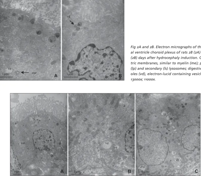
On animals of the control groups, no alterations were seen. On both experimental groups (28 and 35 days after kaolin injection), the following alterations were found: presence of concentric cell membrane fragments, similar to the myelin sheath on the cyto-plasm (Fig 2A), larger number of primary and second-ary lysossomes (Figs 2A and 2B), vacuoles (Fig 2B), and cytoplasmic vesicles of different sizes, containing an electron-lucid material (Figs 2A, 3A and 3B). On ani-mals 35 days after kaolin injection, an enlargement of the intercellular space and between the basolater-al interdigitation of the choroid epithelium was basolater-also
Fig 2A and 2B. Electron micrographs of the later-al ventricle choroid plexus of rats 28 (2A) and 35 (2B) days after hydrocephaly induction. Concen-tric membranes, similar to myelin (me); primary (lp) and secondary (ls) lysosomes; digestive vacu-oles (vd), electron-lucid containing vesicles (ve). 13000x; 11000x.
Arq Neuropsiquiatr 2007;65(4-A)
977
Hydrocephalus: choroid plexus ultrastructure Tirapelli et al.
seen (Figs 3A and 3C).
DISCUSSION
Among the main functions of the choroid plexus is the CSF production. Nevertheless, it is well known the role of the choroid plexus in the nutrition and protection of the CNS since the CSF is a chemically stable fl uid19,21. The choroid plexus is able to transfer
nutrients to the CSF, actively regulating the molecules concentration of this fl uid and determining the se-lective characteristics of the blood-CSF barrier21. The
choroid plexus also present a role on the CSF clearance of drugs and substances formed on the brain tissue due to the metabolic reactions, thus presenting sev-eral active-transport systems9,21. The fi rst structure to
suffer on the hydrocephalus process in high CSF pres-sure is the ependymal epithelium, with compression, distension and isolated disruption10,11.
Go e tal.12 and Go and Molenaar22 observed the
in-festation of Kolmer fagocytic cells on the ependymal cells and choroid cells surface, as well as a degenera-tion of ependymal cilia. The fragments observed on the choroid plexus surface of hydrocephalic animals are consequence of an impaired ciliar movement due to the ciliar degeneration described previously12,22. In
this way, an altered CSF dynamics and the CSF accu-mulation on an enlarged ventricular system impairs the correct elimination of the metabolic products23.
Del Bigio et al.10, Madhavi and Jacob13 and
Kief-er et al.11 have observed alterations on the choroid
epithelium such as microvilii distortion, atypical cilia, fl atten cells with and vacuoles, intracellular inclusions and enlargement of intercellular space. Some of these alterations were also seen on the present study. In this way, the vesicles containing electron-lucid material observed on the choroid cells might be the result of a fl uid penetration into the cytoplasm. The presence of a large amount of lysossomes, secondary lysosomes and digestive vesicles might suggest that there is a se-cretion and absorption impairment of CSF products or the cytoplasmic organelles death. In a morphometric study, Madhavi and Jacob24,25 have shown a signi
fi -cant decrease on the volume and area of the choroid epithelium in hydrocephalic animals. Also, these au-thors shoed a reduction of the mitochondrial internal membrane area, including the crests, which was at-tributed to a decreased activity of the choroid cells, followed by a reduction on the CSF secretion on those animals.
In conclusion, the choroid plexus is the main struc-ture responsible for production and maintenance of
the CSF homeostasis, an essential factor for the de-velopment and normal functioning of the CNS. In this way, the alterations observed in this structure are as-sociated to an increase of the ventricular pressure, inducing morpho-functional effects on the choroid plexus integrity, contributing to several morpho-func-tional alterations of the CNS26.
REFERENCES
1. Sato O, Oi S, Yamada S. Hydrocephalus: experimental considerations and clinical analyses. In Choux M, Di Rocco C, Hockley AD, Walker ML (Eds). Pediatric neurosurgery. London: Churchill Livingstone, 1999:237-252. 2. Del Bigio MR, Massicot e EM. Protective eff ect of nimodipine on
be-havior and white matter of rats with hydrocephalus. J Neurosurg 2001;94:788-794.
3. Khan OH, Enno TL, Del Bigio MR. Brain damage in neonatal rats follow-ing kaolin induction of hydrocephalus. Exp Neurol 2006;200:311-320. 4. Kondziella D, Ludemann W, Brinker T, Sletvold O, Sonnewald U.
Al-terations in brain metabolism, CNS morphology and CSF dynamics in adult rats with kaolin-induced hydrocephalus. Brain Res 2002;8:35-41. 5. Kuchiwaki H, Nagasaka M, Inao S, Sugita K. Progression of kaolin-in-duced hydrocephalus and changes in performance of operant tasks by rats. J Neurol Sci 1994;121:32-38.
6. Matsumoto S, Hirayama A, Yamasaki S, Shirataki K, Fujiwara K. Com-parative study of various models of experimental hydrocephalus. Childs Brain 1975;1:236-242.
7. Tashiro Y, Chakrabort y S, Drake JM, Hat ori T. Progressive loss of glu-tamic acid decarboxylase, parvalbumin, and calbindin D28K immuno-reactive neurons in the cerebral cortex and hippocampus of adult rat with experimental hydrocephalus. J Neurosurg 1997;86:263-271. 8. Del Bigio MR, MCallister JP. Hydrocephalus: pathology. In Choux M, Di
Rocco C, Hockley AD, Walker ML (Eds). Pediatric neurosurgery. Lon-don: Churchill Livingstone, 1999:217-236.
9. Machado A. Neuroanatomia funcional. 2.Ed. Rio de Janeiro: Editora Atheneu, 2000.
10. Del Bigio MR, Bruni JE, Fewer HD. Human neonatal hydrocephalus: an electron microscopic study of the periventricular tissue. J Neurosurg 1985;63:56-63.
11. Kiefer M, Eymann R, Von Tiling S, Muller A, Steudel I, Booz KH. The ependyma in chronic hydrocephalus. Child’s Nerv Syst 1998;14:263-270.
12. Go KG, Stokroos I, Blaauw EH, Zuiderveen F, Molenaar I. Changes of ventricular ependyma and choroids plexus in experimental hydroceph-alus, as observed by scanning electron microscopy. Acta Neuropathol 1976;34:55-64.
13. Madhavi C, Jacob M. Atypical cilia in the choroid plexus of guineapig. Indian J Med Res 1989;90:484-489.
14. Lopes LS, Machado HR, Lachat JJ. Study of corpus callosum in experi-mental hydrocephalic wistar rats. Acta Cirurg Bras 2003;18:10-14. 15. Lopes LS, Machado HR, Lachat JJ. Hidrocefalia experimental. In
Cas-tro e Silva O Jr., Zucoloto S, Beer A Jr. (Eds). Modelos experimentais de pesquisa em cirurgia. São Paulo: Robe Editorial, 1998:753-772. 16. Junqueira LCU, Carneiro J. Histologia básica. 9.Ed. Rio de Janeiro:
Ed-itora Guanabara Koogan SA, 1999.
17. Meek WJ. A study of the choroid plexus. J Comp Neurol Psychol 1907;17:286-306.
18. Zimman L. Investigaciones sobre la estractura de los plexos coroideos em estado normal y patológico. Arch Histol 1943;1:278-328.
19. Dohrmann GJ. The choroid plexus: a historical review. Brain Res 1970; 18:197-218.
20. Peters A. The surface fi ne structure of the choroid plexus and ependy-mal lyning of the rat lateral entricle. J Neurocytol 1974;3:99-108. 21. Spector R, Johanson CE. Plexos coroideos de los mamíferos. Invest
Cien-cia 1989;226:44-51.
22. Go KG, Molenaar I. Some applications of scanning electron microsco-py for the study of biopsies in central nervous system pathology. Scan Electron Microsc 1983;143-150.
23. Castro-Gago M, Rodriguez IN, Rodriguez-Nunez A, Guitian JP, Ro-camonde SJ, Roodriguez-Segade S. Therapeutic criteria in hydrocepha-lic children. Child´s Nerv Syst 1989;5:361-363.
24. Madhavi C, Jacob M. Morphometry of choroid plexus in hydrocephal-ic guineapigs. Indian J Med Res 1990;92:89-94.
25. Madhavi C, Jacob M. Morphometry of mitochondria in the choroidal ependyma of hydrocephalic guineapigs. Indian J Med Res 1992;96:72-77.