Arq Neuropsiquiatr 2007;65(4-A):951-954
951
MOTOR LEARNING PROCESSES
An electrophysiologic perspective
Bruna Velasques
1,6, Camila Ferreira
1, Silmar Silva Teixeira
5,
Vernon Furtado
5, Elizabeth Mendes
5, Luis Basile
2, Mauricio Cagy
1,4,5,
Roberto Piedade
1,6, Pedro Ribeiro
1,3,4,5,6ABSTRACT - The goal of the present study was to investigate electrophysiologic, qEEG, changes when indi-viduals were exposed to a motor task. Subjects’ brain electrical activity was analyzed before and after the typewriting training task. For the neurophysiological variable asymmetry, a paired t-test was performed to compare each moment, pre and post-task, in the beta bands. The findings showed a change for the qEEG variable in each scalp site, F3/F4; C3/C4 and P3/P4. These results suggest an adaptation of pre-frontal, senso-ry-motor and parietal cortex, as a consequence of the typewriting training.
KEY WORDS: sensory-motor integration, procedural learning, qEEG.
Processos de aprendizagem motora: uma perspectiva eletrofisiológica
RESUMO - O objetivo do presente estudo foi investigar mudanças eletrofisiológicas através do EEGq quan-do indivíduos são expostos a uma tarefa motora. A atividade elétrica no córtex quan-dos sujeitos foi analisada antes e após o treinamento da tarefa motora. Para a variável neurofisiológica assimetria, um teste t foi im-plementado para comparar cada momento, pré e pós-tarefa, na banda beta. Os achados demonstraram mudança em assimetria para as seguintes regiões no escalpo: F3/F4, C3/C4 e P3/P4. Estes resultados suge-rem uma adaptação das regiões pré-frontal, somatosensorial e parietal como conseqüência do treinamen-to de datilografia.
PALAVRAS-CHAVE: integração sensório-motora, memória de procedimento, EEGq.
1Laboratório de Mapeamento Cerebral e Integração Sensório-Motora, Programa de Pós-Graduação em Psiquiatria e Saúde Mental
(PROPSAM), Instituto de Psiquiatria da Universidade do Brasil (IPUB), Universidade Federal do Rio de Janeiro, Brasil (UFRJ); 2Divisão
de Neurocirurgia Funcional, Instituto de Psiquiatria, Escola de Medicina, Universidade de São Paulo, Brasil (USP); 3Departamento
de Biociências da Atividade Física, Escola de Educação Física e Desportos (EEFD), UFRJ, Brasil; 4Departamento de Epidemiologia
e Bioestatística, Instituto de Saúde da Comunidade, Universidade Federal Fluminense, Rio de Janeiro, Brasil (UFF); 5Programa de
Pós-Graduação Strictu Sensu em Ciência da Motricidade Humana (PROCIMH), Universidade Castelo Branco, Rio de Janeiro, Brasil;
6Instituto Brasileiro de Biociências Neurais (IBBN), Rio de Janeiro, Brasil.
Received 16 January 2007, received in fi nal form 6 June 2007. Accepted 6 August 2007.
Dra. Bruna Brandão Velasques - Rua Paula Brito 350 / 1102 - 20541-190 Rio de Janeiro RJ - Brasil. E-mail: [email protected] To maintain stability at a highly dynamic
environ-ment, the central nervous system (CNS) is in constant activity. It continuously receives external sensory stim-uli, many specifi cally required to maintain motor per-formance1-3. Many studies have demonstrated that
pre-cision during the motor gesture is increased as conse-quence of motor learning4,5. Motor learning promotes
a gradual minimization of task errors, an increase in coordination, agility and movement execution6.
Different mechanisms take part in the complexi-ty of motor learning which involves various levels of cortical structures, such as: pre frontal areas related to decision making, contralateral primary motor cortex7,
ipsilateral primary motor cortex, supplementary motor area, pre motor area, primary sensory areas8, and the
parietal region responsible for information integra-tion processes. The different funcintegra-tional components and the plastic reorganization of the CNS have lead to investigations objecting the examination of neu-rofunctional phenomena involving motor learning9.
Hence, this study aimed at investigating how par-ticipative is the learning of a motor task in the cortex organizational mapping. For that, we used quantita-tive electroencephalography (qEEG) to detect neural changes during the motor learning process10. Beta
ac-tivity was specifi cally investigated, since it is respon-sive to movements and electro-stimulation of limbs11,12.
METHOD
Arq Neuropsiquiatr 2007;65(4-A)
952
Motor learning processes Velasques et al.
in several other studies13-15. Thus, the methods will be
sum-marized below.
The sample was composed for 29 healthy individuals, both sexes, with ages varying between 20 to 40 years, ab-sence of mental and physical illness (previous anamnese), right handed (Edinburgh)16, and do not making use of any
psychoactive or psychotropic substance during the whole time of the study. The experiment consisted of a task of a typewriting method of progressive learning, in which train-ing was performed on a strain-ingle day. The exercise was made up of four blocks, each block represented by twelve lines. Each line had fi ve sequences of letters for each hand.
Spatial electrode localization and frequency bands – Three areas of interest were investigated: pre frontal, cen-tral and parietal. Pre frontal area is related to motivation, planning and decision making. Central area is associated
with sensory reports of motor gesture and execution of vol-untary movements, corresponding to the somatosensory and primary motor cortex. The parietal region, including the posterior-parietal cortex relates to sensory and attention in-tegration processes. The beta band (13–25 Hz) was than se-lected due to its relation to somatomotor processes.
Statistical analysis – As electrodes have different scalp (spatial) positions, we have chosen an independent statis-tical analysis. A t-test was employed for each electrode at beta (F3-F4/C3-C4/P3-P4).
RESULTS
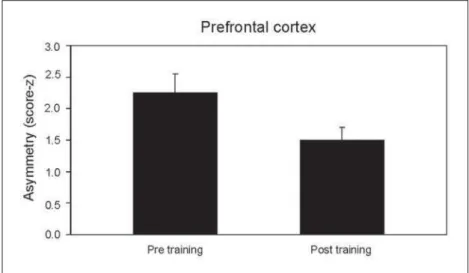
Neurophysiological variables – Figure1 describes
the variation in asymmetry between pre and post training times at F3/F4. Statistical analysis has
dem-Fig 1. Asymmetry differences in beta between the pre and post training times (cen-tral cortex).
Arq Neuropsiquiatr 2007;65(4-A)
953
Motor learning processes Velasques et al.
onstrated a signifi cant difference between two ex-perimental times (p=0.003).
Figure 2 displays the variation in asymmetry be-tween the pre and post training times at C3/C4, with a signifi cant difference of (p=0.019).
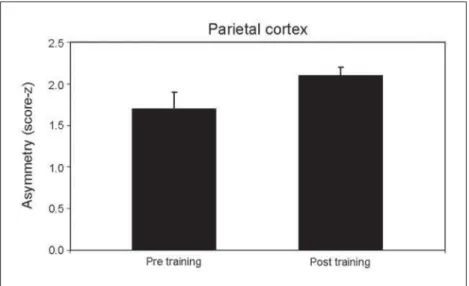
Figure 3 represents the oscillation in asymmetry between pre and post training times P3/P4. Statistical difference was also signifi cant (p=0.025).
DISCUSSION
This investigation aimed at examining electro-physiological alterations produced by a learning task through quantitative electroencephalography. The discussion will be presented following the results ap-pearance. Hence, the fi rst section elaborates on the participation of the prefrontal cortex in planning and decision making processes. The second section conjec-tures over the possible plastic alterations occurring in the somatosensory cortex as a consequence of the motor task. The third section focus on the electroen-cephalographic outcomes regarding changes in the parietal cortex.
Prefrontal cortex: memory and planning – The
pre-frontal cortex is responsible for anticipation of conse-quences, planning and organizing strategies17,18. Our
results show an increased hemispheric asymmetry at prefrontal regions following the two-hour typewrit-ing task. Since all individuals had a prior experience with typewriting, it was assumed that they were all at the so-called “controlled stage” of learning19. This
stage is associated to initial periods of learning where subjects divide the attention focus with differentiated elements of the task and the environment2, leading
to reduced motor coordination, increased number of mistakes and execution time. As observed in the results, such increase in symmetry suggests changes in the representation of neuronal activity at the pre-frontal cortex, as noticed by other investigations18,20.
The prefrontal cortex integrates with the limbic asso-ciative cortex, connecting directly to limbic structures as the amigdala and the cingulate cortex. Therefore, the results imply changes in structures associated with procedural memory, in particular the way informa-tion is registered21,22.
Somatosensorycortex: plastic alterations as a
conse-quence of the task – Results demonstrate that the
two-hour typewriting training produced asymmetry chang-es at C3/C4, suggchang-esting a reorganization of neuronal activity at the somatosensory cortex. Previous studies have observed such alterations as a consequence of sequential repeated fi nger movements23,24. It is
impor-tant to remind that these investigations used animals and that they trained for months. Experimental pro-portions must be considered since training mode in pri-mates (monkeys) is different from humans and gesture specifi city between species is a key factor in cortical representation25. The reason between cortical areas,
as expressed in asymmetry (C3/C4), detects changes in the relation between the two hemispheres after the typewriting task. This allegedly means that increased symmetry between regions suggests a reorganization of the supposed interaction between the two hemi-spheres13. It is essential, however, to replicate these
fi ndings employing other neuroimaging techniques since the spatial resolution of EEG does not allow a precise cortical identification of hands and fingers. Fig 3. Asymmetry differences in beta between the pre and post training times
Arq Neuropsiquiatr 2007;65(4-A)
954
Motor learning processes Velasques et al.
Posterior parietal cortex: attention and sensory
in-tegration processes – Our results show reduced
asym-metry at P3/P4. Beta activity is related to stimulation processes and voluntary movements26,27. Posterior
pa-rietal cortex (Brodmann areas 5 and 7) is located next to the somatosensory primary area (S-1) and possesses neurons with great receptive fi elds, which allows this region to specialize in differentiated and complex ac-tivities. The parietal cortex has a convergence site of simple and segregated sensory stimuli, functioning as a multiple integration organization28,29. Therefore,
the parietal region is associated to visual and motor information, also waking and attention mechanisms as well30. The reduced asymmetry values suggest
pos-sible changes in somatosensory and visual integra-tion processes. Particularly, neurons in the area 5 col-lect information from different articulations or arm muscle groups; and neurons in the area 7 integrate tactile and visual stimuli, and participate actively in the eye-hand coordination31. The posterior parietal
cortex also receives visual communication regarding the representation of the visual world and movement planning. Consequently, such variation in asymmetry might represent a task automaticity process32.
REFERENCES
1. Teasdale N, Simoneau M. At entional demands for postural control: the eff ects of aging and sensory reintegration. Gait Posture 2001;4:203-210. 2. Schmidt R, Wrisberg C. Aprendizagem e performance motora: uma
abordagem da aprendizagem baseada no problema. 2.Ed. Porto Ale-gre: Artmed Editora, 2001.
3. Ladewing I. A importância da atenção na aprendizagem de habilidades motoras. Rev Paul Educ Fis 2000;2(Supl):S62-S71.
4. Jenkins I, Passingham R, Brooks D. The eff ect of movement frequen-cy on cerebral activation: positron emission tomography. J Neurol Sci 1997;151:195-205.
5. Jones E. Cortical and subcortical contributions to activity-depen-dent plasticity in primate somatosensory cortex. Annu Rev Neurosci 2000;23:1-17.
6. Karni A, Meyer G, Jezzard P, Adams MM, Turner R, Ungerleider LG. Functional MRI evidence for adult motor cortex plasticity during mo-tor skill learning. Nature 1995;377:155-158.
7. Sommer M, Classen J, Cohen LG, Hallet M. Time course of determina-tion of movement direcdetermina-tion in the reacdetermina-tion time task in humans. J Neu-rophysiol 2001;86:1195-1201.
8. Georgiadis M, Cramon D. Motor-learning-related changes in piano play-ers and non-musicians revealed by functional magnetic-resonance sig-nals. Exp Brian Res 1999;125:417-425.
9. Schmidt RA. A schema theory of discrete motor skill learning. Psychol Rev 1975;84:225-260.
10. Gong P, Nikolaev A, Leeuwen C. Scale-invariant fl uctuations of
dynam-ical synchronization in human brain electrdynam-ical activity. Neurosci Let 2003;336:33-36.
11. Alegre M, Labarga A, Gurtubay IG, Iriarte J, Malanda A, Artieda J. Movement-related changes in cortical oscillatory activity in ballistic, sustained and negative movements. Exp Brain Res 2003;148:17-25. 12. Pfurtsh eller G, Graimann B, Huggins JE, Levine SP, Sh uh LA.
Spa-tiotemporal pat erns of beta desynchronization and gamma synchroni-zation in corticographic data during self-paced movement. Clin Neu-rophysiol 2003;114:1226-1236.
13. Cunha M, Bastos VH, Veiga H, et al. Alterações na distribuição de potên-cia cortical em função da consolidação da memória no aprendizado de datilografi a. Arq Neuropsiquiatr 2004;62:662-668.
14. Portella CE, Silva JG, Bastos VH, et al. Procedural learning and anxiolyt-ic eff ects: electroencephalographic, motor and at entional measures. Arq Neuropsiquiatr 2006;64:478-484.
15. Cunha M, Machado D, Bastos VH, et al. Neuromodulatory eff ect of bromazepam on motor learning: an electroencephalographic approach. Neurosci Let 2006;407:166-170.
16. Oldfi eld R. The assessment and analysis of handedness: the Edinburgh
inventory. Neuropsychology 1971;9:97-113.
17. Miller EK. The prefrontal cortex: complex neural properties for complex behavior. Neuron 1999;22:15-17.
18. Miller A, Tomarken A. Task-dependent changes in frontal brain asym-metry: eff ects of incentive cues, outcome expectancies, and motor re-sponses. Psychophysiology 2001;38:500-511.
19. Mars RB, Coles MG, Grol MJ, et al. Neural dynamics of error process-ing in medial frontal cortex. Neuroimage 2005;4:1007-1013.
20. Gotlib IH, Rosenfeld CJP. Frontal EEG alpha asymmetry, depression and cognitive functioning. Cogn Emotion 1998;12:449-478.
21. Sakai K, Hikosaka O, Miyauchi S, Takino R, Sasaki Y, Putz B. Transition of brain activation from frontal to parietal areas in visuomotor sequence learning. J Neurosci 1998;5:1827-1840.
22. Boet iger CA, D’Esposito M. Frontal networks for learning and executing arbitrary stimulus-response associations. J Neurosci 2005;25:2723-2732. 23. Fitzgerald PJ, Lane JW, Thakur PH, Hsiao SS. Receptive fi eld properties
of the macaque second somatosensory cortex: representation of orienta-tion on diff erent fi nger pads. J Neurosci 2006;26:6473-6484.
24. Hluschuk Y, Hari R. Transient suppression of ipsilateral primary somatosen-sory cortex during tactile fi nger stimulation. J Neurosci 2006;26:5819-5824.
25. Gandolfo F, Li C, Benda B, Schioppa C, Bizzi E. Cortical correlates of learning in monkeys adapting to a new dynamical environment. Proc Natl Acad Sci 2000;29:2259-2263.
26. Neuper C, Pfurtscheller G. Event-Related dynamics of cortical rhythms: frequency specific and functional correlates. Int J Psychophysiol 2001;43:41-58.
27. Stancak A Jr, Pfurtscheller G. Event-related desynchronisation of cen-tral beta rhythms during brisk and slow self-paced fi nger movements
of dominant and nondominant hand. Brain Res Cogn 1996;4:171-183. 28. Iacoboni M, Zaidel E. Interhemispheric visuo-motor integration in humans:
the role of the superior parietal cortex. Neuropsychology 2004;42: 419-425. 29. Assmus A, Marshall JC, Ritzl A, Noth J, Zilles K, Fink GR. Let inferi-or parietal cinferi-ortex integrates time and space during collision judgments. Neuroimage 2003;20:82-88.
30. Schubert T, Von Cramon DY, Niendorf T, Pollmann S, Bublak P. Corti-cal areas and the control of self-determined fi nger movements: an fMRI
study. Neuroreport 1998; 9:3171-3176.
31. Inoue K, Kawashima R, Sugiura M, et al. Activation in the ipsilater-al posterior parietipsilater-al cortex during tool use: a PET study. Neuroimage 2001;14:1469-1475.