BRAIN TUMORS IN THE FIRST THREE YEARS OF LIFE
A review of twenty cases
Adriana Ávila de Espíndola
1, Hamilton Matushita
1, Juliana Martins Pimenta
2,
Ana Cristina da Silva Fernandes
2, Sérgio Rosemberg
3, Umbertina Conti Reed
1ABSTRACT - Objective and Method: To review the clinical and neuropathological findings as well as the type of therapy and outcome in 20 infants under 3 years-old with central nervous system (CNS) tumor. They were treated at the Department of Neurology, “Hospital das Clínicas” University of São Paulo Medical School, from January 1997 to May 2001. Results: Astrocytoma was the most common histological type (n=7), fol-lowed by ependymoma (n=3), medulloblastoma (n=2), craniopharyngioma (n=2) and desmoplastic ganglio-glioma (n=2). The location of the tumor was predominantly supratentorial. Mean follow-up time was 20.2 months with recurrence in 7 cases. For each type of tumor we have emphasized the treatment currently recommended. Conclusion: Although follow-up time is not sufficient for analyzing survival, a trend of im-provement in prognosis was noted, compared to another series of cases from our Institution that had been evaluated before 1990.
KEY WORDS: brain tumors, children, central nervous system tumors, neuro-oncology treatment.
Tumores do sistema nervoso central nos primeiros três anos de vida: revisão de vinte casos
RESUMO - Objetivo e Método: Avaliar os aspectos clínicos e histopatológicos, o tipo de tratamento e a evo-lução de 20 crianças menores de três anos de idade, com o diagnóstico de tumor de sistema nervoso cen-tral, que foram tratadas em nossa Instituição no período de janeiro de 1997 a maio de 2001. Resultados: O astrocitoma foi o tumor mais comum (n=7), seguido pelo ependimoma (n=3), meduloblastoma (n=2), cra-niofaringioma (n=2) e ganglioglioma desmoplásico infantil (n=2). A localização do tumor foi predominan-temente supratentorial. A média de seguimento foi 20,2 meses e houve recidiva em sete casos. Para cada tipo de tumor enfatizamos o tipo de tratamento recomendado na atualidade. Conclusão: Embora o tem-po de seguimento não seja suficiente, ainda, para analisar a sobrevida, foi notada nítida tendência a me-lhor prognóstico em comparação com a casuística proviniente de nossa Instituição que analisou casos abor-dados antes da década de 90.
PALAVRAS-CHAVE: tumores cerebrais, crianças, tumores do sistema nervoso central, tratamento neuroon-cológico.
1Neurology Department, University of São Paulo Medical School, São Paulo SP, Brazil (FMUSP); 2Medical Student, FMUSP; 3 Pathol-ogy Department, FMUSP.
Received 10 November 2006, received in fi nal form 20 July 2007. Accepted 23 August 2007.
Dra. Adriana Ávila de Espíndola - Avenida Dr. Enéas de Carvalho Aguiar 255 / 5º andar / sala 5031 - 05403-000 São Paulo SP - Brasil. E-mail: [email protected]
Nearly 8 to 20% of central nervous system (CNS) tumors occur in children under three years of age1.
The prognosis is usually poor2 due to diffi culties in
surgical technique, the malignant histology most of these tumors and the impossibility of treating with radiotherapy3,4. The rate of survival is low, around 20
to 40%4,5; however, after 1980, some institutions have
obtained better rates of prognosis, reporting 56% of survival at 5 years and normal neurological develop-ment after treatdevelop-ment in 50% of the patients6-8. In
small infants the most common histological types are ependymoma, medulloblastoma, choroid plexus pap-illoma, astrocytoma and teratoma. Supratentorial
tu-mors are almost twice as frequent, particularly in the fi rst year of life9,10.
Clinical signs and symptoms are usually not spe-cifi c, therefore causing diagnostic delay. The most common clinical manifestations depend on in-creased intracranial pressure, vomiting, macroceph-aly, drowsiness/lethargy and delay in neurological development9,11. Focal neurological changes are more
commonly observed in the second year of life10.
METHOD
From January 1997 to May 2001, 20 children under three years of age at diagnosis among 84 with CNS tumor were treated in the Department of Neurology, “Hospital das Clíni-cas”, University of São Paulo Medical School (FMUSP).
The diagnosis in all patients was established by mag-netic resonance imaging (MRI). The following items were analyzed: patient’s age and sex, topography and histolo-gy of the tumors, clinical features until the diagnosis, re-sults achieved by surgeries, surgical complications, adjuvant therapies, management of recurrences, and follow-up of the patients.
Tables 1 and 2 exhibit these variables
This study was approved by the Ethics Committee for re-search projects of our Institution protocol number 015/99.
RESULTS
Patients´ages ranged from 6 months to 35 months and the group included 13 boys and 7 girls. In 15 cases
(75%) the tumor was supratentorial, in 4 (20%) it was infratentorial and in one (5%) it was spinal.
The most common histological type was astrocyt-ic in 7 cases, followed by ependymoma in three cas-es and two of each of the following: medulloblas-toma, craniopharyngioma and desmoplasic infantile ganglioglioma. The other tumors were neurofi bro-ma, choroid plexus carcinobro-ma, choroid plexus papil-loma and atypical teratoid/rhabdoid tumor, one case of each. The astrocytic tumors were classifi ed as: 3 pilocytic astrocytomas, 2 low grade astrocytomas, one subependymal giant cell astrocytoma in a pa-tient with tuberous sclerosis and one mixed oligoas-trocytoma.
The length of clinical complaints before diagnosis varied from one day to 17 months (mean 4.36 months). The fi rst clinical manifestations were: epileptic
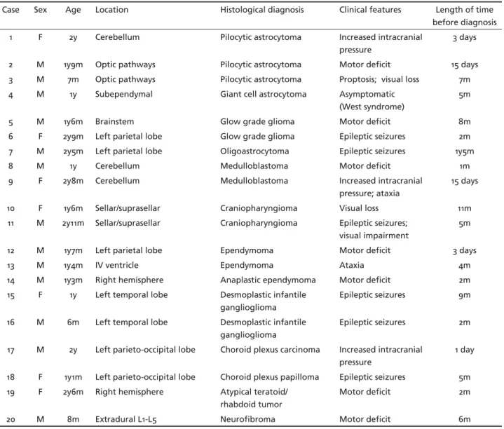
sei-Table 1. Central nervous system tumors: location, histological diagnosis and clinical features in 20 children under 3 years of age.
Case Sex Age Location Histological diagnosis Clinical features Length of time before diagnosis 1 F 2y Cerebellum Pilocytic astrocytoma Increased intracranial
pressure
3 days
2 M 1y9m Optic pathways Pilocytic astrocytoma Motor defi cit 15 days 3 M 7m Optic pathways Pilocytic astrocytoma Proptosis; visual loss 7m 4 M 1y Subependymal Giant cell astrocytoma Asymptomatic
(West syndrome)
5m
5 M 1y6m Brainstem Glow grade glioma Motor defi cit 8m
6 F 2y9m Left parietal lobe Glow grade glioma Epileptic seizures 2m 7 M 2y5m Left parietal lobe Oligoastrocytoma Epileptic seizures 1y5m
8 M 1y Cerebellum Medulloblastoma Motor defi cit 1m
9 F 2y8m Cerebellum Medulloblastoma Increased intracranial pressure; ataxia
15 days
10 F 1y6m Sellar/suprasellar Craniopharyngioma Visual loss 11m
11 M 2y11m Sellar/suprasellar Craniopharyngioma Epileptic seizures; visual impairment
5m
12 M 1y7m Left parietal lobe Ependymoma Motor defi cit 3 days
13 M 1y4m IV ventricle Ependymoma Ataxia 4m
14 M 1y3m Right hemisphere Anaplastic ependymoma Motor defi cit 2m 15 F 1y Left temporal lobe Desmoplastic infantile
ganglioglioma
Epileptic seizures 9m
16 M 6m Left temporal lobe Desmoplastic infantile ganglioglioma
Epileptic seizures 2m
17 M 2y Left parieto-occipital lobe Choroid plexus carcinoma Increased intracranial pressure
1 day
18 F 1y1m Left parieto-occipital lobe Choroid plexus papilloma Epileptic seizures 5m 19 F 2y6m Right hemisphere Atypical teratoid/
rhabdoid tumor
Motor defi cit 2m
20 M 8m Extradural L1-L5 Neurofi broma Motor defi cit 6m
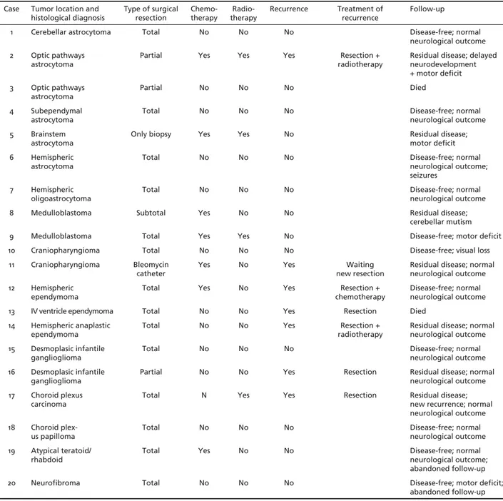
Table 2. Central nervous system tumors: type of treatment and follow-up in 20 children under 3 years of age.
Case Tumor location and
histological diagnosis Type of surgical resection Chemo-therapy therapyRadio- Recurrence Treatment ofrecurrence Follow-up
1 Cerebellar astrocytoma Total No No No Disease-free; normal neurological outcome 2 Optic pathways
astrocytoma
Partial Yes Yes Yes Resection + radiotherapy
Residual disease; delayed neurodevelopment + motor defi cit 3 Optic pathways
astrocytoma
Partial No No No Died
4 Subependymal
astrocytoma Total No No No Disease-free; normal neurological outcome 5 Brainstem
astrocytoma
Only biopsy Yes Yes No Residual disease; motor defi cit 6 Hemispheric
astrocytoma
Total No No No Disease-free; normal
neurological outcome; seizures
7 Hemispheric oligoastrocytoma
Total No No No Disease-free; normal
neurological outcome 8 Medulloblastoma Subtotal Yes No No Residual disease;
cerebellar mutism 9 Medulloblastoma Total Yes Yes No Disease-free; motor defi cit 10 Craniopharyngioma Total No No No Disease-free; visual loss 11 Craniopharyngioma Bleomycin
catheter
Yes No Yes Waiting
new resection
Residual disease; normal neurological outcome 12 Hemispheric
ependymoma
Total Yes No Yes Resection + chemotherapy
Disease-free; normal neurological outcome 13 IV ventricle ependymoma Total No No Yes Resection Died
14 Hemispheric anaplastic
ependymoma Total No No Yes radiotherapyResection + Residual disease; normal neurological outcome 15 Desmoplasic infantile
ganglioglioma
Total No No No Disease-free; normal
neurological outcome 16 Desmoplasic infantile
ganglioglioma Partial No No Yes Resection Residual disease; normal neurological outcome 17 Choroid plexus
carcinoma
Total N Yes Yes Resection Residual disease; new recurrence; normal neurological outcome 18 Choroid
plex-us papilloma Total No No No Disease-free; normal neurological outcome 19 Atypical teratoid/
rhabdoid
Total Yes No No Disease-free; normal
neurological outcome; abandoned follow-up 20 Neurofi broma Total No No No Disease-free; motor defi cit;
abandoned follow-up
zures in 7 patients, motor defi cit in 7, severe visual defi cit in three, increased intracranial pressure in two and ataxia in two patients. The diagnosis in the pa-tient with tuberous sclerosis was made during inves-tigation for West syndrome.
All patients underwent surgery for tumor removal. Total excision was achieved in 14 patients (70%) and a subtotal removal (at least 90%) in one patient with medulloblastoma (5%). Partial removal, (i.e. from 50 to 90%), was obtained in three patients (15%), two of them with optic pathways astrocytoma and one with desmoplasic infantile ganglioglioma. Biopsy, cor-responding to less than 50% of tumor resection was performed in one patient with low grade brainstem
astrocytoma (5%) and in one patient with craniophar-yngioma (5%) a catheter was employed for bleomy-cin infusion.
Surgical complications occurred in 5 pa-tients (25%): two papa-tients with medulloblas-toma had cerebellar mutism; one patient with craniopharyngioma manifested panhypopitu-itarism; one patient with ependymoma devel-oped meningitis, and one patient with desmo-plasic infantile ganglioglioma had hemiparesis.
suffered from medulloblastoma (case 8), craniophar-yngioma (case 11), ependymoma (case 12) and atypical teratoid/rhabdoid tumor (case 19). The patient with choroid plexus carcinoma (case 18) was treated with radiotherapy, and the patients treated with both, chemotherapy and radiotherapy had optic pathways astrocytoma (case 2), brainstem glioma (case 5) and medulloblastoma (case 9).
Follow-up ranged from one month to 3 years and 8 months, mean of 20.2 months.
Tumor recurrence at follow-up was present in 7 patients whose tumor type was: optic pathways pilo-cytic astrocytoma (case 2), craniopharyngioma (case 11), ependymoma (cases 12, 13 and 14), desmoplasic infantile ganglioglioma (case16) and choroid plexus carcinoma (case 17).
The patient with optic pathways glioma under-went a new surgery and radiotherapy; at follow-up residual disease was encountered and delay in neuro-logical development (already noticed prior to diagno-sis). The patient with craniopharyngioma had a nor-mal neurological outcome; the cyst disappeared af-ter bleomycin therapy but during follow-up the sol-id part of the tumor has grown and the patient is currently awaiting a surgical procedure. All three pa-tients with ependymoma (cases 12, 13 and 14) under-went a new surgery. Case 12 was also treated with chemotherapy and at the moment of this report he has no apparent disease and shows normal neurolog-ical development. Case 13 died due to a late surgneurolog-ical complication (respiratory distress). Case 14 received radiotherapy, and is developing normally although with residual tumor.
The patient with desmoplasic infantile ganglio-glioma (case 16) was submitted to a new attempt of tumor resection but removal was partial once again. However, he no longer has epileptic seizures and shows normal neurological development.
The tumor in the patient with choroid plexus car-cinoma (case 17) was totally removed again but de-spite chemotherapy a new recurrence was detected by neuroimaging at the last examination.
Two patients were lost to follow-up: case 19 had atypical teratoid/rhabdoid tumour and after receiving chemotherapy, went back to his home town to start radiotherapy and was lost to follow-up; case 20, with spinal neurofi broma located between L1-L5 also aban-doned the follow-up.
DISCUSSION
In a previous study on brain tumors , which in-volved 460 children aged 0 to 15 years treated at our
Institution from 1962 to 198912, 40 children (8.7%),
were less than two years of age. That study indi-cated a slight predominance of infratentorial tu-mors (IT), particularly of medulloblastoma followed by ependymoma. Among the supratentorial tumors (ST), choroid plexus tumors, astrocytoma and primi-tive neuroectodermal tumors constituted the three most frequent tumors. In the present study, we ob-served a change in the epidemiological distribution of the tumors, in patients under age two. ST was more predominant than IT and the histological distribution was more varied: two cases of ependymoma, chor-oid plexus tumor, infantil desmoplasic ganglioglioma and optoquiasmatic pilocytic astrocytoma, one case of craniopharyngioma and one case of subependy-mal giant cell astrocytoma in a boy with tuberous sclerosis.
We believe that the advances in neurodiagnostic imaging, particularly of MRI, permitted more precise and earlier diagnosis of some low density parenchy-mal tumors, most of them detected during epilep-tic seizures (7/20 patients had epilepepilep-tic seizures), and more facilities of our citizens in getting healthcare, contributed to the changes in tumor distributions. An additional observation was the higher incidence (23.8%) of CNS tumors among children younger than three in the present study, compared to that found in the literature, which reports a range from 8 to 20%13.
Very few institutions in our country are referral cen-ters for pediatric neurosurgery and neurology, par-ticularly when medical care for infants is necessary. Related to a greater incidence of ST in this popula-tion study group, it seems there is also a trend in the literature13.
Age influences some of the clinical features of these very young patients. It is a well known fact that due to cranial and cerebral plasticity of young chil-dren and the diffi culty they have in referring symp-toms, the intracranial tumor may grow silently with-out producing signifi cant neurological changes14,15.
Regarding topographic distribution, midline mors were present in 7/19 patients and lateralized tu-mors in 12/19 patients. Three patients out of 7 (42.8%) with midline tumors and 2 patients out 12 (16.6%) with lateralized tumors had symptoms for longer than 6 months. Symptoms related to increased intrac-ranial pressure (ICP) were present in only three chil-dren; patients with posterior fossa tumor in two, and choroid plexus tumor in one case. IT has been agreed to be a cause of increased ICP, through obstruction of CSF leading to hydrocephalus. Mechanisms of in-creased ICP in patients with choroid plexus tumors, has been controversial. Tumor size and disturbance of CSF production or obstruction are the most accept-able mechanisms16.
Other signs and symptoms exhibited in our pa-tients were closely related to the functional anatomy of the site where the tumors originated. For example: case 3 with optoquiasmatic glioma had nystagmus and proptosis observed in the neonatal period.
Brain tumors may be found, incidentally and very early in the patient’s life, as in children harboring some neurocutaneus diseases. This was the situation with case 4, who presented with a tumor in the later-al ventricle, in the neighborhood of the foramem of Monro, during investigation of an epileptic syndrome (West syndrome), as a part of tuberous sclerosis.
The most important goal in the treatment of brain tumors in infants is the complete surgical resection of the lesion. The malignant histological nature of the tumor at this age may also affect the prognosis but not as signifi cantly as the extension of tumor resec-tion9. In a recent study with 39 children younger than
three years of age, the extension of tumor removal was the most important prognostic factor10. The only
exception was the optic pathways gliomas17. Adjuvant
chemotherapy can be indicated depending on the his-tological diagnosis and the amount of the surgical re-moval. In the last 20 years chemotherapy has become a meaningful resource in the therapeutic plan for a brain mass in young children in whom irradiation can cause cognitive and endocrine side effects3.
Chemo-therapy may postpone radioChemo-therapy until the child is older or may even defer radiotherapy3.
REFERENCES
1. Keene DL, Hsu E, Ventureyra E. Brain tumors in childhood and adoles-cence. Pediatr Neurol 1999;20:198-203.
2. Cohen ME, Duff ner PK. Brain tumors in children: principles of diagno-sis and treatment. New York: Raven Press; 1994.
3. Suc E, Kalifa C, Brauner R, et al. Brain tumors under the age of three: the price of survival. Acta Neurochir 1990;106:93-98.
4. Davis FG, McCarthy BJ. Epidemiology of brain tumors. Curr Opin Neu-rol 2000;13:635-640.
5. Kieran MW. Advances in pediatric neuro-oncology. Curr Opin Neurol 2000;13:627-634.
6. Raimondi AJ, Tomita T. Brain tumors during the fi rst year of life. Child’s Brain 1983;10:193-207.
7. Sala F, Colarusso E, Mazza C, Talacchi A. Brain tumors in children un-der 3 years of age. Pediatr Neurosurg 1999;31:16-26.
8. Pollack IF. Pediatric brain tumors. Semin Surg Oncol 1999;16:73-90. 9. Reed UC, Rosemberg S, Gherpelli J, Matushita H, Almeida G, Diament
A. Brain tumors in the fi rst two years of life: a review of forty cases. Pe-diatr Neurosurg 1993;19:180-185.
10. Furata T, Tabuchi A, Adachi Y, et al. Primary brain tumors in children under age 3 years. Brain Tumor Pathol 1998;15:7-12.
11. Silva MM, Goldman S, Keating G, Marymont MA, Kalapurakal J, Tomi-ta T. Optic pathway hypothalamic gliomas in children under three years of age: a role of chemotherapy. Pediatr Neurosurg 2000;33:151-158. 12. Rickert CH. Epidemiological features of brain tumors in the fi rst 3 years
of life. Childs Nerv Syst 1998;14:547-550.
13. Packer RJ. Childhood tumors. Curr Opin Pediatr 1997;9:551-557. 14. Siff ert J, Greenleaf M, Mannis R, Allen J. Pediatric brain tumors. Child
Adolesc Psychiatry Clin N Am 1999;8:879-903.
15. Palmer CA. The neuropathology of pediatric brain tumors. Pediatric Neuropathology. Annual Meeting of the American Academy of Neu-rology. Philadelfi a, 2001.
16. Pianet i G, Fonseca LF. Tumores do plexo coróide. Arq Neuropsiquiatr 1998;56:223-231.