Case Report
Introduction
The pericardium can be affected by virtually any type of disease1.Some of these conditions require specific therapy, and, consequently, an accurate etiologic diagnosis is sometimes imperative. Clinical investigation and the current imaging methods have made the diagnosis of pericardial effusion and tamponade a routine practice. However, the same is not true in regard to determining its etiology2. Therefore, pericardial biopsy has an important role in the evaluation of indeterminate pericardial effusions.
The ideal operative biopsy technique should confirm the etiology of the effusion, be therapeutic and minimally invasive. Video-assisted pericardioscopy is a safe procedure, allows full inspection of the pericardial surface and selection of the biopsy site, thus increasing the diagnostic accuracy.
Case Report
An 18-year-old man was evaluated due to a three-week progressive dyspnea. He denied fever, night sweats and weight loss. Physical examination revealed no peripheral
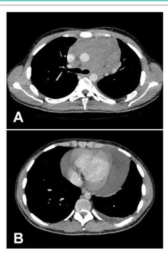
adenopathy. The patient was tachycardic and the systemic blood pressure was 100 x 70 mmHg. He had narrow pulse pressure accompanied by pulsus paradoxus, distant heart sounds and an elevated central venous pressure. Lung sounds were clear. The Computed Tomography depicted a bulky mediastinal mass associated with a large pericardial effusion (Figures 1A and 1B). The echocardiogram showed signs of right atrial and ventricular compression. We decided to perform a video-assisted pericardioscopy in order to drain, inspect and biopsy the pericardium.
The patient was operated through a small midline subxiphoid incision, under general anesthesia. Our operative technique has been described elsewhere3. Pericardiocentesis allowed withdrawal of 1500 ml of clear pericardial fluid. The video-assisted pericardioscopy was performed and depicted gross neoplastic invasion at the top portion of the pericardium (Figure 2). Guided biopsies were performed and sent for histological analysis. A broad anterior pericardial resection was also made, aiming at reducing recurrence. The pericardial space was drained, using a thoracostomy tube. The patient had an uneventful recovery and the final diagnosis was Hodgkin`s lymphoma.
Discusion
Invasive diagnostic methods are needed in order to identify the etiologies of pericarditis. The pericardial biopsy is the most commonly performed procedure4. The access route for pericardial biopsy may be transthoracic, median sternotomy, or subxiphoid2. The examination of the effusion is troublesome, as there are cytological and microbiological limitations in the analysis of the pericardial fluid3,4. In regard to pericardial biopsy, the major limitation is the difficulty in obtaining tissue adequate for analysis. The traditional subxiphoid approach allows very limited exposure of the pericardium, resulting in a low index of etiologic diagnosis and in rates of recurrence of pericardial effusion of up to 20%5. With the development of endoscopic equipment, it was possible to introduce new techniques, such as video-assisted pericardioscopy2,3 and thoracoscopic pericardial window6,7.
Thoracoscopic pericardial window is an effective technique for pericardial drainage6,7. It has the advantage of allowing the concurrent performance of additional procedures, such as biopsy of the mediastinal masses or management of a pleural effusion6,7. However, the need for single-lung ventilation, general anesthesia and the need for lateral positioning (which prevents easy access to the pericardium) may limit the role of the procedure in hemodynamically unstable patients8. O`Brien et al8 compared 15 thoracoscopic
Key words
Pericardium, pericardial effusion, pericardial biopsy, video-assisted pericardioscopy.
The pericardium can be affected by infectious, neoplastic, inflammatory and metabolic diseases. Many conditions require specific treatment and, consequently, an accurate diagnosis is important. Therefore, the pericardial biopsy has an important role in the evaluation of pericardial effusions. The pericardioscopy offers the advantage of the traditional subxiphoid approach, as it allows access to parts of the pericardium that would not be reached by digital palpation, as well as direct visualization through the subxiphoid window or thoracoscopy.
We report the case of a patient with a large pericardial effusion and an undiagnosed mediastinal mass, in which video-assisted pericardioscopy was fundamental in the diagnosis and treatment of the disease.
Usefulness of Pericardioscopy in the Diagnosis of Pericardial
Effusion
Fernando Conrado Abrão, Benoit Jacques Bibas, Paulo Manuel Pêgo-Fernandes, Fabio Biscegli Jatene
Instituto do Coração do Hospital das Clínicas da Faculdade de Medicina da USP, São Paulo, SP - Brazil
Mailing address: Fernando Conrado Abrão •
Rua Souza Ramos, 144 apt. 11 - Vila Mariana - 04120-080 - São Paulo, SP - Brazil E-mail: [email protected]
Manuscript received June 04, 2009; revised manuscript received June 16, 2009; accepted September 21, 2009.
Case Report
Abrão et al Diagnostic usefulness of pericardioscopy
Arq Bras Cardiol 2010; 94(5) : e65-e67
pericardial windows with 71 subxiphoid approaches. Procedural morbidity was low overall, but was significantly higher in the pericardial window group and related mainly to complications associated with the accessing of the pleural space (particularly pneumothoraces). The higher morbidity
Figures 1A and 1B -Computed tomography of the thorax depicting (A): Bulky anterior mediastinal mass measuring 13 x 10 cm, and (B): Large pericardial effusion.
Figure 2 - Video-assisted pericardioscopic image showing extensive neoplastic iniltration of the posterior portion of the pericardium. Arrow indicates selected biopsy site.
seemed to reflect the greater complexity of the procedure. Recurrence was observed in 1 patient after thoracoscopy (8%) and in 5 patients after the subxiphoid approach (10%), and anesthesia time was significantly longer at thoracoscopy.
The video-assisted pericardioscopy offers the advantages of the traditional subxiphoid approach: 1) the patient remains in the supine position and access is readily available for pericardiocentesis if instability occurs after induction, 2) it can be performed under local anesthesia, 3) it does not need single-lung ventilation, 4) it does not penetrate the pleural space and 5) the abdominal incision confers no risk of prolonged neuralgia that can occur after thoracoscopy2,3,8. Additionally, it permits direct visualization of the pericardial surface and guided biopsies of suspicious areas or intra-pericardial deposits. Accordingly, it allows access to parts of the pericardium that would not be reached by digital palpation, direct visualization through the subxiphoid window or thoracoscopy3.
In this case, the objective was a surgical procedure aimed to resolve the risk of cardiac tamponade and provide etiologic diagnostic. The echocardiogram revealed signs of right atrial and ventricular compression; for that reason we decided to keep the patient in the supine position and the surgical approach was the video-assisted pericardioscopy. Another approach would be the traditional subxiphoid biopsy; however, the tumor would not be reached because it was at the top of the pericardium at the emergence of the aorta.
Porte et al9 evaluated 114 patients with pericardial effusions and history of cancer. In 22.7%, pericardioscopy improved the results of the pericardial fluid cytology and surgical pericardial biopsy. The sensitivities of the cytological studies of the pericardial fluid, pathological examinations of pericardial biopsy and the pericardioscopic pericardial biopsy were 75%, 65% and 97%, respectively. Perioperative mortality was 3.5%, and post-operative morbidity was 6.1%.
We have analyzed 91 patients with indeterminate pericardial effusions that were submitted to video-assisted pericardioscopy during a 9-year period.(3) The diagnosis was established as follows: nonspecific inflammation in 50 (54.94%) cases, neoplastic disorders in 22 (24.17%) cases, tuberculosis in 11(12.08%) cases, bacterial inflammatory processes in 3 (3.29%) cases, chylopericardium in 2 (2.19%) cases, fungal infections in 2 (2.19%) cases and viral infection in 1 (1.09%) case. Video-assisted guided biopsies of the pericardium established the diagnosis in 36.26% of the cases, diagnosis through the fluid analysis was observed in 13.18% and association of both methods guaranteed 45.05% of the definitive diagnosis in the study. Overall morbidity was 4.3%, and the most common complication was arrhythmias due to intra-operative manipulation. There was 1 (1.09%) death in the perioperative period due to cardiac tamponade during the induction of anesthesia, despite immediate drainage of the effusion.
Conclusion
Video-assisted pericardioscopy is a safe and efficient method to attain a definite diagnosis and satisfactory therapeutic results in cases of indeterminate pericardial effusions. Excellent visualization of the pericardial surface
Case Report
Abrão et al
Diagnostic usefulness of pericardioscopy
Arq Bras Cardiol 2010; 94(5) : e65-e67
permits guided biopsies of suspicious areas. It also allows access to parts of the pericardium that would not be reached by the other available biopsy techniques.
Potential Conflict of Interest
No potential conflict of interest relevant to this article was reported.
Sources of Funding
There were no external funding sources for this study.
Study Association
This study is not associated with any post-graduation program.
References
1. Hoit BD. Pericardial disease and pericardial tamponade. Crit Care Med. 2007; 35 (8 Suppl): S355-64.
2. Pêgo-Fernandes PM, Fernandes F, Ianni BM, Rohr SS, Bernardelli IM, Jatene FB, et al. Video-assisted pericardioscopy: how to improve diagnostic efficacy in pericardial effusions. Arq Bras Cardiol. 2001; 77 (5): 399-406. 3. Pêgo-Fernandes PM, Mariani AW, Fernandes F, Ianni BM, Stolf NG, Jatene
FB. The role of videopericardioscopy in evaluating indeterminate pericardial effusions. Heart Surg Forum. 2008; 11 (1): E62-5.
4. Fernandes F, Ianni BM, Arteaga E, Benvenutti L, Mady C. Value of pericardial biopsy in the etiologic diagnosis of pericarditis. Arq Bras Cardiol. 1998; 70 (6): 393-5.
5. Naunheim KS, Kesler KA, Fiore AC, Turrentine M, Hammell LM, Brown JW, et al. Pericardial drainage: subxiphoid vs transthoracic approach. Eur J Cardiothorac Surg. 1991; 5 (2): 99-104.
6. Georghiou GP, Stamler A, Sharoni E, Fichman-Horn S, Berman M, Vidne BA, et al. Video-assisted thoracoscopic pericardial window for diagnosis and management of pericardial effusions. Ann Thorac Surg. 2005; 80 (2): 607-10. 7. Neragi-Miandoab S, Linden PA, Ducko CT, Bueno R, Richards WG,
Sugarbaker DJ, et al. VATS pericardiotomy for patients with known malignancy and pericardial effusion: survival and prognosis of positive cytology and metastatic involvement of the pericardium: a case control study. Int J Surg. 2008; 6 (2): 110-4.
8. O’Brien PK, Kucharczuk JC, Marshall MB, Friedberg JS, Chen Z, Kaiser LR, et al. Comparative study of subxiphoid versus video-thoracoscopic pericardial “window”. Ann Thorac Surg. 2005; 80 (6): 2013-9.
9. Porte HL, Janecki-Delebecq TJ, Finzi L, Métois DG, Millaire A, Wurtz AJ. Pericardoscopy for primary management of pericardial effusion in cancer patients. Eur J Cardiothorac Surg. 1999; 16 (3): 287-91.