2 7 3
Arq Bras Oftalmol. 2012;75(4):273-6
Artigo Original |
Original articleABSTRACT
Purpose: To report the response of choroidal neovascularizationto intravitreal ranibi-zumab or bevaciranibi-zumab treatment in the setting of age-relatedmacular degeneration with extensive pre-existing geographicatrophy of the retinal pigment epithelium.
Methods: This is a retrospective case series of 11 eyes in ten consecutivepatients retrieved from a photographic database. The patients were treated with ranibizumab or bevacizumab for neovascular age-relatedmacular degeneration withpre-exis ting geographicatrophy. Patients were included if they had geographicatrophy at or adjacent to the foveal center of at least 1 disc area in size that was present before the developmentof choroidal neovascularization. The best corrected visual acuity and optical coherencetomography analysis of the central macular thickness were recorded for each visit. Serial injections of ranibizumab or bevacizumab were admi-nistered until there was complete resolution of subretinal luidon optical coherence tomography. Data over the entire follow-up periodwere analyzed for overall visual and optical coherencetomography changes.
Results: The patients received an average of 7 ± 3 intravitrealinjections over the treatment period. Seven of 11 eyes had reducedretinal thickening on optical co-herencetomography. On average, the central macular thicknesswas reduced by 72 ± 115 µm. Six of these 7 eyes had improvementof one or more lines of vision and one had no change. The average acuity change for all patientswas -0.04 ± 0.46 logMAR units, which corresponded toa gain of 0.2 ± 4.4 lines of Snellen acuity. The treatmentresulted in a good anatomic response with resolution of the subretinal luid and overall stable visual acuity.
Conclusions: The results of this case series suggest that the use of anintravitreal anti-vascular endothelial growth factor (VEGF)agent (ranibizumab or bevacizumab) for choroidal neovascularization in age-relatedmacular degeneration with pre-exis tinggeographicatrophy is efective. Our results are not as striking as published results from large-scale trials of anti-vascular endothelial growth factor therapy for subfovealchoroidal neovascularization, presumably due to the limitation in the baseline visualacuity caused by the underlying geographicatrophy. The favorable anatomic responsewith the resolution of subretinal luid and stable acuity were consistentwith other ranibizumab and bevacizumab studies.
Keywords: Macular degeneration; Geographic atrophy; Retinal pigment epithelium, Choroidal neovascularization; Antibodies, monoclonal/therapeutic use; Retina; Anti-bodies, monoclonal, humanized; Tomography, optical coherence
Intravitreal ranibizumab and bevacizumab therapy for choroidal neovascularization
in age-related macular degeneration with extensive pre-existing geographic atrophy
Avaliação da resposta da injeção intravítrea de ranibizumab e bevacizumab em pacientes com
neovascularização de coroide da degeneração macular relacionada à idade com
atroia geográica extensa pré-existente
Miguel Hage aMaro1, aaron Brock roller2
Submitted for publication: November 11, 2010 Accepted for publication: June 21, 2012
Study carried out at the Retina Service, Department of Ophthalmology. University of Iowa, USA.
1 Physician, Instituto de Olhos e Laser de Belém - Belém (PA), Brazil.
2 Physician, Department of Ophthalmology. University of Iowa Hospitals and Clinics - Iowa City, IA,
USA.
Funding: No specific financial support was available for this study.
Disclosure of potential conflicts of interest: M.H.Amaro, None; A.B.Roller, None.
Correspondence address: Aaron Brock Roller.University of Iowa, Department of Ophthalmology & Visual Sciences - 200 Hawkins Drive - Iowa City, Iowa - USA - 52242-1091
E-mail: [email protected]
RESUMO
Objetivo: Avaliação dos resultados da injeção intravítrea de ranibizumab e bevacizu -mab em pacientes com neovascularização de coróide da degeneração macular relacio-nada a idade, com atrofia geográfica extensa, pré-existente.
Métodos: Este é um estudo retrospectivo de 10 pacientes, 11 olhos com neovasculari-zação de coroide da degeneração macular relacionada à idade, com atrofia geográ-fica ex ten sa, pré-existente. Os pacientes incluídos apresentaram atrofia geográgeográ-fica, envol vendo a fóvea ou adjacência, antes do desenvolvimento da neovascularização de coroide. A melhor correção visual e o exame de tomografia de coerência óptica com análise da espessura macular foram registrados em cada visita. As injeções de ranibizumab e bevacizumab intravítrea foram feitas até a resolução do líquido sub--retiniano pela tomografia de coerência óptica e clinicamente. Todos os pacientes tinham seguimento de 6 meses do diagnóstico a 2 anos, com média de 16 meses.
Resultados: Onze olhos de 10 pacientes incluídos receberam uma média de 7 ± 3
injeções intravítreas de ranibizumab e bevacizumab, sendo que 7 apresentaram redu-ção do espessamento macular pelo tomografia de coerência óptica. A mácula teve o espessamento reduzido entre 72 ± 115 µm, 6 olhos tiveram melhora de 1 ou mais linhas de visão, um olho teve acentuada diminuição da visão e um outro não teve alteração. A média do resultado do tratamento em logMAR era -0,04 ± 0,46 correlacionando um ganho de visão na tabela de Snellen entre 0,2 ± 4,4 linhas de visão.
Conclusões: Estes resultados sugerem que o uso do ranibizumab e bevacizumab intra-vítrea para neovascularização de coroide da degeneração macular relacionada à idade em extensa atrofia geográfica pré-existente é eficaz. Há, entretanto dificuldades na ava-liação da acuidade visual destes pacientes em virtude da extensa atrofia geográfica que apresentavam e sobre esta ainda a neovascularização de coroide, se comparados a casos em que a neovascularização de coroide não ocorre em atrofia geográfica pré-existente.
IntravItrealranIbIzumabandbevacIzumabtherapyforchoroIdalneovascularIzatIonInage-relatedmaculardegeneratIon wIthextensIvepre-exIstInggeographIcatrophy
2 7 4 Arq Bras Oftalmol. 2012;75(4):273-6 INTRODUCTION
Geographic atrophy (GA) of the retinal pigment epithelium is a form of advanced age-related macular degeneration (AMD) that tends to progress slowly into the center of the macula and cause vision loss. It is deined in the Age-Related Eye Disease Study (AREDS) as a well--deined patch with sharp borders, usually more or less circular shape, and depigmentation of the retinal pigment epithelium (RPE) with ex-posure of underlying large choroidal blood vessels(1, 2). GA associated
with age-related macular degeneration (AMD) is estimated to afect nearly 1% of the US population, with this prevalence expected to increase by 50% by the year 2020(3).
Histopathological sections of GA show thinning or absence of the RPE, closure of the choriocapillaris, and degeneration of the overlying photoreceptors. The site of the initial appearance of GA is initially occupied by drusen, which are large (>125 μm in diameter) in 96% of cases(4-6). The drusen are usually conluent, with at least
two in contact, and sometimes extensive enough to form plaques of drusenoid material. In addition, the GA is nearly always preceded by the appearance of hyperpigmentation around or overlying dru-sen, followed by regression of the drusen and the appearance of hypo pigmentation, sometimes accompanied by refractile deposits. Research has shown that the strongest predictor of the subsequent spread of GA is growth in the previous 2 years(7, 8). Increased fundus
autoluorescence outside atrophic patches of GA may also be an important predictor of subsequent progression(9).
Both GA and choroidal neovascularization (CNV) are advanced forms of age-related macular degeneration(2,10). Although pigmentary
abnormalities of the RPE and drusen are precursors of neovascula-rization and GA, the pathophysiologic relationship between these two forms of advanced AMD is unclear. Both are commonly seen simultaneously, and the coexistence of GA and CNV has been proven histopathologically(4,5,11).
Many studieshave shown that the monoclonal antibodies to vascular endothelial growth factor (VEGF), ranibizumab and bevaci-zumab, improve the visual acuity in patients with AMD and subfo-veal CNV(12-16). In the MARINA and ANCHOR trials with ranibizumab,
94.6% and 96.4% of the patients avoided a 15-letter acuity decrease, respectively. Moreover, 34% and 43% of patients gained at least 15 letters of acuity, respectively. The inal mean visual acuity improved by 7.2 and 11.3 letters in the MARINAand ANCHOR trials, respecti-vely(12,14). Treatment of CNV with of-label use of the anti-VEGF agent
bevacizumab has been shown to be equally efective(16). The more
recently introduced anti-VEGF agent alibercept is also efective in the treatment of CNV, with comparative studies pending(17).
The utility of anti-VEGF intravitreal therapy in the treatment of CNV associated with AMD is well established. The aim of this study is to investigate the role of ranibizumab or bevacizumab intravitreal monotherapy in the treatment of CNV from AMD in eyes with exten-sive pre-existing GA.
METHODS
This is a retrospective case series study from a photographic database center of 11 eyes in 10 consecutive patients under active treat ment with ranibizumab monotherapy for neovascular AMD in the setting of preexisting geographic atrophy. Patients were included if they had an area of GA at oradjacent to the foveal center that was at least 1 disc area in size that was present before or at the time of the developmentof CNV. Patients were excluded if they had any prior history of or prior treatment for CNV in the study eye. Baseline color fundus photography and luorescein angiography was performed in all patients. The best corrected Snellen visual acuity and Heidelberg spectral-domain optical coherence tomography (OCT) were obtai-ned at all visits.
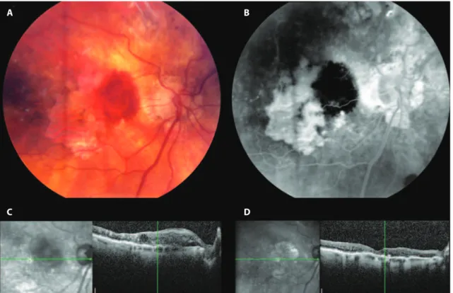
Figure 1. Fundus photographs of study eye 1 with subretinal hemorrhage resulting from occult choroidal neovascularization at the edge of pre-existing geographic atrophy (A and B), and optical coherence tomography of the central macula before (C) and two months after (D) treatment with bevacizumab, with improvement in vision from 20/100 to 20/70.
A B
Amaro MH, Roller AB
2 7 5
Arq Bras Oftalmol. 2012;75(4):273-6
Ranibizumab or bevacizumab were injected intravitreally every 4-6 weeks until there was resolution of the subretinal or intraretinal luid by OCT (Figure 1), or therapy was continued in the case of persis-tent luid. After resolution of luid, a treat-and-extend approach was taken. Data on the overall visual and OCT changes over the entire available follow-up period for each patient were analyzed.
RESULTS
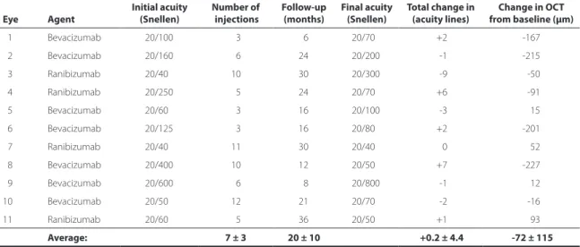
The study included 11 eyes from 10 patients. Four eyes were trea-ted with ranibizumab monotherapy and seven eyes with bevacizu-mab monotherapy. The data for the ranibizubevacizu-mab and bevacizubevacizu-mab treated eyes were considered together for the purpose of this study given the small sample numbers. All patients were followed for at least 6 months (mean 20 months) and up to 3 years after diagnosis. The patients received an average of 7 ± 3 intravitreal injections over the treatment period. Seven eyes had reduced retinal thickening on OCT. On average, the central macular thickness was reduced by 72 ± 115 µm. Six eyes had an improvement of one or more lines of Snellen acuity, three eyes lost three or fewer lines, one eye lost three lines, one eye had dramatic vision loss with hand motions due to macular hemorrhage and one eye had no change in vision (Table 1). In the eyes with mild vision loss, there was visible expansion of GA over the follow-up period. The CNV developed at the edge of the GA or in spared areas of retinal pigment epithelium within the GA in all but one eye. The average treatment outcome for all patients was -0.04 ± 0.46 logMAR units, which corresponded to a gain of 0.2 ± 4.4 lines of Snellen acuity.
DISCUSSION
Choroidal neovascularization and GA are advanced forms of AMD as deined by the AREDS study(2). When CNV develops in eyes
with GA, it can cause both an abrupt drop in visual acuity and pro-gression to central vision loss(11). The AREDS data indicate that about
one-third of the participants had central GA at the time when GA was irst identiied. There was a median time to progression from non-central GA to non-central GA of 2 years. Visual acuity is often decreased before the development of central GA; for those with central GA who do not develop CNV, vision is expected to decline an additional 22 letters on average during the next 5 years. Eyes that developed subsequent CNV have an even worse prognosis(1).
CNV in eyes with GA is typically noted to develop at the edge of GA lesions or in new areas previously uninvolved by GA. In a case report on a patient with GA with histopathological corre-lation in whom the existence of CNV was not initially suspected and a fluorescein angiogram had not revealed CNV, the resear-chers noted that CNV developed in one eye with GA in areas of residual choriocapillaris and pigment epithelium, while no CNV developed in the fellow eye with GA despite breaks in Bruch’s membrane. This was presumably because the breaks occurred in areas of GA, without residual choriocapillaris and RPE(5). This
finding is similar to the recent study by Sunness et al, in which no CNV was noted to develop within areas of GA(11). Our case series
also mirrors this finding (Figure 1).
The MARINA and ANCHORstudies proved the efficacy of anti- VEGF therapy with ranibizumab in the treatment of subfoveal CNV(12,14). The CATT study confirmed these results and
demonstra-ted the equivalent efficacy of ranibizumab and bevacizumab(16).
In our study of ranibizumab or bevacizumab treatment for CNV in AMD with pre-existing GA, 63% of eyes had improvement of one or more lines of vision, with an average gain of 0.2 lines. In com-parison to the Macular Photocoagulation Study, with only 57% of patients with extrafoveal CNV and 26% of patients with juxtafoveal CNV demonstrating stable or improved vision, our patients had improved outcomes as well(18-20). Given that our patients tended
to have poorer vision and central vision loss at baseline due to pre-existing atrophy, our results compare favorably to the other ranibizumab and bevacizumab studies, with disappearance of the subretinal fluid and overall stable visual acuity. Therefore GA, of itself, should not be a contraindication to anti-VEGF treatment for eyes with CNV.
CONCLUSION
This small case series demonstrates that the use of the intra-vitreal anti-VEGF agents ranibizumab and bevacizumab for CNV in AMD with significant preexisting GA was largely effective for the patients in our series. Our results are not as striking as the results of large-scale trials of anti-VEGF therapy for subfoveal CNV, presu-mably due to the limitation of improvement of visual acuity over baseline caused by the underlying GA. Our study is limited by a very small sample size, and a larger prospective study with longer follow-up is needed to confirm our results.
Table 1. Visual acuity at baseline and inal follow-up, anti-VEGF agent used, number of injections, total follow-up, inal acuity and total change in acuity and central macular thickness compared to baseline exam at initiation of treatment along with averaged results for all subject eyes
Eye Agent
Initial acuity (Snellen)
Number of injections
Follow-up (months)
Final acuity (Snellen)
Total change in (acuity lines)
Change in OCT from baseline (μm)
01 Bevacizumab 20/100 3 6 20/70 +2 -167
02 Bevacizumab 20/160 6 24 20/200 -1 -215
03 Ranibizumab 20/40 10 30 20/300 -9 -50
04 Ranibizumab 20/250 5 24 20/70 +6 -91
05 Bevacizumab 20/60 3 16 20/100 -3 15
06 Bevacizumab 20/125 3 16 20/80 +2 -201
07 Ranibizumab 20/40 11 30 20/40 0 52
08 Bevacizumab 20/400 10 12 20/50 +7 -227
09 Bevacizumab 20/600 6 8 20/800 -1 12
10 Bevacizumab 20/50 12 21 20/70 -2 -16
11 Ranibizumab 20/60 5 36 20/50 +1 93
IntravItrealranIbIzumabandbevacIzumabtherapyforchoroIdalneovascularIzatIonInage-relatedmaculardegeneratIon wIthextensIvepre-exIstInggeographIcatrophy
2 7 6 Arq Bras Oftalmol. 2012;75(4):273-6 ACKNOWLEDGEMENTS
We thank Dr. James C. Folk, the Judith (Gardner) and Donald H. Beisner, M.D. Professor of Vitreoretinal Diseases and Surgery at the University of Iowa, Iowa City, Iowa USA for his generous help with the design and completion of this study.
REFERENCES
1. Lindblad AS, Lloyd PC, Clemons TE, eGensler GR, Ferris FL 3rd, Klein ML, Armstrong JR; Age-Related Eye Disease Study Research Group. Change in area of geographic atrophy in the Age-Related Eye Disease Study: AREDS report number 26. Arch Ophthalmol. 2009;127(9):1168-74.
2. Age-Related Eye Disease Study Research Group. A randomized, placebo-controlled, clinical trial of high-dose supplementation with vitamins C and E, beta carotene, and zinc for age-related macular degeneration and vision loss: AREDS report no. 8. Arch Ophthalmol. 2001;119(10):1417-36. Erratum in: Arch Ophthalmol. 2008;126(9): 1251.
3. Friedman DS, O’Colmain BJ, Muñoz B, Tomany SC, McCarty C, de Jong PT, Nemesure B, Mitchell P, Kempen J; Eye Diseases Prevalence Research Group. Prevalence of age-related macular degeneration in the United States. Arch Ophthalmol. 2004; 122(4):564-72. Comment in: JAMA. 2004;291(15):1900-1.
4. Green WR, Enger C. Age-related macular degeneration histopathologic studies. The 1992 Lorenz E. Zimmerman Lecture. Ophthalmology. 1993;100(10):1519-35. 5. Green WR, Key SN 3rd. Senile macular degeneration: a histopathologic study. Trans
Am Ophthalmol Soc. 1977;75:180-254.
6. Klein ML, Ferris FL 3rd, Armstrong J, Hwang TS, Chew EY, Bressler SB, Chandra SR; AREDS Research Group. Retinal precursors and the development of geographic atrophy in age-related macular degeneration. Ophthalmology. 2008;115(6):1026-31. 7. Sunness JS, Margalit E, Srikumaran D, Applegate CA, Tian Y, Perry D, et al. The
long-t erm nalong-tural hislong-tory of geographic along-trophy from age-relalong-ted macular degeneralong-tion: enlargement of atrophy and implications for interventional clinical trials. Ophthalmo-logy. 2007;114(2):271-7.
8. Sunness JS, Applegate CA, Bressler NM, Hawkins BS. Designing clinical trials for age-re lated geographic atrophy of the macula: enrollment data from the geographic atrophy natural history study. Retina. 2007;27(2):204-10.
9. Holz FG, Bindewald-Wittich A, Fleckenstein M, Dreyhaupt J, Scholl HP, Schmitz-Valcken-berg S. Progression of geographic atrophy and impact of fundus auto luorescence patterns in age-related macular degeneration. Am J Ophthalmol. 2007;143(3): 463-72.
10. Gass JD. Drusen and disciform macular detachment and degeneration. Trans Am Ophthalmol Soc. 1972;70:409-36.
11. Sunness JS, Gonzalez-Baron J, Bressler NM, Hawkins B, Applegate CA. The develop-ment of choroidal neovascularization in eyes with the geographic atrophy form of age-related macular degeneration. Ophthalmology. 1999;106(5):910-9.
12. Rosenfeld PJ, Brown DM, Heier JS, Boyer DS, Kaiser PK, Chung CY, Kim RY: MARINA Study Group. Ranibizumab for neovascular age-related macular degeneration. N Engl J Med. 2006;355(14):1419-31. Comment in: N Engl J Med. 2006;355(14):1409-12. N Engl J Med. 2007;356(7):748-9; N Engl J Med. 2006;355(14):1493-5.
13. Rich RM, Rosenfeld PJ, Puliaito CA, Dubovy SR, Davis JL, Flynn HW Jr, et al. Short-term safety and eicacy of intravitreal bevacizumab (Avastin) for neovascular age-related macular degeneration. Retina. 2006;26(5):495-511.
14. Brown DM, Kaiser PK, Michels M, Soubrane G, Heier JS, Kim RY, SY JP, Schneider S; ANCHOR Study Group. Ranibizumab versus verteporin for neovascular age-re-lated macular degeneration. N Engl J Med. 2006;355(14):1432-44. Comment in: N Engl J Med. 2006;355(14):1409-12; N Engl J Med. 2007;356(7):748-9; N Engl J Med. 2007;356(7):747-8; N Engl J Med. 2006;355(14):1493-5.
15. Rosenfeld PJ, Moshfeghi AA, Puliaito CA. Optical coherence tomography indings after an intravitreal injection of bevacizumab (Avastin) for neovascular age-related macular degeneration. Ophthalmic Surg Lasers Imaging. 2005;36(4):331-5. Comment in: Ophthalmic Surg Lasers Imaging. 2005;36(4):270-1.
16. CATT Research Gourp, Martin DF, Maguire MG, Ying GS, Grunwald JE, Fine SL, Jafe GJ. Ranibizumab and bevacizumab for neovascular age-related macular degeneration. N Engl J Med. 19;364(20):1897-908. Comment in: N Engl J Med. 2011;365(23):2237; N Engl J Med. 2011;365(23):2238; Clin Experiment Ophthalmol. 2011;39(7):718-20; N Engl J Med. 2011;364(20):1966-7.
17. Dixon JA, Oliver SC, Olson JL, Mandava N. VEGF Trap-Eye for the treatment of neovascular age-related macular degeneration. Expert Opin Investig Drugs. 2009; 18(10):1573-80.
18. Laser photocoagulation for juxtafoveal choroidal neovascularization. Five-year re-sults from randomized clinical trials. Macular Photocoagulation Study Group. Arch Ophthalmol. 1994;112(4):500-9.
19. Five-year follow-up of fellow eyes of patients with age-related macular degeneration and unilateral extrafoveal choroidal neovascularization. Macular Photocoagulation Study Group. Arch Ophthalmol. 1993;111(9):1189-99. Comment in: Arch Ophthalmol. 1994;112(7):874-5.
20. Krypton laser photocoagulation for neovascular lesions of age-related macular degeneration. Results of a randomized clinical trial. Macular Photocoagulation Study Group. Arch Ophthalmol. 1990;108(6):816-24. Comment in: Arch Ophthalmol. 1991;109(5):614-5.