Cardiac Electrophysiological Evaluation in Patients with Sarcoidosis
Jefferson Curimbaba, Silvia Carla Souza Rodrigues, José Marcos Moreira, Lenine Ângelo Alves Silva,
Carlos Alberto de Castro Pereira, João Pimenta
Hospital do Servidor Público Estadual - IAMSPE - São Paulo, SP - Brazil
Abstract
Background: Sarcoidosis is a multisystem granulomatous disease of unknown origin that can cause sudden death.
Objective: Electrophysiological evaluation of patients with suspected sarcoidosis with cardiac involvement.
Methods: We studied 22 patients with mean age of 55.32 ± 13.13 years, diagnosed with sarcoidosis and suspected cardiac involvement. These patients underwent clinical evaluation, laboratory tests, electrocardiogram, echocardiogram, 24-hour Holter, technetium or gallium scintigraphy and electrophysiological study. In selected cases, we performed positron emission tomography or magnetic resonance imaging. Patients were followed up in the outpatient care service with quarterly visits.
Results: Cardiac involvement was confirmed in four (18.2%) patients. Ventricular extrasystoles with density > 100/24 h were documented in 24-Holter monitoring in 12 (54.5%) patients. Electrophysiological studies revealed an increased HV interval in seven patients (31.8%) and increased Wenckebach point in four (18.2%) patients. There was induction of atrial fibrillation in seven patients (31.8%) and sustained ventricular tachycardia in one patient (4.5%). Four patients with confirmed cardiac sarcoidosis had documented ventricular extrasystoles with density > 100/24 h. Out of these, two had prolonged HV interval and atrial fibrillation was induced in two of them. Sustained ventricular tachycardia was not induced in any of these patients. After mean follow-up period of 20.9 ± 15.7 months, one patient with cardiac sarcoidosis had sudden death.
Conclusion: Patients with sarcoidosis and suspected cardiac involvement have a high prevalence of ventricular extrasystoles (VEs) and conduction system disorders. (Arq Bras Cardiol 2011;96(4):266-271)
Keywords: Sarcoidosis; heart conduction system/abnormalities; heart failure; arrhythmias, cardiac.
Mailing address: Jefferson Curimbaba •
Rua Jacaracanga, 157 - Apto 112 - Vila Formosa - 03358-140 - São Paulo, SP - Brazil
E-mail: jcurimbaba@cardiol.br, jeffery@uol.com.br
Manuscript received August 19, 2010; revised manuscript received November 27, 2010; accepted on December 08, 2010.
Introduction
Sarcoidosis is a multisystem granulomatous disease of unknown origin1 often observed in individuals between the 2nd and 5th decades of life. Its etiology is still not completely understood; infectious, environmental, occupational and genetic causes may be involved, but not totally confirmed2. Almost all systems of the human body may be affected. The most common ones are the respiratory (lung 90%) and lymphatic, often presenting non-caseous granulomas3.
Cardiac involvement occurs in 20 to 50% of patients who may have adverse prognostic and evolution4-7. All cardiac structures may be involved, but the myocardium is the most area8. The three main consequences are heart conduction system abnormalities, arrhythmias and heart failure (HF). Cardiac manifestation is pleomorphic ranging from completely asymptomatic patients to patients with palpitations, chest pain, symptoms of heart failure, syncope and sudden death (SD)9,10.
SD is the most feared cardiac manifestation of sarcoidosis. It is often unpredictable and may occur as the first manifestation of the disease or at any time, and it is more common in the later stages when there is greater heart damage. Incidence varies greatly, reaching up to 67% of cases8 and may be caused by complete AV block and ventricular arrhythmias11,12. Thus, risk stratification is important when we establish the diagnosis of cardiac involvement. Hence, this study was designed to evaluate the atrioventricular (AV) and intraventricular (IV) conduction system and the induction of atrial and ventricular arrhythmias in patients with recent clinical and laboratory diagnosis of sarcoidosis with suspected cardiac involvement.
Patients and methodology
Study design
Prospective, open, started in April 2005 and completed in April 2009, approved by the Ethics Committee of the Institution.
Population
(55.32 ± 13.13) years with newly diagnosed sarcoidosis defined by histopathology. We evaluated patients with suspected cardiac involvement, selected by the symptoms and initial cardiac examinations, without specific standard treatment, not using any medication that could alter the electrophysiological conditions of the heart conduction system. After eligibility for admission into the study, patients were informed and signed a consent form.
Methods
All patients underwent clinical evaluation, routine laboratory evaluation, electrocardiogram (ECG) and 24-hour Holter. Cardiac involvement was documented by chest radiography, two-dimensional echocardiography (ECHO) and one of the following imaging methods: technetium or gallium scanning, positron emission tomography and magnetic resonance imaging. After having agreed to participate in the study, the patients underwent the electrophysiological study (EPS) according to previously described techniques13. Follow-up was conducted with quarterly clinic visits, or as required. We analyzed demographic and clinical data, as well as the pulmonary stage of sarcoidosis (radiological classification of Scadding)14.In the ECG obtained by polygraph TEB with 12 simultaneous and continuous leads, we evaluated the fundamental rhythm, ÂQRS, presence of abnormal Q waves, ST segment changes, presence of AV and IV block, divisional block, QT interval (QTc) corrected by the Bazett formula and QT interval dispersion. The QT interval was measured manually at a speed of 100 mm/s. The baseline rhythm, presence of AV and IV block, pauses, atrial arrhythmias such as supraventricular extrasystoles and tachycardia, ventricular arrhythmias such as ventricular extrasystoles (EV) with densities of < 100/day and > 100/day15 and nonsustained ventricular tachycardia (NSVT), heart rate variability (HRV) in time and frequency were analyzed by 24-hour Holter. The device used was brand DMI Cardios Systems. All tests were reviewed by a single professional.
AH, H, HV, and V intervals were assessed at baseline conditions during the EPS. Sinus node recovery time (SNRT) was corrected under three cycles of stimulation frequencies (600, 500 and 400 ms), the Wenckebach point (WP) and the atrioventricular nodal effective refractory period (AVNERP) were evaluated under programmed atrial stimulation. The ventriculoatrial conduction, effective refractory period and the induction of tachyarrhythmias were analyzed by programmed ventricular stimulation from the apex and RV outflow tract with up to three extrastimulations coupled to each frequency cycle of 600, 500 and 400 ms. Measurements of electrophysiological parameters were performed at the recording speed of 200 mm/s for greater precision.
Statistical analysis was performed using the software SPSS for Windows version 13.0. For the analysis of qualitative data, we performed frequency distributions as mean ± standard deviation.
Results
The characteristics of the population and changes in key examinations (ECG, ECHO and Holter) are presented in
Table 1. No patient had any manifestation of paroxysmal tachycardia, presyncope or syncope before the study. All patients had pulmonary sarcoidosis in the stages of radiological classification of Scadding 0, 1, 2, 3 and 4 (4.5%, 18.2%, 31.8%, 36.4% and 9.1% respectively). The diagnosis of cardiac sarcoidosis was confirmed in four of them (18.2%) through scintigraphy with gallium (Ga-67), positron emission tomography (PET) or magnetic resonance imaging (MRI). The presence of noncaseating granulomas typical of sarcoidosis in the lymph node biopsy and abnormal uptake on PET in the heart and other organs in a patient diagnosed with SC are illustrated in Figure 1.
The main abnormalities revealed by Holter monitoring were the presence of EV and a predominance of sympathetic tone on HRV. All of the four patients with cardiac sarcoidosis showed EV with density > 100/24 h. There was a predominance of the sympathetic tone in 3 (75%) patients.
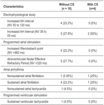
The most relevant abnormalities observed in the EPS were abnormalities in AV node function and HV, PW, PRNAV interval, and induction of atrial fibrillation in a significant number of patients. The main findings are shown in Table 2. Sustained monomorphic ventricular tachycardia (SMVT), unstable, was induced in one patient without a confirmed diagnosis of cardiac involvement. Among the four patients diagnosed with cardiac sarcoidosis, the number 2, aged 60, suffering from hypertension and diabetes, had palpitations and fatigue. Their ECG showed left bundle branch block, 24 h-Holter with 1,707 EV and ejection fraction of 65% with LV diastolic dysfunction. Another patient (number 6), a 46-year-old man, also had fatigue and palpitations, ECG with left ÂQRS, echocardiography with EF 69% and Holter with 17,781 EV/24 h. The third patient (number 14), also male, aged 54, had symptoms of physical exhaustion, normal ECG and echocardiogram with EF 62% with left ventricular hypertrophy and diastolic dysfunction and Holter with 27,572 EV/24 h and periods of NSVT. Finally, the fourth patient (number 16), aged 80, suffering from hypertension, had symptoms of fatigue, ECG with anterior superior divisional block, echocardiogram with EF 69% with left ventricular hypertrophy and diastolic dysfunction and Holter with 1,010 EV/24 h and NSVT. During a mean follow up of 20.9 ± 15.7 months, one patient (number 2) (4.5%) with CS had SD. This patient had manifestation of heart failure, controlled with medication. Four patients (three with SC) were submitted to another EPS as they had worsening of symptoms of palpitation and increased density of VE on Holter. EPS did not change significantly compared to previous studies and treatment was not modified.
Discussion
of cardiac involvement is very difficult due to limited sensitivity and specificity of diagnostic tests, a diagnosis of exclusion is usually adopted, notably by the absence of a specific biological marker. Endomyocardial biopsy, considered the “gold standard”, also has low sensitivity for being a granulomatous disease that predominantly involves the basal region and the left ventricle tip.
Risk stratification for SD is an important issue in patients with sarcoidosis, because of its catastrophic and unpredictable nature. There is a multitude of methods such as 24-hour Holter, heart rate variability, high-resolution ECG, QT dispersion, T wave alternans, EPS, which attempt to predict the occurrence of SD, however, the value of these methods is still questioned in patients with sarcoidosis. The presence of VE in Holter density > 100/day (12,017.7 ± 10,540.56) occurred in 4 patients with documented cardiac involvement, which is consistent with the observations of Suzuki et al15. This author, in a study involving 38 patients, suggested that
the presence of VE density > 100/day would be a predictor of cardiac involvement by sarcoidosis with a sensitivity of 67% and 80% specificity.
The role of EPS in the evaluation of patients with sarcoidosis, although not yet well defined, would be the evaluation of the conduction system, given the high prevalence of granulomatous infiltration in this tissue, with rates of SD for total AV block up to 40%8. In our study, changes in the conduction system were found in two (50%) of the four patients with cardiac involvement. Moreover, electrophysiological changes were also found in a significant percentage of patients without cardiac involvement, which can reinforce the concept that there may be latent myocardial fibrosis before diagnosis of cardiac sarcoidosis16-18.
Induction of monomorphic VT has been established as a predictor of SD in patients with ischemic heart disease, but its value is questionable in patients with nonischemic heart disease. Yasaki et al19 observed that the NYHA class, the LV Table 1 - Demographic and clinical characteristics and major abnormalities in tests of patients without cardiac sarcoidosis (n = 18) and with cardiac sarcoidosis (n = 4)
Characteristics Without CS (n = 18) With CS (n = 4)
Sex
Male/Female 5 (27.8%)/13 (72.2%) 2 (50%)/2 (50%)
Race
White/Black 11 (61.1%)/7 (38.9%) 0 (0%)/4 (100%)
Age, years (x ± SD) 57.72 ± 10.72 60 ± 9.52 Diseases
Diabetes mellitus 3 (16.7%) 1 (25%)
Hypertension 9 (50%) 2 (50%)
Symptoms
Fatigue 18 (100%) 4 (100%)
Palpitations 10 (55.5%) 2 (50%)
Follow-up period - months (x ± SD) 20.9 ± 15.7 20.9 ± 15.7 Electrocardiography
ÂQRS deviation to the left 2 (11.1%) 1 (25%)
Anterosuperior divisional block 3 (16.7%) 1 (25%)
Partial right bundle branch block 4 (18.2%) 0 (0%)
Left bundle branch block 0 (0%) 1(25%)
Increased QTC interval 1 (5.55%) 2 (50%)
24 h Holter
Ventricular extrasystole (> 100/24 h) 8 (44.4%) 4 (100%)
Nonsustained ventricular tachycardia 1 (5.55%) 2 (50%)
HRV (sympathetic predominance) 12 (66.7%) 3 (75%)
Echocardiography
LV ejection fraction.% (x ± SD) 66.23 ± 3.98 66.25 ± 2.45
Left ventricular hypertrophy 3 (16.7%) 2 (50%)
Diastolic function 7 (38.9%) 3 (75%)
Figure 1 - Patient with CS conirmed. A - Inguinal lymph node biopsy by hematoxylin-eosin coloration, demonstrating noncaseating granulomas of epithelioid cells;
B - PET scanning with myocardial 18F-luorodeoxyglucose showing areas with increased metabolism (inlammation) in the left ventricle; C - Mapping of the whole body,
showing uptake in other organs such as spleen and mediastinal lymph nodes.
diastolic diameter and the presence of SVT were important independent predictors of mortality in patients with cardiac sarcoidosis. In our population, only one patient without a definitive diagnosis of cardiac involvement had SVT induced. The patient presented good ventricular function (ECO = 67%) and remained with mild symptoms and palpitations up to the completion of this study, receiving only corticosteroids as a drug therapy. The evolution of patients diagnosed with cardiac sarcoidosis was good, not differing from the others, except for a higher dose of corticoids, and/or other drugs. However, one patient with SC had SD, although she has undergone another EPS without showing induction of tachyarrhythmias or significant changes in the conduction system, suggesting that the electrophysiological evaluation had no major role as a predictor of risk for SD.
Study limitations
The main limitations of this study were the small sample size and the difficulties found in the diagnostic investigation of all patients. The world literature advocates the use of tests such as MRI and PET when there is doubt about the diagnosis, which was not possible in all patients. The short-term follow-up did not enable us to assess properly the risk factors for SD and the presence of hypertension and diabetes may also Table 2 - Main electrophysiological changes found in patients with
(n = 4) and without (n = 18) cardiac sarcoidosis
Characteristics Without CS (n = 18) With CS (n=4)
Electrophysiological study
Increased AH interval
(NV 55 to 120 ms) 4 (22.2%) 0 (0%)
Increased AH interval (NV 35 to
55 ms) 5 (27.8%) 2 (50%)
Programmed atrial stimulation
Increased Wenckebach point
(NV <460 ms) 4 (22.2%) 0 (0%)
Atrioventricular Nodal Effective
Refractory Period (NV <320 ms) 5 (27.7%) 0 (0%)
Atrial arrhythmia
Nonsustained atrial ibrillation 1 (5.55%) 1 (25%)
Sustained atrial ibrillation 4 (22.2%) 1 (25%) Nonsustained atrial tachycardia 1 (4.5%) 0 (0%)
Programmed ventricular stimulation
Sustained ventricular tachycardia 1 (4.5%) 0 (0%)
have caused difficulties in interpreting the abnormalities in the patients studied.
Potential Conflict of Interest
No potential conflict of interest relevant to this article was reported.
Sources of Funding
There were no external funding sources for this study.
Study Association
This article is part of the thesis of master submitted by Jefferson Curimbaba, from Hospital do Servidor Público Estadual - IAMSPE - São Paulo.
References
1. Iannuzzi MC, Rybicki BA, Teirstein AS. Sarcoidosis. N Engl J Med. 2007;357(21):2153-65.
2. B a u g h m a n R P, L o w e r E E , d u B o i s R M . S a r c o i d o s i s . L a n c e t . 2003;361(9363):1111-8.
3. Thomas PD, Hunninghake GW. Current concepts of the pathogenesis of sarcoidosis. Am Rev Respir Dis. 1987;135 (3):747-60.
4. Bernstein M, Konzelmann FW, Sidlick DM. Boeck’s sarcoid: report of a case with visceral involvement. Arch Intern Med. 1929;44:721-34.
5. Longcope WT, Freiman DG. A study of sarcoidosis: based on a combined investigation of 160 cases including 30 autopsies from The Johns Hopkins Hospital and Massachusetts General Hospital. Medicine (Baltimore). 1952;31(1):1-132.
6. Silverman KJ, Hutchins GM, Bulckley BH. Cardiac sarcoid: a clinicopathologic study of 84 unselected patients with systemic sarcoidosis. Circulation. 1978;58(6):1204-11.
7. Sharma OP, Maheshwari A, Thaker K. Myocardial sarcoidosis. Chest. 1993; 103(1):253-8.
8. Roberts WC, McAllister HA Jr, Ferrans VJ. Sarcoidosis of the heart: a clinicopathologic study of 35 necropsy patients (group I) and review of 78 previously described necropsy patients (group II). Am J Med. 1977;63(1):86-108.
9. Corrado D, Thiene G. Cardiac sarcoidosis mimicking arrhythmogenic right ventricular cardiomyophaty/dysplasia: the renaissance of endomyocardial biopsy? J Cardiovasc Electrophysiol. 2009;20(5):477-9.
10. Baughman RP, Winget DB, Bowen EH, Lower EE. Predicting respiratory failure in sarcoidosis patients. Sarcoidosis Vasc Diffuse Lung Dis. 1997;14(2):154-8.
11. Abeler V. Sarcoidosis of the cardiac conducting system. Am Heart J. 1979;97(6):701-7.
12. Furushima H, Chinushi M, Sugiura H, Kasai H, Washizuka T, Aizawa Y. Ventricular tachyarrhythmia associated with cardiac sarcoidosis: its mechanisms and outcome. Clin Cardiol. 2004;27(4):217-22.
13. Pimenta J, Miranda M, Pereira CB. Electrophysiologic findings in long-term asymptomatic chagasic individuals. Am Heart J. 1983;106(2):374-80.
14. Scadding JG. Prognosis of intrathoracic sarcoidosis in England. Br Med J. 1961;2(5261):1165-72.
15. Suzuki T, Kanda T, Kubota S, Imai S, Murata K. Holter monitoring as a noninvasive indicator of cardiac involvement of sarcoidosis. Chest. 1994;106(4):1021-4.
16. Gibbons WJ, Levy RD, Nava S, Malcolm I, Marin JM, Tardif C, et al. Subclinical cardiac dysfunction in sarcoidosis. Chest. 1991;100(1):44-50.
17. Uslu N, Akyol A, Gorgulu S, Eren M, Ocakli B, Celik S, et al. Heart rate variability in patients with systemic sarcoidosis. Ann Noninvas Electrocardiol. 2006;11(1):38-42.
18. Yodogawa K, Seino Y, Ohara T, Takayama H, Kobayashi Y, Katoh T, et al. Non-invasive detection of latent cardiac conduction abnormalities in patients with pulmonary sarcoidosis. Circ Jap. 2007;71(4):540-5.
