Effects of hypertonic sodium chloride
solution on the electrophysiologic
alterations caused by bupivacaine
in the dog heart
Divisão de Pesquisa, Instituto do Coração, Faculdade de Medicina, Universidade de São Paulo, São Paulo, SP, Brasil
A. Scalabrini, F. Corregiari and M. Rocha e Silva
Abstract
The effects of various hypertonic solutions on the intraventricular conduction, ventricular repolarization and the arrhythmias caused by the intravenous (iv) injection of bupivacaine (6.5 mg/kg) were studied in sodium pentobarbital-anesthetized mongrel dogs. Hypertonic solu-tions, given iv 5 min before bupivacaine, were 7.5% (w/v) NaCl, 5.4% (w/v) LiCl, 50% (w/v) glucose (2,400 mOsm/l, 5 ml/kg), or 20% (w/v) mannitol (1,200 mOsm/l, 10 ml/kg). Bupivacaine induced se-vere arrhythmias and ventricular conduction and repolarization distur-bances, as reflected by significant increases in QRS complex duration, HV interval, IV interval and monophasic action potential duration, as well as severe hemodynamic impairment. Significant prevention against ventricular electrophysiologic and hemodynamic disturbances and ventricular arrhythmias was observed with 7.5% NaCl (percent in-crease in QRS complex duration: 164.4 ± 21.8% in the non-pretreated group vs 74.7 ± 14.1% in the pretreated group, P<0.05; percent increase in HV interval: 131.4 ± 16.1% in the non-pretreated group vs 58.2 ± 7.5% in the pretreated group, P<0.05; percent increase in monophasic action potential duration: 22.7 ± 6.8% in the non-pre-treated group vs 9.8 ± 6.3% in the pretreated group, P<0.05; percent decrease in cardiac index: -46 ± 6% in the non-pretreated group vs -28 ± 5% in the pretreated group, P<0.05). The other three hypertonic solutions were ineffective. These findings suggest an involvement of sodium ions in the mechanism of hypertonic protection.
Correspondence A. Scalabrini
Disciplina de Emergências Clínicas Av. Dr. Enéas C. Aguiar, 255 5º andar, Sala 5023 05403-010 São Paulo, SP Brasil
E-mail: [email protected]
Research supported by FAPESP and Fundação E.J. Zerbini.
Received February 25, 2002 Accepted August 20, 2002
Key words
·Local anesthetics ·Cardiotoxicity
·Cardiac electrophysiology ·Intraventricular conduction ·Sodium ion
·Hypertonic solutions
Introduction
Local anesthetics are a large group of drugs sharing the same mechanism of action, i.e., blockade of fast Na+ channels at the
membrane level (1). In the peripheral nerve, this mechanism is responsible for the anes-thetic effect, preventing impulse conduction
and thereby causing anesthesia.
Similar effects may be observed in the cardiac muscle. Local anesthetics affect myo-cardial depolarization by decreasing the in-ward Na+ current. Therefore, the slope of
myocardial repolarization, prolonging the recovery time of the fast sodium channel and consequently increasing the refractory pe-riod (6-8).
Bupivacaine is a local anesthetic widely used in anesthesiology because of its long duration of action. However, its extremely high affinity for the membrane channels can lead to toxic cardiac electrophysiologic ef-fects when the drug is accidentally adminis-tered iv (4,9-11). There are several reports of serious anesthetic accidents and fatalities due to inadvertent iv injection or enhanced absorption of bupivacaine during regional anesthesia (12-15).
Because of the rapid onset and severity of arrhythmias and hypotension, the man-agement of this condition is very difficult. In fact, there is no consensus on how to treat this event (16-20).
Hypertonic sodium solutions effectively correct the hypotension induced by severe hemorrhage (21,22). Prehydration with small amounts of hypertonic saline is also effec-tive to minimize hypotension associated with spinal anesthesia (23). Simonetti et al. (24) studied the effects of hypertonic saline on the hypotension caused by iv bupivacaine and noticed a protective effect on the cardiac arrhythmias as well. This led us to investi-gate the effects of hypertonic solutions on the electrophysiologic and hemodynamic al-terations induced by toxic doses of bupiva-caine in dogs.
Material and Methods
This experiment was conducted accord-ing to NIH guidelines for the use of experi-mental animals and reviewed by the Institu-tional Board. Seventy-three mongrel dogs of either sex weighing 16.7 ± 4.7 kg were anes-thetized with 20 mg/kg iv sodium pentobar-bital and ventilated with a Takaoka model 670 volume ventilator with 100% oxygen. The left femoral artery was cannulated and used for arterial pressure monitoring. The
right femoral vein was cannulated for drug infusion and the right femoral artery for blood sampling. A lead II surface electrocar-diogram was monitored throughout the ex-periment.
Studies on intraventricular conduction
A #5F bipolar electrode was introduced into the left femoral vein of 57 dogs, and positioned by fluoroscopy at the level of the septal leaflet of the tricuspid valve for His bundle electrogram recording. Two other #5F bipolar electrodes were then introduced into the left jugular vein; one was positioned by fluoroscopy in the upper right atrium to record a right atrial electrogram close to the sinus node, and the other in the right ventric-ular apex to record the right ventricventric-ular elec-trogram. Recordings were obtained with a Hewlett-Packard model 8890A polygraph at a bandpass of 50 to 500 Hz and recorded on Kodak Linagraph 2201 photographic paper at a speed of 100 mm/s. Recorded data were: i) QRS complex duration; ii) HV interval, namely the time interval between the His bundle spike and the first deflection of the ventricular electrogram; iii) IV interval, i.e., the time interval between the ventricular electrograms obtained at the His bundle electrogram site and the right ventricular apex.
The dogs were then assigned to five groups according to the pretreatment they received: B6 (17 dogs) - iv injection of 6.5 mg/kg bupivacaine; H5B6 (17 dogs), iv in-jection of 6.5 mg/kg bupivacaine 5 min after pretreatment with 5 ml/kg 7.5% (w/v) NaCl; L5B6 (9 dogs), iv injection of 6.5 mg/kg bupivacaine 5 min after pretreatment with 5 ml/kg 5.4% (w/v) LiCl; G5B6 (7 dogs), iv
injection of 6.5 mg/kg bupivacaine 5 min after pretreatment with 5 ml/kg 50% (w/v) glucose; M10B6 (7 dogs), iv injection of 6.5 mg/kg bupivacaine 5 min after pretreatment with 10 ml/kg 20% (w/v) mannitol.
5 min after pretreatment, immediately after bupivacaine injection, and every 30 s until death or for 3 min after the injection.
Studies on ventricular repolarization
In 16 other dogs, a monophasic action potential electrode was introduced through the right carotid artery and positioned at the level of the left ventricular apex by fluoros-copy. Monophasic action potential record-ings were then obtained with a model ES2000 Gould polygraph at a bandpass of 0.5 to 300 Hz and acquired with a microcomputer us-ing a software developed by the Bioengi-neering Division of the Heart Institute. The ventricular repolarization was analyzed tak-ing into account the duration (ms) of the monophasic action potential at 20 (T20), 50 (T50) and 90% (T90) repolarization. The dogs were then assigned to two groups: B6R (8 dogs), same as group B6; H5B6R (8 dogs), same as group H5B6.
Recordings were obtained at the same times as in the intraventricular conduction studies.
Studies on cardiovascular responses and metabolic changes
In all dogs, mean arterial pressure was monitored throughout the experiment. In 8 dogs of group B6 and 8 dogs of group H5B6,
a #7F Edwards 3-way thermodilution cath-eter was introduced into the right jugular vein and its tip positioned in the pulmonary circulation to record pulmonary pressures and to measure cardiac output. In these dogs, arterial blood samples for blood bupivacaine levels, plasma Na and K levels, plasma os-molarity, and arterial blood gases were drawn at baseline, immediately and 5 min after 7.5% NaCl infusion, and then at 30, 90, and 180 s after bupivacaine injection. At the same times, cardiac output was measured using a model COM-1 American Edwards Instruments Cardiac Output Computer.
Data analysis
Intraventricular conduction and pressure measurements were performed using a Digicon model 1812 digitizer table (Alberta, Canada) coupled to a microcomputer with a dedicated software. Each measurement was the mean of five consecutive readings during sinus rhythm. Intraventricular repolarization measurements were performed using a mi-crocomputer with a dedicated software.
Plasma osmolarity was measured with a model 3D2 Advanced Digimatic Osmom-eter (Needham Heights, MA, USA), with each value representing the mean of two consecutive measurements.
Blood bupivacaine levels were measured by high-performance liquid chromatography.
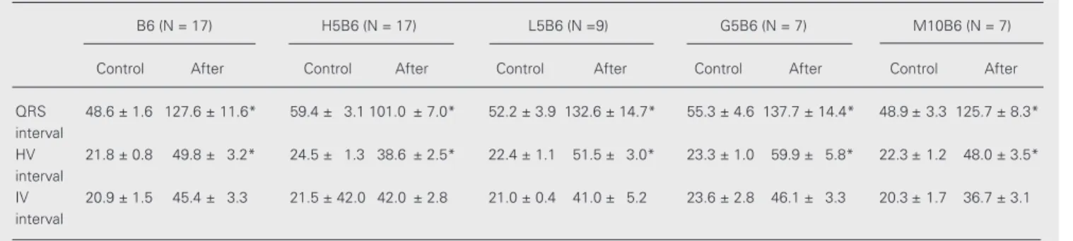
Table 1. Maximal variation of the intraventricular conduction parameters in the groups.
B6 (N = 17) H5B6 (N = 17) L5B6 (N =9) G5B6 (N = 7) M10B6 (N = 7)
Control After Control After Control After Control After Control After
QRS 48.6 ± 1.6 127.6 ± 11.6* 59.4 ± 3.1 101.0 ± 7.0* 52.2 ± 3.9 132.6 ± 14.7* 55.3 ± 4.6 137.7 ± 14.4* 48.9 ± 3.3 125.7 ± 8.3* interval
HV 21.8 ± 0.8 49.8 ± 3.2* 24.5 ± 1.3 38.6 ± 2.5* 22.4 ± 1.1 51.5 ± 3.0* 23.3 ± 1.0 59.9 ± 5.8* 22.3 ± 1.2 48.0 ± 3.5* interval
IV 20.9 ± 1.5 45.4 ± 3.3 21.5 ± 42.0 42.0 ± 2.8 21.0 ± 0.4 41.0 ± 5.2 23.6 ± 2.8 46.1 ± 3.3 20.3 ± 1.7 36.7 ± 3.1 interval
Data are reported as means ± SEM in ms. B6: iv injection of 6.5 mg/kg bupivacaine; H5B6, L5B6, G5B6, and M10B6: pretreatment with 5 ml/kg 7.5% NaCl, 5 ml/kg 5.4%LiCl, 5 ml/kg 50% glucose, and 10 ml/kg 20% mannitol, respectively, followed by iv injection of 6.5 mg/kg bupivacaine.
Figure 1. Recording from a typi-cal B6 dog. Note the widening of all intervals (QRS, HV and IV) starting immediately after bupi-vacaine injection, followed by hy-potension, ventricular tachycardia and death. LII = ECG lead II; HRA = high right atrial electrogram; HBE = His bundle electrogram; RV = right ventricular electro-gram; MAP = mean arterial pres-sure.
ANOVA or the Student t-test for independ-ent samples; if ANOVA showed significant differences, the Student-Newman-Keuls test was applied to determine the differences. The level of significance was set at 0.05.
Results
Intraventricular conduction
Bupivacaine severely prolonged intraven-tricular conduction times (Table 1). Its effect was immediate, reaching a peak within 30 to 90 s and decreasing thereafter.
In group B6, QRS complex duration in-creased by 164.4 ± 21.8%, HV interval by 131.4 ± 16.1% and IV interval by 128.5 ± 18.8%. Among the hypertonic solutions tested, only 7.5% NaCl reduced this effect. In group H5B6, the QRS complex duration increased by 74.7 ± 14.1% (P = 0.005) and the HV interval by 58.2 ± 7.5% (P<0.001). The IV interval increased by 115.5 ± 18.8% (P = 0.573); although this shows a tendency to a lower increase compared to B6, this value was not statistically significant and not different from the other treatments.
The other hypertonic solutions had no effect at all: in group L5B6, QRS complex duration increased by 157.1 ± 27.2%, HV interval by 134.2 ± 17.7% and IV interval by 96.3 ± 25.8%; in group G5B6, QRS complex increased by 149.1 ± 14.4%, HV interval by 156.6 ± 20.7% and IV interval by 102.3 ± 12.3%, and in group M10B6, QRS complex increased by 164.1 ± 25.7%, HV interval by 115.6 ± 10.5% and IV interval by 81.9 ± 9.7%. None of these increases differed from group B6.
Figures 1 and 2 show the electrophysi-ologic recordings of typical dogs, one from group B6 and one from group H5B6. Note that changes in QRS duration, HV, and IV intervals in the H5B6 dog are much less marked than in the B6 dog; also note the development of ventricular tachycardia in the B6 dog.
Figure 2. Recording from a typi-cal H5B6 dog. Note the slight widening of all intervals, with maintenance of mean arterial pressure. For abbreviations, see legend to Figure 1.
LII
HRA
HBE
RV
LII
HRA
HBE
RV
MAP = 123 mmHg MAP = 108 mmHg
30 s 45 s
Control 15 s
MAP = 17 mmHg MAP = 0 mmHg
LII
HRA
HBE
RV
LII
HRA
HBE
RV
MAP = 80 mmHg MAP = 79 mmHg
30 s 45 s
Control 15 s
MAP = 65 mmHg MAP = 65 mmHg
Statistical analysis
Ventricular repolarization
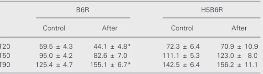
Bupivacaine also affected left ventricu-lar repoventricu-larization analyzed by the monopha-sic action potential (Table 2). The effects on repolarization were also very early, starting immediately after injection of the drug, reach-ing a peak 30 to 90 s later and decreasreach-ing thereafter.
Bupivacaine shortened T20 (the pla-teau phase), but increased T90 (the total action potential duration). Thus, in group B6R, T20 was shortened by -25.0 ± 7.5% (P = 0.03), T50 by -15.5 ± 6.9% (P = 0.15) and T90 increased by 22.7 ± 6.8% (P = 0.003) after the injection of bupivacaine. Conversely, in group H5B6R the effects were much less marked: T20 increased by 4.1 ± 16.3% (P = 0.91), T50 by 10.4 ± 6.8% (P = 0.236) and T90 by 9.8 ± 6.3% (P = 0.302).
Figures 3 and 4 show typical monophasic action potential recordings for one dog in group B6R and another in group H5B6R. Note the differences in the ventricular repo-larization alterations, clearly visible in the B6R dog and much less apparent in the H5B6R dog.
Cardiac arrhythmias and mortality
The arrhythmias observed in this series were sinus node dysfunction (SND) and ven-tricular arrhythmias (Table 3). SND caused severe bradycardia and, in some dogs, led to death due to asystole. Ventricular arrhyth-mias were classified as: 1) nonsustained ven-tricular tachycardia (NSVT), when the epi-sode lasted less than 30 s; 2) sustained ven-tricular tachycardia (SVT), when it lasted more than 30 s or degenerated to ventricular fibrillation (VF), and 3) primary VF. Each arrhythmia occurred alone or in combina-tion in each animal.
In the non-NaCl-pretreated groups (B6 and B6R), SND occurred in 2/25 dogs, NSVT in 2/25 dogs, SVT in 5/25 dogs, and VF in 8/25 dogs, and 12/25 dogs died. When 7.5%
Table 2. Maximal variation of the intraventricular repolarization parameters in the groups.
B6R H5B6R
Control After Control After
T20 59.5 ± 4.3 44.1 ± 4.8* 72.3 ± 6.4 70.9 ± 10.9
T50 95.0 ± 4.2 82.6 ± 7.0 111.1 ± 5.3 123.0 ± 8.0
T90 125.4 ± 4.7 155.1 ± 6.7* 142.5 ± 6.4 156.2 ± 11.1
Data are reported as means ± SEM for N = 8 in each group. T20, T50, T90: duration (ms) of the monophasic action potential at 20, 50 and 90% repolarization, respectively. B6R and H5B6R are the same as B6 and H5B6, respectively (see Table 1).
*P<0.05 compared to respective control (ANOVA).
Figure 3. Monophasic action po-tential recording from a typical B6R dog. Note the typical bu-pivacaine action: shortening of T20 (the plateau phase) with sig-nificant enlargement of T90 (the total duration of the potential).
Control 30 s
117 ms 154 ms
Table 3. Cardiac arrhythmias and mortality.
SND NSVT SVT VF Death
B6 2 2 3 3 5
H5B6 0 1 0 0 0
B6R 0 0 5 5 7
H5B6R 1 0 1 1 1
L5B6 4 3 2 2 3
G5B6 6 5 1 1 3
M10B6 5 3 1 1 2
SND: sinus node dysfunction; NSVT: nonsustained ventricular tachycardia; SVT: sustained ventricular tachycardia; VF: ventricular fibrillation. For group abbreviations, see legends to Tables 1 and 2.
Figure 4. Monophasic action potential recording from a typi-cal H5B6R dog. There was prac-tically no alteration in the po-tential morphology and duration after bupivacaine injection.
Control 120 s
NaCl was used as pretreatment (groups H5B6 and H5B6R), only one dog showed one epi-sode of NSVT, one dog presented SND and another dog VF. The overall mortality was 1/25 dogs. Pretreatment with the other hy-pertonic solutions (groups L5B6, G5B6 and M10B6) had no protective effect, as shown in Table 3.
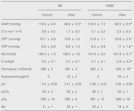
Cardiovascular responses and metabolic changes (Table 4)
Cardiac dynamics changed substantially after bupivacaine injection, with marked de-pression in cardiac function parameters. Thus, in group B6, mean arterial pressure decreased from 110.0 ± 3.4 to 40.8 ± 5.5 mmHg (-62 ± 5%), cardiac index decreased from 3.0 ± 0.2 to 1.7 ± 0.2 l min-1 m-2 (-46 ±
6%), systolic pulmonary pressure increased from 10.1 ± 0.9 to 13.6 ± 1.0 mmHg (38 ± 9%), diastolic pulmonary pressure from 6.5
± 0.9 to 9.8 ± 1.0 mmHg (63 ± 20%), and mean pulmonary pressure from 8.4 ± 0.9 to 12.1 ± 0.9 mmHg (49 ± 11%).
Pretreatment with 7.5% NaCl significant-ly decreased these actions: mean arterial pres-sure was reduced only from 116.3 ± 7.3 to 82.5 ± 6.5 mmHg (-26 ± 6%, P<0.001), cardiac index from 3.1 ± 0.3 to 2.3 ± 0.3 l min-1 m-2 (-28 ± 5%, P = 0.04), systolic
pulmonary pressure increased from 12.9 ± 1.1 to 23.8 ± 2.5 mmHg (85 ± 13%, P = 0.01), diastolic pulmonary pressure from 8.2 ± 0.9 to 17.0 ± 1.8 mmHg (113 ± 16%, P = 0.07), and mean pulmonary pressure from 10.8 ± 1.0 to 21.6 ± 2.4 mmHg (100 ± 15%, P = 0.02). The significant increases in pul-monary pressures in group H5B6 did not lead to clinical pulmonary congestion.
Plasma osmolarity was significantly higher following hypertonic NaCl treatment. Plasma osmolarity increased from 280 ± 2 to 287 ± 3 mOsm/l (3 ± 1%) in group B6 and from 282 ± 3 to 325 ± 10 mOsm/l (15 ± 5%, P = 0.02) in group H5B6.
Plasma sodium levels decreased from 140.3 ± 1.3 to 139.5 ± 1.6 mEq/l (-1 ± 1%) in group B6, but increased from 141.6 ± 2.0 to 161.6 ± 5.7 mEq/l in group H5B6 (14 ± 2%, P<0.001). Plasma potassium levels decreased from 3.5 ± 0.1 to 3.2 ± 0.1 mEq/l (-7 ± 1%) in group B6 and from 3.7 ± 0.1 to 2.9 ± 0.2 mEq/l (-24 ± 4%, P = 0.001) in group H5B6. Blood bupivacaine levels similarly reached toxic levels in groups B6 and H5B6. In group B6, the maximal levels were 15 ± 2 µg/ml 30 s after bupivacaine injection, de-creasing to 5 ± 1 µg/ml 180 s after the injection; in group H5B6, the levels were comparable: the maximal levels were 20 ± 4 µg/ml, decreasing to 10 ± 2 µg/ml, measured at the same times.
The other metabolic parameters for groups B6 and H5B6 did not change significantly.
The mean arterial pressure for group L5B6 changed from 121.6 ± 7.3 to 61.8 ± 10.0 mmHg, group G5B6 from 127.9 ± 6.7 to 74.3 ± 9.7 mmHg and group M10B6
Table 4. Maximal variation of the hemodynamic and metabolic parameters among groups B6 and H5B6.
B6 H5B6
Control After Control After
MAP (mmHg) 110.0 ± 3.4 40.8 ± 5.5* 116.3 ± 7.3 82.5 ± 6.5*
CI (l min-1 m-2) 3.0 ± 0.2 1.7 ± 0.2 3.1 ± 0.3 2.3 ± 0.3
SPP (mmHg) 10.1 ± 0.9 13.6 ± 1.0 12.9 ± 1.1 23.8 ± 2.5*
DPP (mmHg) 6.5 ± 0.9 9.8 ± 1.0 8.2 ± 0.9 17 ± 1.8*
Na (mEq/l) 140.3 ± 1.3 139.5 ± 1.6 141.6 ± 2.0 161.6 ± 5.7*
K (mEq/l) 3.5 ± 0.1 3.2 ± 0.1 3.7 ± 0.1 2.9 ± 0.2*
Osmolarity (mOsm/l) 280 ± 2 287 ± 3 282 ± 3 325 ± 10*
Bupivacaine (µg/ml) 0 15 ± 2 0 20 ± 4
pH 7.4 ± 0.02 7.41 ± 0.02 7.36 ± 0.01 7.35 ± 0.02
pCO2 34 ± 2 30 ± 2 36 ± 1 34 ± 1
pO2 292 ± 16 293 ± 9 281 ± 13 269 ± 18
Bic 21 ± 1 20 ± 1 20 ± 1 18 ± 0
from116.4 ± 9.2 to 71.1 ± 10.6 mmHg.
Discussion
The main finding of our study was that hypertonic NaCl pretreatment effectively protected against the electrophysiologic dis-turbances and ventricular arrhythmias in-duced by bupivacaine intoxication. Toxic blood levels of bupivacaine, above 5 µg/ml (1), were detected in every dog in this study. It is well known that bupivacaine binds to the inactivated Na+ channel, slows phase 0
of the action potential and impairs stimulus conduction in the heart. This was well dem-onstrated in our study: QRS complex dura-tion, HV interval and IV interval consis-tently increased in all dogs. The fact that this anesthetic binds to the Na+ channel in the
inactivated state or during the plateau phase, plus a calcium channel blocking effect (25), may explain the shortening of T20 observed in our data. It has been shown that bupiva-caine blocks delayed rectifier (8) and tran-sient outward K+ currents (7), and prolongs
action potential duration, as seen in our ma-terial by the prolongation of the monophasic action potential in T90.
Among the hypertonic solutions tested, only 7.5% NaCl prevented the electrophysi-ologic effects of the drug both on stimulus conduction and ventricular repolarization. Hyperosmolarity per se cannot explain this protection. The effects of hyperosmolarity on normal electrophysiologic parameters have been studied by other authors. Ehara and Hasegawa (26), using isolated guinea pig ventricular muscle submitted to hyper-tonic electrolytic and non-electrolytic solu-tions, found increases in action potential duration and a decrease in Vmax during phase 0 when a non-electrolytic solution (glucose or sucrose) was used. The same investigators also tested lithium chloride and found an initial increase in Vmax. However, this action only lasted for a short period of time.
Our findings are consistent with these data. Actually, from the studies mentioned above, it would be expected that hypertonic solutions other than Na+ salts might worsen
the electrophysiologic disturbances since their effects tend to decrease phase 0 (and consequently the conduction velocity) and to prolong the action potential duration. This is suggestive of what we found when glucose and mannitol were used: no improvement or even a slight impairment in intraventricular conduction was observed when compared to the non-pretreated group. The results after LiCl infusion, however, were not what might have been expected: a slight protection might have occurred, but our data showed results similar to those obtained when non-electro-lytic solutions were tested.
The mechanism of the protective effect of sodium is unknown. There is evidence that sodium overload by itself has no effect on normal myocardial electrophysiology (27-29). Thus, it probably reverses, at least in part, the bupivacaine sodium channel block-ade. Our results differ from those obtained with lidocaine, which showed no protective effect with hypertonic saline (30,31). Both drugs bind to the sodium channel during the inactivated state; however, different binding sites or properties may prevent competition between extracellular sodium and lidocaine but not bupivacaine. The bupivacaine used in this study is an equimolar mixture of R(+)-and S(-)-bupivacaine (levobupivacaine). Levobupivacaine has been reported to be less arrhythmogenic than bupivacaine (32, 33). Levobupivacaine had less affinity for myocardial sodium channels than dextrobu-pivacaine and dissociation from sodium chan-nels was faster (34,35). Thus, further studies are needed to determine whether sodium overload has different protective effects on the electrophysiologic alterations induced by bupivacaine and levobupivacaine.
ex-cept for plasma sodium levels. Hemodynam-ic findings give little insight into electro-physiologic mechanisms, although NaCl-pre-treated dogs have a much better hemody-namic outcome; we believe this better out-come is simply part of the action of hyper-tonic NaCl.
Differences in the effect of hypertonic NaCl on HV and IV intervals may suggest that protection occurs mainly in the proxi-mal part of the conduction system (HV inter-val). However, the limitations of the IV in-terval as a measure of peripheral conduction
must be considered. It must be remembered that several structures (right bundle branch, Purkinje network, and right ventricular muscle), with different electrophysiologic properties, compose the IV interval.
In conclusion, pretreatment with hyper-tonic NaCl effectively protects against the cardiovascular toxicity of bupivacaine, an effect not seemingly related to hypertonicity alone. Further studies disclosing the mechan-isms of such protection should provide in-sights into the significance and applicability of these findings.
References
1. Covino BG & Vassallo HG (1986). Local Anesthetics: Mechanisms of Actions and Clinical Use. Grunne & Stratton, Inc., New York, NY, USA, 29-55.
2. Vaugham-Williams EM (1984). A classifi-cation of antiarrhythmic actions reas-sessed after a decade of new drugs. Jour-nal of Clinical Pharmacology, 24: 129-147. 3. Campbell TJ (1983). Kinetics of onset of rate-dependent effects of class I antiar-rhythmic drugs are important in determin-ing their effects on refractoriness in guin-ea-pig ventricle, and provide a theoretical basis for their subclassification. Cardio-vascular Research, 17: 334-352. 4. Clarkson CW & Hodeghem LM (1995).
Mechanism for bupivacaine depression of cardiac conduction: fast block of sodium channels during the action potential with slow recovery from block during diastole. Anesthesiology, 62: 396-405.
5. Anderson KP, Walker R, Lux RL, Ershler PR, Menlove R, Williams MR, Krall R & Moddrelle D (1990). Conduction velocity depression and drug-induced ventricular tachyarrhythmias. Circulation, 81: 1024-1038.
6. Kasten GW (1986). Amide local anesthetic alterations of effective refractory period temporal dispersion: relationship to ven-tricular arrhythmias. Anesthesiology, 65: 61-66.
7. Castle NA (1990). Bupivacaine inhibits the transient outward K current but not the inward rectifier in rat ventricular myo-cytes. Journal of Pharmacology and Ex-perimental Therapeutics, 255: 1038-1064. 8. Courtney KR & Kendig JJ (1988). Bupiva-caine is an effective potassium channel
blocker in heart. Biochimica et Biophysica Acta, 939: 163-166.
9. Boettner RB, Dunbar RW, Haley JV & Morrow DH (1972). A comparison of the antiarrhythmic effects of bupivacaine and lidocaine. Southern Medical Journal, 65: 1328-1330.
10. Chapin JC, Kushins LG, Munson ES & Schick LM (1980). Lidocaine, bupivacaine, etidocaine and epinephrine-induced ar-rhythmias during halothane anesthesia in dogs. Anesthesiology, 52: 23-25. 11. Dunbar RW, Boettner RB, Glatz RN,
Pennington RE & Morrow DH (1970). The effect of mepivacaine, bupivacaine and lidocaine on digitalis-induced ventricular arrhythmias. Anesthesia and Analgesia, 49: 761-766.
12. Romanoff ME & Ellis Jr JS (1991). Bupiva-caine toxicity after stellate ganglion block. Anesthesia and Analgesia, 72: 546-548. 13. Gibson P & Murrell D (1991).
Complica-tions of epidural analgesia. Anaesthesia and Intensive Care, 19: 282-284. 14. Salemi S & Gristina E (1991). Toxic effects
of bupivacaine on heart conduction during peridural anesthesia. Minerva Anestesio-logica, 57: 468-469.
15. Wolff AP, Hasenbos MA, Liem TH & Gielen MJ (1992). Accidental overdose of epidural bupivacaine and sufentanil. Re-gional Anesthesia, 17: 237-238. 16. Kinney WW, Kambam JR & Wright W
(1991). Propranolol pretreatment reduces cardiorespiratory toxicity due to plain, but not epinephrine-containing, intravenous bupivacaine in rats. Canadian Journal of Anaesthesia, 38: 533-536.
17. Bernards CM & Artu AA (1991).
Hexa-methonium and midazolam terminate dysrhythmias and hypertension caused by intracerebroventricular bupivacaine in rab-bits. Anesthesiology, 74: 89-96. 18. Hyman SA, Kinney WW, Horn JL, Skelley
CC & Kambam JR (1992). Nimodipine re-duces the toxicity of intravenous bupiva-caine in rats. Anesthesia and Analgesia, 74: 851-855.
19. De Kock M, Le Polain B, Henin D, Vande-walle F & Scholtes JL (1993). Clonidine pretreatment reduces the systemic toxic-ity of intravenous bupivacaine in rats. An-esthesiology, 79: 282-289.
20. Fujita S, Endoh S, Yasukawa T & Sari A (1998). Lidocaine increases the ventricu-lar fibrillation threshold during bupivacaine induced cardiotoxicity in pigs. British Jour-nal of Anaesthesia, 80: 218-222. 21. Rocha e Silva M, Velasco IT, Nogueira da
Silva RI, Oliveira MA, Negraes GA & Oliveira MA (1987). Hyperosmotic sodium salts reverse severe hemorrhagic shock: other solutes do not. American Journal of Physiology, 253: H751-H762.
22. Velasco IT, Pontieri V, Rocha e Silva M & Lopes OU (1980). Hyperosmotic NaCl and
severe hemorrhagic shock. American
Journal of Physiology, 239: H664-H673. 23. Wang BW, Chiou YH, Chen WB, Peng TY
& Leung HK (1997). Intravenous pretreat-ment of hypertonic saline can prevent sys-temic hypotension induced by spinal an-esthesia. Acta Anaesthesiologica Sinica, 35: 85-90.
Revista Brasileira de Anestesiologia, 37 (Suppl 7): 88 (Abstract).
25. Lynch C (1986). Depression of myocardial contractility in vitro by bupivacaine. Anes-thesia and Analgesia, 65: 551-559. 26. Ehara T & Hasegawa JI (1983). Effects of
hypertonic solution on action potential and input resistance in the guinea-pig ventric-ular muscle. Japanese Journal of Physiol-ogy, 33: 151-167.
27. Turgeon J, Wisialowski T, Wong W, Altemeier W, Wikswo J & Roden D (1992). Suppression of longitudinal ver-sus transverse conduction by sodium channel block. Circulation, 85: 221-226. 28. Bajaj A, Woosley R & Roden D (1989).
Acute electrophysiologic effects of so-dium administration in dogs treated with
a-desmethyl encainide. Circulation, 80: 994-1002.
29. Pentel PR, Fifield J & Salermo DM (1990).
Lack of effect of hypertonic sodium bicar-bonate on QRS duration in patients taking therapeutic doses of class IC antiarrhyth-mic drugs. Journal of Clinical Pharmacolo-gy, 30: 789-794.
30. Barber M, Wendt D, Starmer C & Grant A (1992). Blockade of cardiac sodium chan-nels: competition between the permeant ion and antiarrhythmic drugs. Journal of Clinical Investigation, 90: 368-381. 31. Ujhelyi MR, Schur M, Frede T, Bottorff
MB, Gabel M & Markel ML (1997). Hyper-tonic saline does not reverse the sodium channel blocking actions of lidocaine: evi-dence from electrophysiologic and defi-brillation studies. Journal of Cardiovascu-lar Pharmacology, 29: 61-68.
32. Huang YF, Pryor ME, Mather LE & Veer-ing BT (1998). Cardiovascular and central nervous system effects of intravenous levobupivacaine and bupivacaine in
sheep. Anesthesia and Analgesia, 86: 797-804.
33. Gristwood R, Bardsley H, Baker H, Watson N & Nimmo W (1994). Reduced cardiotoxicity of levobupivacaine compared with racemic bupivacaine (Marcaine): new clinical evidence. Expert Opinion on Investigational Drugs, 3: 1209-1212. 34. Valenzuela C, Synders DJ, Bennett PB,
Tamango J & Hondeghem LM (1995). Stereoselective block of cardiac sodium channels by bupivacaine in guinea pig ven-tricular myocytes. Circulation, 92: 3014-3024.