DOI: 10.1590/0004-282X20160065
ARTICLE
Ursolic acid attenuates beta-amyloid-induced
memory impairment in mice
Ácido ursólico atenua a perda de memória induzida por beta-amilóide em ratos
Wenna Liang1, Xiaoyang Zhao1, Jinping Feng1, Fenghua Song2, Yunzhi Pan3
Alzheimer’s disease (AD) is a progressive neurode-generative disease that is associated with global mental dysfunction and cognitive deterioration. Common path-ological features of AD are the accumulation of
intra-neuronal tau and extracellular amyloid β (Aβ) peptide1.
Accumulation of Aβ leads to the deposition of insoluble
neuritic or senile plaques, thereby initiating a pathologi-cal cascade, which results in synaptic dysfunction,
syn-aptic loss, neuronal death, and cognitive impairments2.
Moreover, oxidative stress and inflammation are involved
in Aβ-induced neuronal death and neurotransmitter
deficits and the progression of AD3.
Aβ25-35 peptide is the core fragment of the full-length
Aβ and possesses the biological activity and toxicity of the
full-length Aβ monomer4. Histological and biochemical
al-terations, oxidative damage, inlammatory responses, and
cognitive dysfunction have been induced by intrahippocam-pal or intracerebroventricular (i.c.v.) injections of Aβ25-355,6.
herefore, this animal model is used to investigate the patho -genesis and progression of AD and to screen new candidates for AD therapy5.
Ursolic acid (UA, 3β-hydroxy-urs-12-en-28-oic acid) is a
lipophilic, pentacyclic triterpenoid compound that is
wide-ly present in fruits and many types of herbs, such as Perilla
1The Third People’s Hospital of Liaocheng, Department of Neurology, Shandong, China; 2The Third People’s Hospital of Liaocheng, Department of Pharmacy, Shandong, China;
3The Third Afiliated Hospital of Qiqihar Medical University, Department of Neurology, Heilong Jiang, China.
Correspondence: Yunzhi Pan; 67 West Dong Chang Road, Liaocheng, Shandong, 252000, P.R. China. Liaocheng 252000 China; E-mail: [email protected] Conflict of interest: There is no conlict of interest to declare.
Received 05 October 2015; Received in inal form 11 January 2016; Accepted 26 January 2016.
ABSTRACT
Objective: Increasing evidence demonstrates that oxidative stress and inlammatory are involved in amyloid β (Aβ)-induced memory impairments. Ursolic acid (UA), a triterpenoid compound, has potent anti-inlammatory and antioxidant activities. However, it remains unclear whether UA attenuates Aβ-induced neurotoxicity. Method: The aggregated Aβ25-35 was intracerebroventricularly administered to mice. Results: We found that UA signiicantly reversed the Aβ25-35-induced learning and memory deicits. Our results indicated that one of the potential mechanisms of the neuroprotective effect was attenuating the Aβ25-35-induced accumulation of malondialdehyde (MDA) and depletion of glutathione (GSH) in the hippocampus. Furthermore, UA signiicantly suppressed the upregulation of IL-1β, IL-6, and tumor necrosis-α factor levels in the hippocampus of Aβ25-35-treated mice. Conclusion: These indings suggest that UA prevents memory impairment through amelioration of oxidative stress, inlammatory responseand may offer a novel therapeutic strategy for the treatment of Alzheimer’s disease.
Keywords: amyloid; memory; oxidative stress; ursolic acid.
RESUMO
Objetivo: Há evidências crescentes de que o estresse oxidativo e a inlamação estão envolvidos na perda de memória induzida pelo peptídeo beta-amilóide (βA). O ácido ursólico (AU), um composto triterpenóide, apresenta atividades anti-inlamatórias e antioxidantes potentes. Entretanto, não se sabe ainda se o AU atenua a neurotoxicidade induzida pelo βA. Método: O agregado βA 25-35 foi administrado aos ratos por via intracerebroventricular. Resultados: Observou-se que o AU reverteu signiicativamente os déicits de aprendizado e de memória induzidos pelo βA 25-35. Portanto, um dos potenciais mecanismos do efeito neuroprotetor seria a atenuação do acúmulo de malondialdeído e a depleção de glutationa no hipocampo induzidos pelo βA 25-35. Além disso, o AU suprimiu signiicativamente a supra regulação dos níveis de IL-1β, IL-6 e do fator de necrose tumoral α no hipocampo dos ratos tratados com βA 25-35. Conclusão: Esses achados sugerem que o AU previne a perda de memória através da melhora do estresse oxidativo e da resposta inlamatória, podendoproporcionar uma nova estratégia terapêutica para o tratamento da doença de Alzheimer.
frutescens, Glechoma hederaceae, Rosemarinus oicinalis, and
Eriobotrya japonica7. UA has many pleiotropic biological
ac-tivities, such as anti-oxidant, -inlammatory, -hyperlipidemic,
-ulcer, -microbial, and -tumoral activities7,8,9,10. Moreover, in
vitro studies indicate that it suppresses Aβ-induced injury in PC12 cells11. In particular, UA attenuates oxidative and
in-lammatory injury in the brain and cognitive impairments in
-duced by D-galactose7,12. However, to the best of our
knowl-edge, no study has been conducted to determine whether UA
has an efect against Aβ25-35-induced cognitive deicits and
neurotoxicity in a mouse model. Here, we addressed this is-sue and investigated the potential mechanisms underlying
the neuroprotective efect of UA.
METHOD
Animals
In total, 80 male Kunming mice (10 weeks old) were obtained from the Branch of National Breeder Center of
Rodents (Shanghai). hey were housed in plastic cages and
kept in a regulated environment (22 ± 2°C, 50 ± 5% humid-ity) with a 12:12-h light/dark cycle (lights on from 8:00 h to
20:30 h). he mice were allowed to acclimate for 1 week,
with food and water ad libitum. All experiments were
per-formed as per the Chinese legislation on the use and care of laboratory animals.
Drugs and treatment
UA (Sigma-Aldrich, MO, USA) was suspended in distilled
water containing 0.1% Tween-80 (dH2O/0.1% Tween-80). Aβ25-35
(Sigma-Aldrich, MO, USA) was dissolved in double-distilled
water at a concentration of 1 mg/ml and stored at −20 °C. Aβ25-35
was aggregated, by incubating it in distilled water at 37 °C for 4 days before the injection as described previously5. Mice were
injected with aggregated Aβ25-35 or vehicle (3 μl/3 min, i.c.v.)
into lateral ventricles on day 06 under mild anesthesia with
ether and then returned to their home cages. In addition, mice received UA (10, 20, or 40 mg/kg) or the distilled water con-taining 0.1% Tween-80 was by oral gavage for 11 days after the
treatment with Aβ25-35 and the time of treatment was based
on our pretest study. he dose of UA in the experiments was
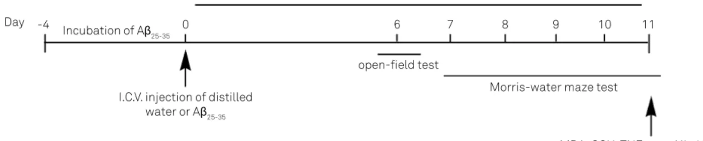
based on previous reports7,12. he schedule of our study is dem
-onstrated in Figure 1.
Open-field test
An open-ield test was conducted on day 6 after the in -jection of Aβ25-35 as described previously
7. Each mouse was
placed in the center of an open-ield apparatus with inter -nal dimensions of 30 cm × 30 cm and walls of height 30 cm.
he open ield was divided into 6 × 6 cm squares marked
with white lines. In the test, mice were individually placed in the center of the arena and permitted for free explora-tions. After 1 min of adaptation, the behavior of each mouse was recorded for 5 min by two observers 1 m away from the
open ield area. Between trials, the mice were returned to their home cages in the same room, and the open ield was
wiped clean with a slightly damp cloth and dried prior to
occupancy by another mouse. he behavioral parameters
were assessed as follows:
• ambulation: the number of grids crossed in adrena during
the observation period;
• rearing/leaning: the number of times the mouse stands
on its hind legs;
• grooming: the number of times the mouse “washes” itself by
licking, wiping, combing, or scratching any part of the body.
Morris water maze test
Following the open-ield test, the Morris water maze
(MWM) task was performed as described previously7 with
minor modiications. he experimental apparatus consist -ed of a circular pool (120 cm in diameter, 60 cm in height) located in a test room with various prominent visual cues on white walls. With its inner surface painted black, the pool
was illed with water to a depth of 40 cm (maintained at 25 ± 1°C). he pool was virtually divided into 4 quadrants and the platform was placed at a ixed position in the center of a
quadrant. An escape platform (black), 10 cm in diameter, was submerged approximately 1.5 cm below the surface of the water and placed at the midpoint of one quadrant. Four dif-ferent start points (NE, SE, SW, and NW) were equally spaced around the circumference of the pool.
Each mouse received 4 training periods per day for 5 consecutive days. For each trail, the mice were placed in the
Figure 1. The experimental design of the study demonstrating subchronic treatment of ursolic acid.
Ursolic acid treatment by oral gavage
open-ield test
Morris-water maze test
MDA, GSH, TNF-α and IL-1β assay I.C.V. injection of distilled
water or Aβ25-35 Incubation of Aβ25-35
water facing the wall at one of the 4 possible starting points, and their latency to the platform was recorded. If a mouse
did not ind the escape platform within 90 s, it was given a
latency score of 90 s. At the end of each session, all animals were dried and returned to their home cages. On day 6, the probe test was conducted by removing the platform and
al-lowing each mouse to swim freely for 90 s. he time spent in
the target quadrant (where the platform was located during hidden platform training) and the swimming path length by the mouse were measured.
Brain homogenate
For biochemical studies, animals were deeply anes-thetized and sacrificed. The hippocampus was promptly isolated after the animals were sacrificed. The hippocam-pus was weighed, homogenized in 9 volumes (1:9 w/v) of cold saline to prepare a 10% tissue homogenate in an ice
bath, and centrifuged at 1699 ×g for 15 min (Eppendorf
Centrifuge 5804R, Hamburg, Germany). Then, the super-natant was collected for the subsequent determination of oxidative alterations in brain.
Determination of oxidative alterations in hippocampus
The contents of malondialdehyde (MDA) and glu-tathione (GSH) in the hippocampus were measured as per their corresponding kits ( Jiancheng Bio-tech, Nanjing, China) as per the manufacturer’s instructions. Measurement of all assays described above was conduct-ed in triplicate. Protein contents were determinconduct-ed by the bicinchoninic assay.
Determination of TNF-α and IL-1β levels in hippocampus
The hippocampus was homogenized in ice-cold nor-mal saline and then centrifuged 12,000 rpm for 5 min at
4°C. TNF-α and IL-1β levels were respectively measured
using an enzyme-linked immunosorbent assay with
mouse TNF-α/IL-1β kits (R&D Systems) as per the
man-ufacturer’s instructions.
Statistical analysis
All statistical analyses were performed using SPSS soft-ware, version 13.0 (SPSS Inc., Chicago, IL, USA). Values are expressed as mean ± SEM. For the MWM task, data were ana-lyzed by Kruskal-Wallis non-parametric analysis of variance
(ANOVA). If the results were signiicant, the intergroup vari
-ance was assessed by Tukey’s post hoc test. For the
biochemi-cal and neuroendocrine assays, diferences between groups
were analyzed by one-way ANOVA followed by the Dunnett’s
post hoc test. he results were considered statistically signii
-cant if p < 0.05.
RESULTS
Effect of UA on the behavioral alterations induced by Aβ25-35 in the open-field test
Figure 2 demonstrated that Aβ25-35 injection produced
be-havioral efects in mice. hese bebe-havioral alterations includ -ed a r-eduction in crossings (p < 0.001), rearings (p < 0.01, and
groomings (p < 0.05). UA treatment markedly suppressed the
decrease in open-ield activity in Aβ25-35-treated mice (p < 0.05
or p < 0.01). here was no signiicant diference in open-ield
activity between the distilled water-injected mice and dis-tilled water/UA group (p > 0.05).
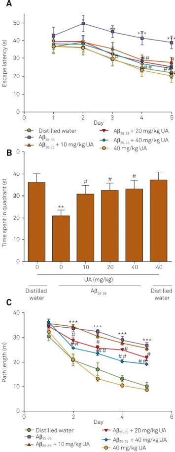
Effect of UA on the behavioral alterations induced by Aβ25-35 in the MWM task
he MWM task is one of the most widely used behav -ioral tasks for the assessment of learning and memory in
mice. he mean escape latency and swimming path length did not difer between any of the groups on the irst day of testing in the MWM task (p < 0.05; Figure 3). From the day
2 onwards, the mean latency and swimming path length in
the Aβ25-35 group were prolonged signiicantly compared with
those of the vehicle group (p < 0.05, p < 0.01, or p < 0.05 or p < 0.001). UA treatment signiicantly decreased the escape
latency and swimming path length in Aβ25-35 treatment mice
(p < 0.05 or p < 0.01). However, there was no signiicant difer -ence between distilled water-injected mice and distilled
wa-ter/UA mice in the time taken to ind the hidden platform or in swimming path length. In the probe test, a signiicant -ly shorter stay in the platform quadrant was observed in the Aβ25-35 group, which was signiicantly suppressed by UA treat
-ment (p < 0.05). However, there was no signiicant diference
in the time UA-treated mice spent in the platform quadrant
when compared with the vehicle group (p > 0.05).
Effect of UA on the MDA and GSH levels in the hippocampus of Aβ25-35-injected mice
Aβ25-35-treated mice demonstrated a signiicant increase
in MDA level in the hippocampus, when compared with the
distilled water group (p < 0.05; Figure 4). Similarly, the GSH
levels in hippocampus of the Aβ25-35 group were signiicant
-ly increased compared with those of distilled water-injected mice, while the decrease in GSH content in the hippocampus
was markedly suppressed by UA treatment (p < 0.05). In ad
-dition, UA treatment signiicantly suppressed the increase in
the MDA level in the hippocampus when compared with the
distilled water group (p < 0.05). However, no signiicant oxi -dative alterations were observed between the distilled water
group and the distilled water/UA group (p > 0.05).
Effect of UA on the TNF-α and IL-1β levels in the hippocampus of Aβ25-35-injected mice
A significant increase in TNF-α and IL-1β
Figure 2. Number of line crossings (A), rearings (B), and groomings (C) of mice in the open-ield test. Data are expressed as mean ± SEM (n = 8). *p < 0.05, **p < 0.01 ***p < 0.001 versus distilled water-injected mice; #p < 0.05, ##p < 0.01 versus only Aβ
25-35-injected mice.
Number of crossings
UA (mg/kg)
Aβ25-35 Distilled water Distilled water 100 80 60 40 20 0
0 0 10 20 40 40
A
Number of rearings/leaning
UA (mg/kg)
Aβ25-35 Distilled water Distilled water 35 30 25 20 15 10 5 0
0 0 10 20 40 40
B
Number of groomings
UA (mg/kg)
Aβ25-35 Distilled water Distilled water 50 40 30 20 10 0 0 0 * # ** # *** # # ## # ## ## ##
10 20 40 40
C
Figure 3. Behavioral performance of mice in the Morris water maze task. (A) Escape latency apparent during the training and probe sessions. (B) Time spent in target quadrant during the probe trial. (C) Path length during the training and probe sessions. Data are expressed as mean ± SEM (n = 8). *p < 0.05, **p < 0.01 ***p < 0.001 versus only Aβ25-35-injected mice; #p < 0.05, ##p < 0.01 versus distilled water-injected mice.
Escape latency (s)
Day 50 40 30 20 10 0
0 1 2 3 4 5
A
Time spent in quadrant (s)
UA (mg/kg)
Aβ25-35 Distilled
water Distilled water 40 30 20 20 20 10 0
0 0 10 20 40 40
B
Path length (m)
40 30 20 10 0 ** # # ## # ## ## ## # #
C
Day0 2 4 6
Distilled water Aβ25-35
Aβ25-35 + 10 mg/kg UA
Aβ25-35 + 20 mg/kg UA
Aβ25-35 + 40 mg/kg UA
40 mg/kg UA
Distilled water Aβ25-35
Aβ25-35 + 10 mg/kg UA
Aβ25-35 + 20 mg/kg UA
Aβ25-35+ 40 mg/kg UA
mice compared with those of the distilled water group (p < 0.001) (Figure 5). UA treatment significantly
attenu-ated the alterations induced by Aβ25-35 in the hippocampus
(p < 0.05, p < 0.01, or p < 0.001). However, UA did not sig
-nificantly affect the TNF-α and IL-1β levels in the
hippo-campus of vehicle-treated mice (p > 0.05).
DISCUSSION
Here, we demonstrated that UA produced a
neuropro-tective efect against Aβ25-35-induced neurotoxicity and
memory impairment in mice. UA treatment attenuated Aβ25-35-induced impairment of memory in the MWM task.
Moreover, we demonstrated that UA suppressed the accu-mulation of lipid peroxide (MDA) and the reduction in GSH
in the hippocampus induced by Aβ25-35. Furthermore, our
data indicated that UA treatment is capable of suppressing the Aβ25-35-induced upregulation of TNF-α and IL-β levels in the hippocampus. Collectively, these results suggest that
re-peated administration of UA attenuates cognitive deicits by regulating oxidative stress and inlammation in the brain.
Oxidative stress and inlammatory responses in the brain
contribute to Aβ-induced neuronal toxicity and cognitive
def-icits6,7. Aβ
25-35 is a proteolytic fragment of Aβ with high
neu-rotoxicity that is produced in the brains of AD patients13,14.
herefore, the Aβ25-35-treated mouse serves as a model has
been used to study Aβ-induced neurotoxicity and memory
Figure 4. Effect of ursolic acid on MDA (A) and GSH (B) levels in the hippocampus of mice. Each value is expressed as mean ± SEM (n = 8). *p < 0.05, ***p < 0.001 versus distilled water-injected mice; #p < 0.05, ##p < 0.01 versus only
Aβ25-35-injected mice.
MDA level (nmol/mg prot)
UA (mg/kg)
Aβ25-35 Distilled water Distilled
water 2.5
2.0
1.5
1.0
0.5
0.0
0 0 10 20 40 40
A
GSH level (nmol/mg prot)
UA (mg/kg)
Aβ25-35 Distilled water Distilled
water 4
3
2
1
0
0 0 10 20 40 40
B
*** # **
#
#
##
# ##
Figure 5. Effect of ursolic acid on the TNF-α (A) and IL-1β (B) levels in the hippocampus of mice. Each value is expressed as mean ± SEM (n = 8). ***p < 0.001 versus distilled water-injected mice; #p < 0.05, ##p < 0.01, ###p < 0.001 versus
only Aβ25-35-injected mice.
TNF-α
(pg/mg)
UA (mg/kg)
Aβ25-35 Distilled water Distilled
water 400
300
200
100
0
0 0 10 20 40 40
A
IL-1
β
(pg/mg)
UA (mg/kg)
Aβ25-35 Distilled water Distilled
water 25
20
15
10
5
0
0 0 10 20 40 40
B
***
# ***
#
##
###
#
References
1. Blennow K, Leon MJ, Zetterberg H. Alzheimer’s disease. Lancet. 2006;368(9533):387-403. doi:10.1016/S0140-6736(06)69113-7
2. Walsh DM, Selkoe DJ. Deciphering the molecular basis of memory failure in Alzheimer’s disease. Neuron. 2004;44(1):181-93. doi:10.1016/j.neuron.2004.09.010
3. Massoud F, Gauthier S. Update on the pharmacological treatment of Alzheimer’s disease. Curr Neurophamarcol. 2010;8(1):69-80. doi:10.2174/157015910790909520
4. Yankner BA, Duffy LK, Kirschner DA. Neurotrophic and neurotoxic effects of amyloid beta protein: reversal by tachykinin neuropeptides. Science. 1990;250(4978):279-82. doi:10.1126/science.2218531
5. Maurice T, Lockhart BP, Privat A. Amnesia induced in mice by centrally administered β-amyloid peptides involves cholinergic dysfunction. Brain Res. 1996;706(2):181-93. doi:10.1016/0006-8993(95)01032-7
6. Lu P, Mamiya T, Lu LL, Mouri A, Zou L, Nagai T et al. Silibinin prevents amyloid β peptide-induced memory impairment and oxidative stress in mice. Br J Pharmacol. 2009;157(7):1270-7. doi:10.1111/j.1476-5381.2009.00295.x
7. Lu J, Zheng YL, Wu DM, Luo L, Sun DX, Shan Q. Ursolic acid ameliorates cognition deicits and attenuates oxidative damage in the brain of senescent mice induced by D-galactose. Biochem Pharmacol. 2007;74(7):1078-90. doi:10.1016/j.bcp.2007.07.007
8. Tokuda H, Ohigashi H, Koshimizu K, Ito Y. Inhibitory effects of ursolic and oleanolic acid on skin tumor promotion by 12-O-tetradecanoylphorbol-13-acetate. Cancer Lett. 1986;33(3):279-85. doi:10.1016/0304-3835(86)90067-4
9. Liu J. Pharmacology of oleanolic acid and ursolic acid. J Ethnopharmacol. 1995;49(2):57-68. doi:10.1016/0378-8741(95)90032-2
impairments6,7,15. Here, the efect of UA on Aβ-induced
neu-rotoxicity was assessed in Aβ25-35-injected mice. Our data in
the open-ield test suggested that injection of Aβ25-35 could
cause motor abnormalities and deicits of novelty-induced
behavioral impairment. Furthermore, Aβ25-35-injected mice
displayed learning and memory deicits, consistent with pre -vious reports16.
Drugs isolated from traditional medicinal plants may provide a promising therapy for brain injuries caused by oxi-dative stress. UA (3β-hydroxy-urs-12-en-28-oic acid) is a
lipo-philic, pentacyclic triterpenoid compound. UA has a signii
-cant anti-oxidant and -inlammatory efect both in vivo and
in vitro17,18. UA also exerts a neuroprotective efect against
D-galactose-induced cognition deicits in mice via the reduc
-tion of its anti-oxidant properties19. Moreover, UA suppresses
NF-κB signaling, thus attenuating the inlammatory respons
-es of PC12 cells18. With this background, the aim of the
pres-ent study was to evaluate whether UA has a protective efect
against cognitive impairment induced by Aβ25-35-injected in
mice. Here, UA signiicantly suppressed the behavioral altera
-tions induced by Aβ25-35. However, no signiicant diference in
behavior was found during either the open-ield test or the
MWM task in UA/vehicle-treated mice as compared with the
vehicle group. hese results suggested that UA could reverse
impaired cognition induced by Aβ25-35 in mice.
Oxidative stress plays a critical role in the
pathogenici-ty and development of AD and mild cognitive impairment6.
Lipid peroxidation has been proposed to be one of the ma-jor outcomes of free radical-mediated injury that directly damages membranes and generates numerous secondary
products, including aldehydes such as MDA6. Furthermore,
accumulation of lipid peroxidation products is found in mul-tiple regions, such as amygdale, hippocampus, and
parahip-pocampal gyrus in the AD brain20. MDA is the most
abun-dant individual aldehyde arising from lipid peroxidation and could therefore be considered a marker of lipid peroxidation.
In this study, Aβ25-35 administration increased MDA levels in
the hippocampus, suggesting that it caused lipid peroxida-tion. Treatment with UA suppressed the accumulation of
MDA induced by Aβ25-35, which suggests a beneicial efect of
UA treatment in reducing lipid peroxidation caused by Aβ
25-35. hese indings suggested that protection from lipid peroxi
-dation is involved in the improving the efects of UA on cog
-nitive deicits.
In addition, Aβ peptide impairs the anti-oxidative defenses in the brain, thereby possibly contributing to the pathogenesis
of AD21. GSH is the most abundant intracellular anti-oxidant,
the dysregulation of which is involved in the pathogenesis of many neurodegenerative diseases. Here, the reduction of GSH
levels induced by Aβ25-35 was suppressed by UA treatment.
hus, the protective efect of UA on Aβ25-35-induced cognitive
deicit involves the activation of antioxidative defenses in the
brain. Moreover, the antioxidant activity of UA has been stud-ied previously7,22. Firstly, UA suppresses O
2
− generation in
xan-thine-xanthine oxidase assay system23. Furthermore, UA can
strengthen anti-oxidative defense against free radicals induced by D-galactose in mice7.
In addition, inflammation occurs in pathologically
vulnerable regions of the AD brain24. Considerable
evi-dence gained over the past decade has indicated that
neuroinflammation is associated with AD pathology25.
Moreover, treatment of microglia or astrocytes with Aβ
leads to the release of inflammatory factors, which may
contribute to neuronal cell damage and eventual death26,27.
Here, increased levels of inflammatory cytokines, such as
TNF-α and IL-1β in the hippocampus were observed in
Aβ25-35-treated mice, consistent with previous reports28,29.
However, these alterations in TNF-α and IL-1β in the
hippocampus were suppressed by UA treatment.
In summary, the present results demonstrate that UA
can alleviate the memory impairments induced by Aβ25-35 in
mice. he efect of UA may be attributed to the prevention of oxidative stress and the inlammatory response induced by
10. Mahato SB, Sarkar SK, Poddar G. Triterpenoid saponins. Phytochemistry. 1988;27(10):3037-67. doi:10.1016/0031-9422(88)80001-3
11. Heo HJ, Cho HY, Hong B, Kim HK, Heo TR, Kim EK et al. Ursolic acid of
Origanum majoranal L. reduces Abeta-induced oxidative injury. Mol Cells. 2002;13(1):5-11.
12. Lu J, Wu DM, Zheng YL, Hu B, Zhang ZF, Ye Q et al. Ursolic acid attenuates D-galactose-induced inlammatory response in mouse prefrontal cortex through inhibiting AGEs/RAGE/NF-κB pathway activation. Cereb Cortex. 2010;20(11):2540-8. doi:10.1093/cercor/bhq002
13. Pike CJ, Walencewicz-Wasserman AJ, Kosmoski J, Cribbs DH, Glabe CG, Cotman CW. Structure-activity analyses of β-amyloid peptides: contributions of the β25-35 region to aggregation and neurotoxicity. J Neurochem. 1995;64(1):253-65. doi:10.1046/j.1471-4159.1995.64010253.x
14. Kubo T, Nishimura S, Kumagae Y, Kaneko I. In vivo conversion of racemized β-amyloid ([D-Ser26]Aβ1-40) to truncated and toxic
fragments ([D-Ser26]Aβ25-35/40) and fragment presence in the
brains of Alzheimer’s patients. J Neurosci Res. 2002;70(3):474-83. doi:10.1002/jnr.10391
15. Kim DH, Jung WY, Park SJ, Kim JM, Lee S, Kim YC et al. Anti-amnesic effect of ESP-102 on Aβ(1-42)-induced memory impairment
in mice. Pharmacol Biochem Behav. 2010;97(2):239-48. doi:10.1016/j.pbb.2010.08.005
16. Alkam T, Nitta A, Mizoguchi H, Itoh A, Nabeshima T. A natural scavenger of peroxynitrites, rosmarinic acid, protects against impairment of memory induced by Abeta (25-35). Behav Brain Res. 2007;180(2):139-45. doi:10.1016/j.bbr.2007.03.001
17. Heo HJ, Cho HY, Hong B, Kim HK, Heo TR, Kim EK et al. Ursolic acid of Origanum majorana L. reduces Abeta-induced oxidative injury. Mol Cells. 2002;13(1):5-11.
18. Yoon JH, Youn K, Ho CT, Karwe MV, Jeong WS, Jun M.
p-Coumaric acid and ursolic acid from Corni fructus attenuated β-amyloid(25-35)-induced toxicity through regulation of the NF-κB signaling pathway in PC12 cells. J Agric Food Chem. 2014;62(21):4911-6. doi:10.1021/jf501314g
19. Lu J, Zheng YL, Wu DM, Luo L, Sun DX, Shan Q. Ursolic acid ameliorates cognition deicits and attenuates oxidative damage
in the brain of senescent mice induced by D-galactose. Biochem Pharmacol. 2007;74(7):1078-90. doi:10.1016/j.bcp.2007.07.007
20. Markesbery WR, Lovell MA. Four-hydroxynonenal, a product of lipid peroxidation, is increased in the brain in Alzheimer’s disease. Neurobiol Aging. 1998;19(1):33-6. doi:10.1016/S0197-4580(98)00009-8
21. Mattson MP, Guo Q, Furukawa K, Pedersen WA. Presenilins, the endoplasmic reticulum, and neuronal apoptosis in Alzheimer’s disease. J Neurochem. 1998;70(1):1-14. doi:10.1046/j.1471-4159.1998.70010001.x
22. Balanehru S, Nagarajan B. Protective effect of oleanolic acid and ursolic acid against lipid peroxidation. Biochem Int. 1991;24(5):981-90.
23. Jiwajinda S, Santisopasri V, Murakami A, Kim OK, Kim HW, Ohigashi H. Suppressive effects of edible Thai plants on superoxide and nitric oxide generation. Asian Pac J Cancer Prev. 2002;3(3):215-23.
24. Akiyama H, Barger S, Barnum S, Bradt B, Bauer J, Cole GM et al. Inlammation and Alzheimer’s disease. Neurobiol Aging. 2000;21(3):383-421. doi:10.1016/S0197-4580(00)00124-X
25. Tuppo EE, Arias HR. The role of inlammation in Alzheimer’s disease. Int J Biochem Cell Biol. 2005;37(2):289-305. doi:10.1016/j.biocel.2004.07.009
26. Garwood CJ, Pooler AM, Atherton J, Hanger DP, Noble W. Astrocytes are important mediators of Aβ-induced neurotoxicity and tau phosphorylation in primary culture. Cell Death Dis. 2011;2(6):e167. doi:10.1038/cddis.2011.50
27. Capiralla H, Vingtdeux V, Zhao H, Sankowski R, Al-Abed Y, Davies P, et al. Resveratrol mitigates
lipopolysaccharide- and Aβ-mediated microglial inlammation by inhibiting the TLR4/NF-κB/STAT signaling cascade. J Neurochem. 2012;120(3):461-72. doi:10.1111/j.1471-4159.2011.07594.x
28. Ji C, Song C, Zuo P. The mechanism of memory impairment induced by Aβ chronic administration involves imbalance between cytokines and neurotrophins in the rat hippocampus. Curr Alzheimer Res. 2011;8(4):410-20. doi:10.2174/156720511795745366
29. Lu P, Mamiya T, Lu LL, Mouri A, Ikejima T, Kim HC et al. Xanthoceraside attenuates amyloid β peptide25-35-induced
ERRATUM DOI: 10.1590/0004-282X20160065err
Erratum
Ursolic acid attenuates beta-amyloid-induced memory impairment in mice
Arq Neuropsiquiatr 2016;74(6):482-488. DOI: http://dx.doi.org/10.1590/0004-282X20160065
he correspondence adress:
Yunzhi Pan; 67 West Dong Chang Road, Liaocheng, Shandong, 252000, P.R. China. Liaocheng 252000 China
Should be: