DOI: 10.1590/0004-282X20160138
VIEW AND REVIEW
Neurological manifestations of Chikungunya
and Zika infections
Manifestações neurológicas das infecções pelos vírus Zika e Chikungunya
Talys J. Pinheiro1, Luis F. Guimarães1, Marcus Tulius T. Silva2, Cristiane N. Soares1
he pandemic of Chikungunya virus (CHIKV) and Zika vi -rus (ZIKV) infection throughout South and Central America is the most important epidemiological occurrence nowadays. High temperatures rates, vector abundance, and inability to control it makes Latin America a high-risk area for the estab -lishment and spread of arbovirus diseases and, as a conse -quence, their neurological complications. his article focuses on the reemergence of these diseases, their neurological man -ifestations, and possible treatments.
CHIKUNGUNYA INFECTION
History and epidemiology
Chikungunya virus is a member of the Togaviradae family, belonging to the genus alphavirus. It was irst isolated from a febrile patient during an outbreak in 1952-1953 in south -ern Tanzania1. he name Chikungunya is derived from the
Kimakonde language, meaning “to become contorted” or “that which bends up”.
1Hospital dos Servidores do Estado, Serviço de Neurologia, Rio de Janeiro RJ, Brasil;
2Laboratório de Pesquisa Clínica em Doenças Neuroinfecciosas, Instituto Nacional de Doenças Infecciosas Evandro Chagas/ Fundação Oswaldo Cruz,
RJ, Brasil.
Correspondence: Cristiane N. Soares; Rua Santa Clara, 50 / sala 1217; 22060-000 Rio de Janeiro RJ, Brasil; E-mail: [email protected]
Conflict of interest: There is no conlict of interest to declare.
Received 08 July 2016; Accepted 26 July 2016. ABSTRACT
The epidemics of Chikungunya virus (CHIKV) and Zika virus (ZIKV) infections have been considered the most important epidemiological occurrences in the Americas. The clinical picture of CHIKV infection is characterized by high fever, exanthema, myalgia, headaches, and arthralgia. Besides the typical clinical picture of CHIKV, atypical manifestations of neurological complications have been reported: meningo-encephalitis, meningoencephalo-myeloradiculitis, myeloradiculitis, myelitis, myeloneuropathy, Guillain-Barré syndrome and others. The diagnosis is based on clinical, epidemiological, and laboratory criteria. The most common symptoms of ZIKV infection are skin rash (mostly maculopapular), fever, arthralgia, myalgia, headache, and conjunctivitis. Some epidemics that have recently occurred in French Polynesia and Brazil, reported the most severe conditions, with involvement of the nervous system (Guillain-Barré syndrome, transverse myelitis, microcephaly and meningitis). The treatment for ZIKV and CHIKV infections are symptomatic and the management for neurological complications depends on the type of affliction. Intravenous immunoglobulin, plasmapheresis, and corticosteroid pulse therapy are options.
Keywords: Guillain-Barre syndrome; Zika virus; Chikungunya virus.
RESUMO
As epidemias provocadas pelo vírus Chikungunya (CHIK) e Zika vírus (ZIKV) têm sido consideradas as ocorrências epidemiológicas mais importantes da América. O quadro clínico da infecção por CHIK caracteriza-se por febre alta, exantema, mialgia, cefaléia e artralgia. Além do quadro clínico típico, manifestações atípicas como complicações neurológicas foram relatadas: meningo-encefalite, mielorradiculopatia, mielorradiculite, mielite, mieloneuropatia, síndrome de Guillain-Barre (GBS), entre outras. O diagnóstico é baseado em critérios clínicos, epidemiológicos e laboratoriais. Em relação aos sinais e sintomas da infecção pelo ZIKV, erupção cutânea (principalmente maculopapular), febre, artralgia, mialgia, cefaléia e conjuntivite são os mais comuns. Algumas epidemias que ocorreram recentemente na Polinésia Francesa e Brasil relataram condições mais severas, com envolvimento do sistema nervoso (GBS, mielite transversa, microcefalia e meningite). O tratamento para ZIKV e CHIK é sintomático, e o manejo das complicações neurológicas dependerá do tipo da afecção. Imunoglobulina venosa, plasmaférese, e pulsoterapia com corticosteróides são opções.
hree genotypes of CHIKV have been deined: the West African, East/Central/South African, and Asian. he virus can be transmitted in a human-mosquito-human transmis -sion cycle and can be spread by viremic humans. his fact is important as in dense human populations with lack of im -munity there is a great probability of an explosive CHIKV epidemic2. Aedes (Ae.) aegypti and Ae. albopictus mosquitoes
are the vectors for this virus and are found throughout the Americas, including parts of the United States.
Chikungunya virus outbreaks have previously been doc -umented in the countries in Africa, Asia, Europe, India and the Paciic islands3. In 2013, the irst conirmed autochtho -nous cases in the Americas were reported on St Martin Island, in the Caribbean. Before that, only imported cases of CHIKV had been detected4
. As most people in that region are not immune, 3.6 billion persons in 124 countries are estimated to be at risk.
In 2015, several southern American countries conirmed local transmission: Bolivia, Ecuador, Colombia, Paraguay, Venezuela, and Argentina5. Brazil had its irst indigenous
transmission in the state of Amapa in 2014.
New cases of Chikungunya in the Americas were re -ported during April 2016 making a total count of 41,116 conirmed and suspected cases. Included in the recent in -creases, Bolivia reported 1,725 new infections, Colombia re -corded 967 new cases (10,415 this year), followed by Brazil and Guatemala6 (Figure).
Clinical and neurological manifestations
he incubation period for CHIKV ranges from one to 12 days and is followed by high fever, exanthema, myalgia, headaches,
and arthralgia. his last symptom is usually symmetrical and, almost always, afects more than one joint. Although the acute symptoms do not last more than one to two weeks, arthralgia can persist for months or years. Asymptomatic infections rates range from 3% to 25% of cases7.
Beside the typical clinical picture of CHIKV, atypical man -ifestations such as neurological complications have been re -ported. In these cases, the clinical spectrum between adults and children has been similar8
. Encephalopathy was the most common complication among CHIKV-infected neonates af -ter mother-to-child transmission. During the delivery period, the rate of transmission for viremic women was close to 50%. Fifty percent of infected infants had pathological MRI ind -ings, such as white matter lesions, swelling of the brain, and cerebral hemorrhages, sometimes progressing to permanent disabilities or death9.
Meningoencephalitis has been reported in outbreaks in India and Réunion Island10. Neurological manifestations sec -ondary to CHIKV infection have been described, ranging from 16% of a total of 300 cases, with encephalitis being the most common neurological presentation11,12. Brainstem encephali -tis post-Chikungunya infection had also been reported10.
Neurological complications described in the recent epidemics include: meningo-encephalitis, meningoencephalo-myeloradicu -litis, myeloradicu-litis, mye-litis, myeloneuropathy, Guillain-Barré Syndrome (GBS), external ophthalmoplegia, facial palsy, sensori -neural deafness, and optic neuritis13,14,15,16,17,18,19,20,21,22,23. Optic nerve
involvement in CHIKV infection includes papillitis, retrobulbar neuritis, and neuroretinitis17. Encephalitis occurs either simul -taneously or within a few days of onset of systemic symptoms,
Figure. Conirmed cases of Chikungunya and Zika infections.
Confirmed cases of Chikungunya and Zika
during the period of viremia. A delay of more than two weeks has been reported with other complications like myelitis, GBS, and optic neuritis13,17,21.
A prospective study, performed in India from August to October 2006, of 20 hospitalized patients, found the occur -rence of neurological symptoms and signs early in the course of the disease in cases of Chikungunya. All patients showed a disturbance in their level of consciousness, such as confu -sion, drowsiness and delirium. Six patients had psychosis and six had focal or generalized seizures with normal EEG. Total blindness due retro-bulbar neuritis occurred in two pa -tients. One patient had right hemiparesis, with diminished deep tendon relexes and lexor plantar response, and mild papilloedema. A brain CT scan revealed a ring-enhancing le -sion in the left basal ganglia19.
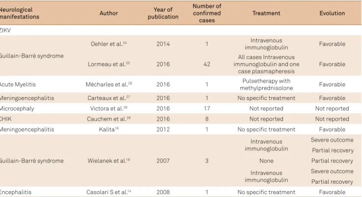
Analysis of the cerebrospinal luid of patients in this study revealed raised protein in 17 patients; glucose was nor -mal in all 20 patients. Nine patients had a cytology count of > 5 cells/mm3, predominantly lymphocytes or mononu -clear cells, indicating that there was no speciic correlation between neurological indings and abnormalities in the cere -brospinal luid (CSF)19 (Table 1).
Neuropathogenesis
he CHIKV can be detected in the brain within two days of experimental infection30. Microgliosis and perivascular
cufs were found in the brain parenchyma of mice infected by CHIKV, as well as neuronal degeneration in the hippocampus and multifocal lymphocytic leptomeningitis. Also, CHIKV seems to targets ependymal cells, progenitor and stem cells
in the subventricular zone. his would impair neurogenesis and neuronal migration, and is an hypothesis for the neuro -pathogenesis of encephalomyelitis related to CHIKV9.
Recent studies have demonstrated that cultured as -trocytes and oligodendrocytes are highly susceptible to CHIKV infection. Glial cells express several pattern recog -nition receptors involved in detecting viral particles as well as damage-associated molecular patterns. hese cells can then be induced to express high levels of cytokines and che -mokines in response to CHIKV infection. he astrocytes’ response to this virus would alter the number and distri -bution of synapses that each astrocyte would be capable of forming. hese results provide the irst evidence that CHIKV infection induces morphometric and innate im -mune activation of astrocytes in vivo31.
Laboratory diagnosis
he diagnosis of CHIKV is based on clinical, epidemi -ological, and laboratory criteria. he detection of viral nu -cleic acid or the infectious virus in serum samples is useful during the initial viremic phase, at the onset of symptoms, and usually for the following ive to 10 days, when CHIKV RNA reaches very high levels. After this, the diagnosis de -pends on the detection of speciic immune responses by se -rological methods32,33,34.
he molecular assays are a rapid and sensitive technique for the diagnosis of CHIKV infection during the early stag -es of illn-ess. Conventional reverse transcription polymerase chain reaction (RT-PCR)32 are available, along with other
RT-PCR real-time assays33 More recently, a one-step SYBR
Neurological
manifestations Author
Year of publication
Number of confirmed
cases
Treatment Evolution
ZIKV
Guillain-Barré syndrome
Oehler et al.24 2014 1 Intravenous
immunoglobulin Favorable
Lormeau et al.25 2016 42
All cases Intravenous immunoglobulin and one
case plasmapheresis
Favorable
Acute Myelitis Mécharles et al.26 2016 1 Pulsetherapy with
methylprednisolone Favorable Meningoencephalitis Carteaux et al.27 2016 1 No specific treatment Favorable
Microcephaly Victora et al.28 2016 17 Not reported Not reported
CHIK Cauchem et al.29 2016 8 Not reported Not reported
Meningoencephalitis Kalita16 2012 1 No specific treatment Favorable
Guillain-Barré syndrome Wielanek et al.18 2007 3
Intravenous immunoglobulin
Severe outcome
Partial recovery
None Partial recovery
Intravenous immunoglobulin
Severe outcome
Partial recovery
green-based real-time assay targeting the non-structural nsp2 gene has been described34.
Virus isolation from the serum of infected patients can also be performed. In the early phase of the disease, the viral load is very high and the immune response is not yet detect -able. he presence of an early antibody seems to prevent iso -lation of the virus, since virus iso-lation has been shown to be successful largely in antibody-negative samples, obtained on, or before, day two of illness35.
Immunological techniques such as IFA and ELISA tests are rapid and sensitive for the detection of speciic antibod -ies for the CHIKV. he IgM antibod-ies are detectable two to three days after the onset of symptoms and persist for sev -eral weeks to three months30. Rarely, IgM can be detected
for longer periods, up to one year. Type-speciic IgG anti -bodies appear after IgM anti-bodies (two to three days) and persist for years.
Another test that can be used is the plaque reduction neutralization test. It is quite speciic for alphaviruses and is the gold standard for conirmation of serologic results. Its positivity may be more than eight days after the onset of ill -ness36 (Table 2).
ZIKA VIRUS
History and epidemiology
he ZIKV was irst reported on April 18, 1947, when a mysterious fever developed in a rhesus monkey in the Zika Forest of Uganda37. he ZIKV was then isolated, using Rhesus
serum inoculation in mouse brains.
he virus was isolated from humans in Nigeria during studies conducted in 1968 and during 1971–197528. From 1951 through
1981, serologic evidence of human ZIKV infection was reported from other African countries such as Uganda, Tanzania, Egypt, Central African Republic, Sierra Leone39, Gabon, and in parts of
Asia39,40. he ZIKV disease was detected outside of Africa and
Asia following an outbreak on Yap Island in 200741,42.
More recently, in October 2013, ZIKV was detected in French Polynesia43. Several cases of ZIKV infections have
been reported in travelers to southeast Asia44,45 and French
Polynesia43,46. In early 2015, the records of patients presenting
with a “dengue-like syndrome” appeared in the public health
service in the city of Natal, Rio Grande do Norte, Brazil47.
he same virus was recognized in February 2015, in Bahia48
and São Paulo. Later, ZIKV was also described in Alagoas, Maranhão, Pará, and Rio de Janeiro, showing its ability to dis -perse41. he virus had spread to at least 14 Brazilian states49
by December 2015. By March 2016, a total of 51,473 suspected cases of ZIKV had been reported (Figure).
In fact, the real incidence of Zika fever is unknown due clinical symptoms that mimic dengue infection, and the lack of simple laboratory tests. However, in endemic areas, epidemiological studies have shown a high prevalence of antibodies against Zika38. An example of this is the Yap epi
-demic that occurred in 2007, resulting in an attack rate of 14.6/1000 inhabitants and a seroprevalence of 75% after the epidemic. his prevalence is certainly overestimated due to cross-reactivity between antibodies directed against Zika and other arboviruses41,50.
he ZIKV is an RNA virus, belonging to the Flaviviridae
family. It has been isolated from mosquitoes Ae. africanus, Ae. apicoargenteus, Ae. luteocephalus, Ae. aegypti, Ae vitattus,
and Ae. Furcifer38,51. Boorman, et al. showed that the extrinsic
incubation period for ZIKV in mosquitoes is approximately 10 days and the ZIKV is transmitted through mosquito bites52.
Clinical and neurological manifestations
he most common signs and symptoms of ZIKV infec -tion are skin rash (mostly maculopapular), fever, arthralgia, myalgia, headache, and conjunctivitis. Other symptoms such as articular oedema, sore throat, cough, and vomiting are also reported45,53.
Despite the generally benign course of the disease, some epidemics that recently occurred in French Polynesia and Brazil reported the most severe conditions with involve -ment of the nervous system (GBS, transverse myelitis, microcephaly and meningitis). During the Zika outbreak in French Polynesia, the irst case of GBS developed seven days after a lu-like illness, bringing to mind Zika infection. Since then, the incidence of GBS has multiplied 20-fold in French Polynesia, raising the possibility of a potential im -plication of ZIKV24.
It has been found that the simultaneous epidemics of dengue virus type 1 and 3 could also be a predisposing factor for the development of GBS during Zika fever, since infection by the dengue virus had also been associated with GBS54,55.
his fact raised the possibility of an immune stimulation by sequential arboviruses that could be responsible for such an unusual clustering of GBS cases during simultaneous Zika in -fection and two dengue serotypes. he risk of GBS develop -ment would be increased by a speciic sequence of dengue vi -rus infections and Zika. Hence, in endemic areas, physicians should be aware of the risk of demyelinating diseases in cases of Zika infection.
In a Polynesia Epi Bulletin there were neurologi -cal or autoimmune complications found in 70 patients
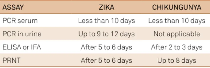
ASSAY ZIKA CHIKUNGUNYA
PCR serum Less than 10 days Less than 10 days
PCR in urine Up to 9 to 12 days Not applicable
ELISA or IFA After 5 to 6 days After 2 to 3 days
PRNT After 5 to 6 days Up to 8 days
Table 2. Diagnostics methods according to disease duration.
studied between November 2013 and February, 201456.
hirty-eight patients had GBS after ZIKV infection and 25 had other neurological complications such as encepha -litis, meningoencephalitis-encepha-litis, facial paralysis, and myelitis. Among the GBS patients, there were 73% males, with a mean age of 45.9 years, and almost all cases were eth -nic Polynesians. All the patients with GBS developed neu -rological symptoms after the development of a disease with symptoms consistent with ZIKV infection in previous days. One of these patients had been laboratory-conirmed as ZIKV by RT-PCR at the initial infection, and other prelimi -nary results showed ZIKV IgG positive after the occurrence of neurological signs56.
A GBS case-controlled study performed at the Hospital of Polynésie Française during the outbreak period (between October 2013 and April 2014), suggested a possible associ -ation between the ZIKV and GBS. According to the study, 42 patients were diagnosed with GBS during the period, where 41 (98%) of these patients had ZIKV anti-IgM or IgG. Patients with GBS had electrophysiological indings consis -tent with acute motor axonal neuropathy, characterized by the distal involvement of motor nerves. he recovery was faster than normally observed in typical GBS25.
he ZIKV was also found in the cerebrospinal luid of a 15-year-old patient, hospitalized in Pointe-à-Pitre, Guadaloupe, in January 2016. He presented with an acute myelitis, suggest -ing that the virus is neurotropic and it should be considered in patients living in, or traveling to, endemic areas26.
Carteaux, et al. described a case of an 81-year-old man who presented with a decreased level of consciousness, left hemi -plegia and paresis of the right upper limb. here was a history of a transient rash 48 hours before the neurological symptoms. Brain MRI and CSF analysis were suggestive of meningoen -cephalitis. he PCR for ZIKV was positive in the CSF, support -ing the diagnosis of ZIKV-associated men-ingoencephalitis27.
he Brazilian Ministry of Health, in November 2015, declared a public health emergency concerning an abnor -mal increase of children born with microcephaly during 2015, in the state of Pernambuco. Until that date, 141 cases of microcephaly in newborns had been reported in the same state, compared with an average of 10 cases per year from 2010-201457. An increase was also reported in the states of
Paraíba and Rio Grande do Norte. Later, in the same year, the Ministry of Health reported the presence of ZIKV RNA de -tected by RT-PCR in amniotic luid samples, collected from two pregnant women. heir babies presented with micro -cephaly57 and these women had symptoms compatible with
ZIKV infection during pregnancy. Between mid-2015 and January 2016, there were 4,783 cases of suspected microceph -aly, 387 cases had brain abnormalities found on imaging, and ZIKV was detected in 17 babies28.
During a retrospective analysis of a large Zika outbreak in French Polynesia, between 2013 and 2014, the authors found that the risk of malformation was about 1% when
women were infected by the ZIKV during the irst trimes -ter of pregnancy29. It had become a major public health
concern, as the incidence of Zika virus in the general pop -ulation may be very high during outbreaks. hus, these indings highlight the need to inform pregnant women to protect themselves from mosquito bites and avoid traveling to afected countries29.
Diagnostic methods
he ZIKV infection can be diagnosed by PCR, which can detect viral RNA, in the acute phase. Samples obtained with -in 10 days after the onset of the disease should have PCR per -formed. In general, the diagnostic tests for laviviruses should include an acute phase serum sample, taken as soon as pos -sible after the onset of disease and a second sample taken two to three weeks after the irst58.
Other diagnostic methods include serologic tests (ELISA or immunoluorescence) to detect IgM or IgG antibodies against ZIKV, which may be positive after ive to six days af -ter the onset of symptoms. his should show an increase of antibody titer in paired samples with an interval of about two weeks. Conirmation of the positive results should be con -irmed with the plaque reduction neutralization test, show -ing at least a fourfold increase in titer of neutraliz-ing anti -bodies to ZIKV. It is known that cross-reactivity with other laviviruses can occur, especially dengue, yellow fever and, less frequently, with the West Nile virus. herefore, the results should be interpreted with caution37,38. Urine samples for the
detection of viral genomes by RT-PCR may be the diagnostic method of choice, since the disappearance of the genome in serum has been shown to be within 10 days for ZIKV, but in urine samples it can be detected until ifteen days after the onset of infection58,59,60 (Table 2).
Appropriate diagnostic specimens for RT-PCR testing in -clude plasma/serum, urine, CSF, amniotic luid and placental tissue. Serology is usually performed on serum; however, viral antibodies may also be detected in the CSF58,59,60.
Chikungunya and Zika treatment
Treatment for Zika and CHIKV is symptomatic, and is very similar to dengue fever. It includes antipyretic, analge -sic, anti-inlammatory drugs to reduce joint and muscle pain, three to six eye drops twice daily as a lubricant, anti-allergy drugs, among others. In the case of pruritic eruptions, anti-histamines may be considered. Medicines containing aspirin should not be used, as well as in cases of dengue fever, be -cause they may increase the risk of bleeding61.
he treatment of neurological manifestations will depend on the type of aliction, such as intravenous immunoglobulin or plasmapheresis in cases of GBS25, and corticosteroid pulse
therapy/intravenous immunoglobulin in cases of myelitis26.
here is no vaccine against these diseases. he Aedes
References
1. Gutierrez-Saravia E, Gutierrez CE. Chikungunya virus in the Caribbean: a threat for all of the Americas. J Pediatric Infect Dis Soc. 2015;4(1):1-3. doi:10.1093/jpids/piv002
2. Morrison TE. Reemergence of chikungunya virus. J Virol. 2014;88(20):11644-7. doi:10.1128/JVI.01432-14
3. Garg M, Alcalde V. Update on emerging infections: news from the centers for disease control and prevention. Ann Emerg Med. 2014;64(5):553-5. doi:10.1016/j.annemergmed.2014.07.456
4. Halstead SB. Reappearance of chikungunya, formerly called dengue, in the Americas. Emerg Infect Dis. 2015;;21(4):557-61. doi:10.3201/eid2104.141723
5. Carbajo AE, Vezzani D. Waiting for chikungunya fever in Argentina: spatio-temporal risk maps. Mem Inst Oswaldo Cruz. 2015;110(2):259-62. doi:10.1590/0074-02760150005
6. Pan American Health Organization – PAHO. Cases of Chikungunya Fever in the Americas, 2016 (for week). Washington, DC: Pan American Health Organization; 2016 [acess 2016 Apr 8]. Available from: http://www.paho.org/hq/index.php?option=com_ docman&task=doc_view&Itemid=&gid=34132&lang=en
7. Burt FJ, Rolph MS, Rulli NE, Mahalingam S, Heise MT.
Chikungunya: a re-emerging virus. Lancet. 2012;379(9816):662-71. doi:10.1016/S0140-6736(11)60281-X
8. Azevedo RS, Oliveira CS, Vasconcelos PF. Chikungunya risk for Brazil. Rev Saúde Pública. 2015;49:58. doi:10.1590/S0034-8910.2015049006219
9. Das T, Jaffar-Bandjee MC, Hoarau JJ, Krejbich Trotot P, Denizot M, Lee-Pat-Yuen G et al. Chikungunya fever: CNS infection and pathologies of a re-emerging arbovirus. Prog Neurobiol. 2010;91(2):121-9. doi:10.1016/j.pneurobio.2009.12.006
10. Gauri LA, Ranwa BL, Nagar K, Vyas A, Fatima Q. Post chikungunya brain stem encephalitis. J Assoc Physicians India. 2012;60:68-70.
11. Rampal SM, Meena H. Neurological complications in Chikungunya Fever. J Assoc Physicians India. 2007;55:765-9.
12. Chandak NH, Kashyap RS, Taori GM, Daginawala HF.
Neurological complications in Chikungunya infection. BMC Proc. 2008;2 Suppl 1:11. doi:10.1186/1753-6561-2-s1-p11
13. Wadia RS. A neurotropic virus (chikungunya) and a neuropathic aminoacid (homocysteine). Ann Indian Acad Neurol. 2007;10:198-213.
14. Casolari S, Briganti E, Zanotti M, Zauli T, Nicoletti L, Magurano F et al. A fatal case of encephalitis associated with Chikungunya virus infection. Scand J Infect Dis. 2008;40(11-12):995-6. doi:10.1080/00365540802419055
15. Lalitha P, Rathinam S, Banushree K, Maheshkumar S, Vijayakumar R, Sathe P. Ocular involvement associated with an epidemic outbreak of chikungunya virus infection. Am J Ophthalmol. 2007;144(4):552-6. doi:10.1016/j.ajo.2007.06.002
16. Kalita J, Kumar P, Misra UK. Stimulus-sensitive myoclonus and cerebellar ataxia following chikungunya meningoencephalitis. Infection. 2013;41(3):727-9. doi:10.1007/s15010-013-0406-2
17. Mittal A, Mittal S, Bharati MJ, Ramakrishnan R, Saravanan S, Sathe PS. Optic neuritis associated with chikungunya virus infection in south India. Arch Ophthalmol. 2007;125(10):1381-6. doi:10.1001/archopht.125.10.1381
18. Wielanek AC, Monredon JD, Amrani ME, Roger JC, Serveaux JP. Guillain-Barré syndrome complicating a Chikungunya virus infection. Neurology. 2007;69(22):2105-7. doi:10.1212/01.wnl.0000277267.07220.88
19. Rampal S, Sharda M, Meena H. Neurological complications in Chikungunya fever. J Assoc Phys India. 2007;55:765-9.
20. Robin D, Le Seach F, Jaffar-Bandjee MC, Rigou G, Alessandri JL. Neurologic manifestations of pediatric chikungunya infection. J Child Neurol. 2008;23(9):1028-35. doi:10.1177/0883073808314151
21. Chandak NH, Kashyap RS, Kabra D, Karandikar P, Saha SS, Morey SH et al. Neurological complications of Chikungunya virus infection. Neurol India. 2009;57(2):177-80. doi:10.4103/0028-3886.51289
22. Lemant J, Boisson V, Winer A, Thibault L, André H, Tixier F et al. Serious acute chikungunya virus infection requiring intensive care during the Reunion Island outbreak in 2005-2006. Crit Care Med. 2008;36(9):2536-41. doi:10.1097/CCM.0b013e318183f2d2
23. Bhavana K, Tyagi I, Kapila RK. Chikungunya virus induced sudden sensorineural hearing loss. Int J Pediatr Otorhinolaryngol. 2008;72(2):257-9. doi:10.1016/j.ijporl.2007.09.022
24. Oehler E, Watrin L, Larre P, Leparc-Goffart I, Lastere S, Valour F et al. Zika virus infection complicated by Guillain-Barre syndrome: case report, French Polynesia, December 2013. Euro Surveill. 2014;19(9):20720. doi:10.2807/1560-7917.ES2014.19.9.20720
25. Cao-Lormeau VM, Blake A, Mons S, Lastère S, Roche C, Vanhomwegen J et al. Guillain-Barré Syndrome outbreak associated with Zika virus infection in French Polynesia: a case-control study. Lancet. 2016;387(10027):1531-9. doi:10.1016/S0140-6736(16)00562-6
26. Mécharles S, Herrmann C, Poullain P, Tran TH, Deschamps N, Mathon G et al. Acute myelitis due to Zika virus infection. Lancet. 2016;387(10026):1481. doi:10.1016/S0140-6736(16)00644-9
27. Carteaux G, Maquart M, Bedet A, Contou D, Brugières P, Fourati S et al. Zika virus associated with meningoencephalitis. N Engl J Med. 2016;374(16):1595-6. doi:10.1056/NEJMc1602964
28. Victora CG, Schuler-Faccini L, Matijasevich A, Ribeiro E, Pessoa A, Barros FC. Microcephaly in Brazil: how to interpret reported numbers? Lancet. 2016;387(10019):621-4. doi:10.1016/S0140-6736(16)00273-7
29. Cauchemez S, Besnard M, Bompard P, Dub T,
Guillemette-Artur P, Eyrolle-Guignot D et al. Association between Zika virus and microcephaly in French Polynesia, 2013-15: a retrospective study. Lancet. 2016;387(10033):2125-32. doi:10.1016/S0140-6736(16)00651-6
30. Fraisier C, Koraka P, Belghazi M, Bakli M, Granjeaud S, Pophillat M et al. Kinetic analysis of mouse brain proteome alterations following Chikungunya virus infection before and after appearance of clinical symptoms. PLoS One. 2014;9(3):e91397. doi:10.1371/journal.pone.0091397
31. Inglis FM, Lee KM, Chiu KB, Purcell OM, Didier PJ, Russell-Lodrigue K et al. Neuropathogenesis of Chikungunya infection: astrogliosis and innate immune activation. J Neurovirol. 2016;22(2):140-8. doi:10.1007/s13365-015-0378-3
32. Hasebe F, Parquet C, Pandey BD, Mathenge EG, Morita K, Balasubramaniam V et al. Combined detection and genotyping of Chikungunya virus by a specific reverse transcription-polymerase chain reaction. J Med Virol. 2002;67(3):370-4. doi:10.1002/jmv.10085
33. Carletti F, Bordi L, Chiappini R, Ippolito G, Sciarrone MR, Capobianchi MR et al. Rapid detection and quantification of Chikungunya virus by a one-step reverse transcription polymerase chain reaction real- time assay. Am J Trop Med Hyg. 2007;77(3):521-4.
34. Ho PS, Ng MM, Chu JJ. Establishment of one-step SYBR green-based real time-PCR assay for rapid detection and quantification of chikungunya virus infection. Virol J. 2010;7(1):13. doi:10.1186/1743-422X-7-13
35. Panning M, Grywna K, Esbroeck M, Emmerich P, Drosten C. Chikungunya fever in travelers returning to Europe from the Indian Ocean region, 2006. Emerg Infect Dis. 2008;14(3):416-22. doi:10.3201/eid1403.070906
37. Dick GW, Kitchen SF, Haddow AJ. Zika virus. I. Isolations and serological specificity. Trans R Soc Trop Med Hyg. 1952;46(5):509-20. doi:10.1016/0035-9203(52)90042-4
38. Fagbami AH. Zika virus infections in Nigeria: virological and seroepidemiological investigations in Oyo State. J Hyg (Lond). 1979;83(2):213-9. doi:10.1017/S0022172400025997
39. Robin Y, Mouchet J. [Serological and entomological study on yellow fever in Sierra Leone]. Bull Soc Pathol Exot Filiales. 1975;68(3):249-58. French.
40. Olson JG, Ksiazek TG. Suhandiman T. Zika virus, a cause of fever in Central Java, Indonesia. Trans R Soc Trop Med Hyg. 1981;75(3):389-93. doi:10.1016/0035-9203(81)90100-0
41. Lanciotti RS, Kosoy OL, Laven JJ, Velez JO, Lambert AJ, Johnson AJ et al. Genetic and serologic properties of Zika virus associated with an epidemic, Yap State, Micronesia, 2007. Emerg Infect Dis. 2008;14(8):1232-9. doi:10.3201/eid1408.080287
42. Duffy MR, Chen TH, Hancock WT et al. Zika virus outbreak on Yap Island, Federated States of Micronesia. N Engl J Med. 2009;360(24):2536-43. doi:10.1056/NEJMoa0805715
43. Baronti C, Piorkowski G, Charrel RN, Boubis L, Leparc-Goffart I, Lamballerie X. Complete coding sequence of zika virus from a French polynesia outbreak in 2013. Genome Announc. 2014;2(3):e00500-14. doi:10.1128/genomeA.00500-14
44. Fonseca K, Meatherall B, Zarra D, Drebot M, MacDonald J, Pabbaraju K et al. First case of Zika virus infection in a returning Canadian traveler. Am J Trop Med Hyg. 2014;91(5):1035-8. doi:10.4269/ajtmh.14-0151
45. Kwong JC, Druce JD, Leder K. Zika virus infection acquired during brief travel to Indonesia. Am J Trop Med Hyg. 2013;89(3):516-7. doi:10.4269/ajtmh.13-0029
46. Wæhre T, Maagard A, Tappe D, Cadar D, Schmidt-Chanasit J. Zika virus infection after travel to Tahiti, December 2013. Emerg Infect Dis. 2014;20(8):1412-4. doi:10.3201/eid2008.140302
47. Zanluca C, Melo VC, Mosimann AL, Santos GI, Santos CN, Luz K. First report of autochthonous transmission of Zika virus in Brazil. Mem Inst Oswaldo Cruz. 2015;110(4):569-72. doi:10.1590/0074-02760150192
48. Campos GS, Bandeira AC, Sardi SI. Zika virus outbreak, Bahia, Brazil. Emerg Infect Dis. 2015;21(10):1885-6. doi:10.3201/eid2110.150847
49. World Health Organization. Zika virus outbreaks in the Americas. Wkly Epidemiol Rec. 2015;90(45):609-10.
50. Faye O, Dupressoir A, Weidmann M, Ndiaye M, Alpha Sall A. One-step RT-PCR for detection of Zika virus. J Clin Virol. 2008;43(1):96-101. doi:10.1016/j.jcv.2008.05.005
51. Marchette NJ, Garcia R, Rudnick A. Isolation of Zika virus from Aedes aegypti mosquitoes in Malaysia. Am J Trop Med Hyg. 1969;18(3):411-5.
52. Boorman JP, Porterfield JS. A simple technique for infection of mosquitoes with viruses; transmission of Zika virus. Trans R Soc Trop Med Hyg. 1956;50(3):238-42. doi:10.1016/0035-9203(56)90029-3
53. Heang V, Yasuda CY, Sovann L, Haddow AD, Travassos da Rosa AP, Tesh RB et al. Zika virus infection, Cambodia, 2010. Emerg Infect Dis. 2012;18(2):349-51. doi:10.3201/eid1802.111224
54. Carod-Artal FJ, Wichmann O, Farrar J, Gascón J. Neurological complications of dengue virus infection. Lancet Neurol. 2013;12(9):906-19. doi:10.1016/S1474-4422(13)70150-9
55. Oehler E, Le Henaff O, Larre P Ghawche F. [Guillain-Barre syndrome following type 4 dengue in Polynesia]. Med Trop (Mars). 2011;71(2):203-4. French.
56. Polynesie Française. Surveillance de la dengue et du zika en Polynésie Française 2014 [acess 2014 Feb 7]. Available from: http:// www.hygiene-publique.gov.pf/spip.php?article120
57. Ministério da Saúde (BR). Microcefalia: Ministério da Saúde divulga boletim epidemiológico. Brasília: Ministério da Saúde; 2015 [acess 2015 Nov 17]. Available from: http://portalsaude.saude.gov.br/index. php/cidadao/principal/agencia-saude/20805-ministerio-da-saude-divulga-boletim-epidemiologico
58. Kutsuna S, Kato Y, Takasaki T, Moi M, Kotaki A, Uemura H et al. Two cases of Zika fever imported from French Polynesia to Japan, December to January 2013. Eurosurveillance. 2014;19(4):20683. doi:10.2807/1560-7917.ES2014.19.4.20683
59. Domingo C, Yactayo S, Agbenu E, Demanou M, Schulz AR, Daskalow K et al. Detection of yellow fever 17D genome in urine. J Clin Microbiol. 2011;49(2):760-2. doi:10.1128/JCM.01775-10
60. Barzon L, Pacenti M, Franchin E, Pagni S, Martello T, Cattai M et al. Excretion of West Nile virus in urine during acute infection. J Infect Dis. 2013;208(7):1086-92. doi:10.1093/infdis/jit290