www.reumatologia.com.br
REVISTA BRASILEIRA DE
REUMATOLOGIA
Original article
Articular manifestations in patients with atypical
rheumatic fever
Teresa Cristina Martins Vicente Robazzi
a,*, Simone Rocha de Araújo
b, Silas de Araújo Costa
b,
Amaurí Batista de Oliveira Júnior
b, Lívia Souza Nunes
b, Isabel Guimarães
aa Department of Pediatry, Medicine School, Universidade Federal da Bahia, Salvador, BA, Brazil
b Medicine School, Universidade Federal da Bahia, Salvador, BA, Brazil
a r t i c l e i n f o
Article history:
Received 18 September 2013 Accepted 10 February 2014
Keywords:
Rheumatic fever Jones criteria
Atypical joint involvement Children
Adolescents
a b s t r a c t
Objectives: To describe the clinical characteristics and the occurrence of atypical arthritis in children diagnosed with rheumatic fever (RF) and followed in tertiary care clinics in Salvador, Bahia, Brazil.
Methodology: A descriptive study of a case series, of the initial clinical presentation, and of recurrence in 41 children diagnosed with RF.
Results: Of the patients studied (n=41), 61% were male, mean age of 9.2 years, and mean age at diagnosis between 5 and 16 years. Arthritis was present in 75.6% of patients; carditis in 75.6%; chorea in 31.7%; erythema marginatum in 14.6%; and subcutaneous nodules in 4.9%. An atypical pattern was observed in 22 of 31 cases of arthritis (70.9%): involvement of small joints and/or axial skeleton in 12 cases (38.7%); >3 weeks of duration in 9 (29%); inadequate response to NSAIDs in 2 (6.5%); oligoarthritis (≤4 joints) in 22/31 (71%), with monoarthritis in 6/31 (1 in the foot, 1 in the ankle, and 4 in the knee). Fever was present in 78% of the cases, and 82.9% of patients were regularly on secondary prophylaxis.
Conclusion: Atypical arthritis was present in most patients presenting with joint involve-ment, being a confounding factor against a proper diagnosis and of therapeutic delay.
© 2014 Sociedade Brasileira de Reumatologia. Published by Elsevier Editora Ltda. All rights reserved.
* Corresponding author.
E-mail: [email protected] (T.C.M.V. Robazzi).
0482-5004/$ - see front matter. © 2014 Sociedade Brasileira de Reumatologia. Published by Elsevier Editora Ltda. All rights reserved. http://dx.doi.org/10.1016/j.rbr.2014.02.006
Manifestações articulares atípicas em pacientes com febre reumática
Palavras-chave:
Febre reumática Critérios de Jones
Acometimento articular atípico Crianças
Adolescentes
r e s u m o
Objetivos: Descrever as características clínicas e a ocorrência de artrite atípica em crianças com diagnóstico de febre reumática (FR) acompanhadas em ambulatórios terciários em Salvador, Bahia.
Metodologia: Estudo descritivo, de uma série de casos, do quadro clínico inicial ou recorrên-cia de 41 crianças com diagnóstico de FR.
presente em 75,6% dos pacientes; cardite em 75,6%; coreia em 31,7%; eritema marginado em 14,6% e nódulos subcutâneos em 4,9%. Um padrão atípico foi observado em 22 dos 31 casos com artrite (70,9%): envolvimento de pequenas articulações e/ou esqueleto axial em 12 casos (38,7%); duração maior que três semanas em nove (29%); resposta inadequada ao AINH em dois (6,5%), oligoartrite (≤ quatro articulações) em 22/31 (71%), sendo monoartrite em 06/31 (uma em pés, uma em tornozelo e quatro em joelho). A febre esteve presente em 78% dos casos e 82,9% dos pacientes utilizavam a proilaxia secundária de forma regular.
Conclusão: Artrite atípica esteve presente na maioria dos pacientes que cursaram com aco-metimento articular, constituindo um fator de confundimento diagnóstico e atraso tera-pêutico adequado.
© 2014 Sociedade Brasileira de Reumatologia. Publicado por Elsevier Editora Ltda. Todos os direitos reservados.
Introduction
Rheumatic fever (RF) is a late, inlammatory, non-suppurative complication of the infection of the upper airways by the group A beta-hemolytic streptococci.1-4 It can affect different
tissues, including the heart, the joints and the central ner-vous system,4 and occurs mainly among genetically
predis-posed children and adolescents between the ages of 5 and 15.4-6
The diagnosis of RF remains one of the most dificult prob-lems in the paediatric area, thanks to the polymorphism of its clinical presentation and to the lack of speciic or pathog-nomonic laboratory tests for the disease.7-10
The Jones criteria were established in 1944, with the goal of reducing the occurrence of diagnostic errors and delays.11
Despite its usefulness in clinical practice, these criteria do not include the dificulties in establishing the diagnosis of RF, es-pecially in the presence of atypical articular manifestations, which may lead to errors or to delays in diagnosis.7 This
diag-nostic dificulty is accentuated in patients who present with arthritis as the only clinical manifestation of the disease.4,12,13
The classic description of arthritis of RF consists of a picture of migratory polyarthritis, mainly of large joints of the lower limb, and with its onset around 2-3 weeks after a streptococcal infection of the oropharynx. The typically in-tense pain responds well to the use of non-steroidal anti-in-lammatory drugs (NSAIDs), and usually has no more than 3 weeks in duration.2,8 The most commonly affected joints are
knees and ankles.
Atypical joint manifestations have been increasingly de-scribed in the literature, being characterized by a monoar-ticular picture lasting longer than six weeks, poor response to salicylates, and an additive arthritis, as well as an unusual involvement of joints such as hips, cervical spine, and small joints.1,7,9 This possible pattern of joint involvement in RF was
irst mentioned in 1975 by Stollerman, when this author not-ed that 32% of children with rheumatic fever did not show the classical pattern of joint involvement.14
Terreri et al. rated the articular pattern in 93 patients with RF. The arthritis was additive in 27% of cases; small joints, such as the metacarpophalangeal and proximal and distal interphalangeal joints, were affected with a frequency of 2%-8%; arthritis with more than six weeks duration was observed in 10%; no response to acetylsalicylic acid occurred in 15% of the cases, and monoarthritis was found in 6% of patients.7
These atypical manifestations further hamper the diagno-sis of RF, especially if the doctor is not aware of the possibility of this kind of event.
The present study was conducted aiming to evaluate the characteristics of joint involvement and the occurrence of atypical arthritis in children during the initial outbreak and in recurrence of RF.
Patients, materials and methods
Forty-one medical records of 41 children and adolescents di-agnosed with RF and treated at the Paediatric Rheumatology Service, Hospital Professor Edgard Santos, Universidade Federal da Bahia, and at the Paediatric Cardiology Service, Hospital Ana Nery, both in the city of Salvador, Bahia, were retrospectively analyzed. The study covered a period of three years, from March 2009 to December 2012. The inclusion criteria were: diagnosis of RF based on modiied Jones criteria (1992) and patients aged up to 21 years-old. The variables used in the study were: gen-der; age; presence and characteristics of major and minor Jones criteria; characteristics of articular involvement; information on the evidence of prior infection by streptococcus (ASLO), and laboratory tests. To take into consideration the presence of an atypical pattern for the joint involvement, at least one of the fol-lowing features was needed: more than three weeks in dura-tion; involvement of small joints and/or cervical spine and/or hip joint; monoarthritis; and poor response to salicylates.
Data processing and analysis were performed with the help of the Statistical Package for Social Sciences (SPSS®) program,
version 12.0, with descriptive data analysis. The mean age of pa-tients and the absolute and relative frequencies of the following variables were analyzed: gender; major and minor Jones criteria; number of joints involved; atypical articular manifestations; de-scription of each atypical feature, and regularity of secondary prophylaxis.
The project was approved by the ethics committee, HUPES (071/2009, Resolution CNS 196/96).
Results
The frequency of Jones major signs was: 31 patients with arthritis; 31 with carditis; 13 with chorea; two with subcutane-ous nodules; six with erythema marginatum (Fig. 1). Arthritis was isolated in six of 31 cases presenting with this symptom (19.4%).
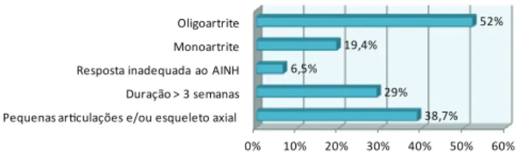
As to the characteristics of joint involvement, the atypi-cal pattern was observed in a considerable number of cases with arthritis: 22/31 (70.9%). Involvement of small joints and/ or axial skeleton occurred in 12 (38.7%); a period greater than three weeks in nine (29%); inadequate response to NSAIDs in two (6.5%), oligoarthritis (≤4 joints) in 22/31 (71%), with mono-arthritis in 06/31 (foot, 1; ankle, 1; and knee, 4) (Fig. 2). The pres-ence of polyarthritis (≥5 joints) occurred in 9/31 patients (29%). Among these 22 unusual cases, isolated arthritis was found in six patients (19.4%); arthritis associated with carditis was found in 12 cases (57.1%); arthritis associated with cardi-tis and chorea in four cases (19%); arthricardi-tis, cardicardi-tis, erythema marginatum and subcutaneous nodules in one case (4.8%); and arthritis, carditis, chorea and erythema marginatum in one case (4.8%).
Fever was present in 78% of cases, and 82.9% of patients were regularly on secondary prophylaxis.
Discussion
In 1982, Goldsmith and Long highlighted the presence of a clinical picture of arthritis with unusual characteristics (sym-metric, longer duration, short latency period after strepto-coccal infection, and poor response to salicylates),15 and
sug-gested an immune response change to some kind of antigenic modii cation of Group A-beta-haemolytic streptococcus.
Since then, many authors are referring to clinical pictures of arthritis after infection with Group A-beta-haemolytic streptococcus with the characteristics above mentioned, not usual to the pattern described by Jones criteria.
The studies in the literature on this form of presentation of arthritis after streptococcal infection are relatively scarce
and heterogeneous, being often based on reports or series of cases, which limit the clear knowledge of the causation of this form of presentation.
Given the conl icting literature, we proposed, out of curi-osity, to map the proi le of patients diagnosed with RF at our service, with greater emphasis on the pattern of presenta-tion of articular involvement.
In the present study, patients’ age at diagnosis ranged from 5 to 16 years, with a mean age of 9.2 years, a i nding similar to results previously described in other regions in Brazil.1,9 According to literature, the incidence of RF is higher
between 5-15 years, both for the i rst outbreak and for re-lapses.16 Therefore, there were no variations in age of onset
for RF with atypical joint pattern.
Among the patients in this study, 61% were male (ratio, 1,6:1). This i nding contradicts the results of most authors, which show a higher prevalence of RF in females (55% to 60.5%).1,2,17
As for Jones major criteria, there was no predominance of arthritis over carditis, as in most studies in the literature, with similar prevalences for both clinical manifestations (75.6%). This can be explained by the inclusion of patients from a center of reference in paediatric cardiology (Fig. 1).
The prevalence of arthritis varies in the literature, but our results were similar to the studies of Motta and Meira and of Terreri et al.,1,18 which found arthritis in 71.4% and 70,
5% of their patients, respectively.
In the present study, 51.6% of our patients had involve-ment of more than one, and up to i ve joints; 19.4% in just one joint; and 29% in more than i ve joints. This result re-vealed a high prevalence of oligoarticular presentation in patients with RF in our community, and these presentations undoubtedly generate diagnostic difi culties and therapeu-tic delays.
Table 1 illustrates the frequency of monoarthritis in RF in different studies, drawing attention to this possibility in the clinical comparison of RF, in contrast to what was set by Jones criteria.12,19-22 An example in clinical practice is
the suspicion of septic arthritis in cases of monoarthritis accompanied by fever, causing loss of time with diagnos-tic procedures and with invasive and unnecessary thera-peutic interventions. Mataika et al. described three cases of monoarthritis initially treated as septic arthritis, with subsequent diagnosis of RF in the presence of a developing endocarditis.23
Among the more involved joints in patients with single joint involvement, the knee joints have prevailed, followed by axial skeleton and ankle joints. Harlan et al.21 evaluated
92 patients with RP, and three of these had monoarthritis
Figure 1 – Frequency of the biggest signs of Jones diagnostic criteria in 41 patients with rheumatic fever diagnosis
0% 10% 20% 30% 40% 50% 60% 70% 80% Artrite
Cardite Coreia Eritema Nódulos subcutâneos
75,6% 75,6% 31,7%
14,6% 4,9%
Figure 2 – Percentage of atypical articular manifestations in 41 patients with rheumatic fever diagnosis
0% 10% 20% 30% 40% 50% 60% Pequenas ariculações e/ou esqueleto axial
Duração > 3 semanas Resposta inadequada ao AINH Monoartrite Oligoartrite
38,7% 29% 6,5%
19,4%
52% Table 1 – Percentage of monoarthritis in rheumatic fever according to different authors
Study Percentage of monoarthritis
Khriesat I, 200312 32%
Carapetis, 200119 17%
Ralph A, 200620 5.5%
Harlan GA, 200621 3.3%
Olgunturk, 200622 16%
(hips in one patient, knees in two), similarly to Pileggi et al.,9 who also found three cases of monoarthritis, two in the
knee and one in the hip joint. In our study, single joint in-volvement occurred in the following joints: knee (4/6), foot (1/6), and ankle (1/6).
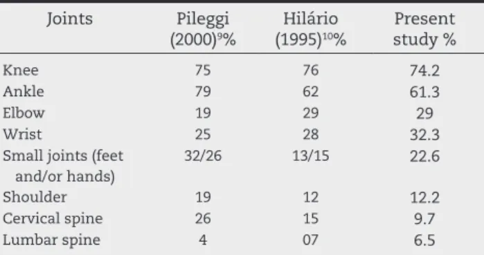
Some manifestations described are the involvement of small joints (such as metacarpophalangeal and metatar-sophalangeal joints), and of the axial skeleton (spine and sacroiliac and hip joints),9,10 and this may cause diagnostic
difi culties with juvenile idiopathic arthritis and the juve-nile spondyloarthritides (Table 2). The involvement of small joints was present in 18% of cases analyzed by Lin Chen et al.24 In 38.7% of our patients, the involvement of small
joints occurred mainly in feet and hands and, in lesser ex-tent, in axial skeleton joints.
In some patients diagnosed with RF, a favourable clini-cal response after appropriate use of NSAIDs does not oc-cur. This implies a long-term use of these drugs, due to the presence of an arthritis of prolonged evolution. In the pres-ent study, despite the early use of this medication in the i rst days after the diagnosis of arthritis, 4.9% of patients were poorly responsive to NSAIDs. In the study of Ferriani et al.,2 this poor response occurred in 19.56% of patients.
The presence of arthritis as the only clinical manifesta-tion is still considered as an unusual clinical manifestamanifesta-tion that complicates and delays the i nal diagnosis of RF; this occurred in 19.4% of patients (6/31). Harlan et al.21
demon-strated that the time for diagnosis of RF was superior to 4 weeks in 59% of patients with atypical arthritis, as com-pared to 35% in other patients.21
Another aspect that complicates the diagnosis of RF is the laboratory diagnosis. In addition to being non-specii c, laboratory abnormalities may not be present in a signii -cant percentage of patients due to various factors: the pe-riod of collection, a previous administration of antibiotics, and accessibility to these tests in a timely manner, among others. Looking specii cally at patients presenting with atypical manifestations, evidence of inl ammatory activity was positive in only 56%, and ASLO was present in 44% of patients.
Thus, the results reveal a considerable percentage of pa-tients with atypical articular manifestations, corroborating observations made for some time by other authors (Fig. 3), and again calling attention to the need to keep in mind this possibility of articular presentation in patients with RF.
Conclusion
From the results of this study, we may stress the impor-tance of the recognition, by rheumatologists, paediatricians and even internists, of atypical articular presentations in the clinical picture of RF, thus avoiding unnecessary diag-nostic delays and, consequently, therapeutic delays, with risk of irreversible cardiac sequelae.
This diagnostic suspicion should be addressed in the case of a patient with evidence of infection with Group A-beta-haemolytic streptococcus; who do not fuli l modii ed Jones criteria for the diagnosis of RF; and who develop a clinical picture of acute outcome, oligo or single joint, sym-metrical or asymsym-metrical, usually non-migratory arthritis that may affect any joint and with a poor response to ace-tylsalicylic acid.
Confl icts of interest
The authors declare no conl icts of interest.
R E F E R E N C E S
1. Terreri MTR, Caldas AM, Len CL, Ultchak F, Hilário MOE. Características clínicas e demográi cas de 193 pacientes com febre reumática. Rev Bras Reumatol. 2006;46:385-90.
2. Ferriane VPL. Manifestações articulares da febre reumática. Rev Soc Cardiol Estado de São Paulo. 2005;15:18-27. 3. Habib GS, Saliba WR, Mader R. Rheumatic fever in the
Nazareth area during the last decade. Isr Med Assoc J (IMAJ). 2000;433-7.
4. Carcellera A, Tapiero B, Rubin E, Miró J. Fiebre reumática aguda: 27 años de experiência em los hospitales pediátricos en Montreal. An Pediatr (Barc). 2007;67:5-10.
5. Carapetis JR, Currie BJ. Rheumatic fever in a high incidence population: the importance of monoarthritis and low grade fever. Arch Dis Child. 2001;85:223-7.
6. Vinker S, Zohar E, Hoffman R, Elhayany A. Incidence and clinical manifestations of rheumatic fever: A 6 Year Community-Based. Isr Med Assoc J (IMAJ). 2010;12:78-81. 7. Terreri MTR, Hilário MOE. Diagnóstico da febre reumática:
os critérios de Jones continuam adequados? Rev Soc Cardiol Estado de São Paulo. 2005;28-33.
8. Kiss MHB. Tratamento clínico da febre reumática. Rev Soc Cardiol Estado de São Paulo. 2005;15:53-8.
9. Pileggi GCS, Ferriane VPL. Manifestações articulares atípicas em crianças com febre reumática. J Pediatr (Rio J). 2000;76:49-54.
Table 2 – Joints affected in patients with rheumatic fever according to different authors
Joints Pileggi (2000)9%
Hilário (1995)10%
Present study %
Knee 75 76 74.2
Ankle 79 62 61.3
Elbow 19 29 29
Wrist 25 28 32.3
Small joints (feet and/or hands)
32/26 13/15 22.6
Shoulder 19 12 12.2
Cervical spine 26 15 9.7
Lumbar spine 4 07 6.5
Figure 3 – Comparison of percentage of atypical manifestations among various authors
0% 10% 20% 30% 40% 50% 60% 70% Stollerman (1975)
Pileggi(2000) Robazzi (2012)
32,00%
47,00%
10. Hilário MO, Len C, Goldenberg J. Febre reumática: manifestações articulares atípicas. Rev Assoc Med Bras 1995;8:214-6.
11. Lilic N, Kumar P. A timely reminder-rheumatic fever. N Z Med J. 2013. 19:126:88-90.
12. Khriesat I, Najada A, Al-Hakim F, Abu-Haweleh A. Acute rheumatic fever in Jordanian children. La Revue de Santé de La Méditerrané e orientale. 2003;9:981-7.
13. Keitzer R. Akutes Rheumatisches Fieber (ARF) und Poststreptokokken reactive arthritis (PSRA). Z Rheumatol. 2005;64:295-307.
14. Stollerman GH. Rheumatic fever and streptococcal infection. New York: Grune and Stratton. 1975. 15. Goldsmith DF, Long SS. Poststreptococcal disease of
childhood – changing syndrome. Arthritis Rheum. 1982;25:S18.
16. Taranta A, Markowitz M. Rheumatic fever. 2 ed. Boston: Kluwer Academic Publishers, 1989.
17. Ozkutlu S, Hallioglu O, Ayabakan C: Evaluation of subclinical valvar disease patients with rheumatic fever. Cardiol Young. 2003;13:495-9.
18. Meira ZMA, Goulard EMA, Colosimo EA, Mota CCC. Long-term follow up of rheumatic fever and predictors of
severe rheumatic valvar disease in Brazilian children and adolescents. Heart. 2005;91:1019-22.
19. Carapetis JR, Currie BJ. Rheumatic fever in a high incidence population: the importance of monoarthritis and low grade fever. Arch Dis Child. 2001;85:223-7.
20. Ralph A, Jacups S, McGough K, McDonald M, Currie BJ. The challenge of acute rheumatic fever diagnosis in a high-incidence population. Heart Lung and Circulation. 2006;15:111-13.
21. Harlan GA, Tani LY, Byington CL. Rheumatic Fever Presenting as Monoarticular Arthritis. Pediatr Infect Dis J. 2006;25:743-6.
22. Olgunturk R, Olgunturk R, Canter B, Tunaoglu FS, Kula S. Review of 609 patients with rheumatic fever in terms of revised and updated Jones criteria. Int J Cardiol. 2006;112:91-8.
23. Mataika R, Carapetis JR, Kado J, Steer AC. Acute Rheumatic fever an importance differential diagnosis of septic arthritis. J Trop Pediatr. 2008;54:205-7.