Histopathology of cotton bollworm midgut infected with
Helicoverpa armigera
cytoplasmic polyhedrosis virus
Rasoul Marzban
1,2,Qian He
2, Qingwen Zhang
2, Xiao Xia Liu
21
Department of Biological Control, Iranian Research Institute of Plant Protection, Tehran, Iran. 2
Department of Entomology, College of Agronomy and Biotechnology, China Agricultural University, Beijing, China.
Submitted: July 3, 2012; Approved: April 4, 2013.
Abstract
This research was carried out to examine cytopathological effects ofHelicoverpa armigera Cyto-plasmic polyhedrosis virus (HaCPV) on infected midgut cotton bollworm (Helicoverpa armigera) using transmission and scanning electron microscope. The symptoms on infected host larvae of the host, compared with healthy ones, were getting swollen with milky-white and fragile Histo-pathological examinations showed infection with HaCPV small polyhedral inclusion bodies (PIB) after 1 or 2 days which were observed in columnar cells of midgut. Virions were partially or com-pletely occupied in a polyhedral matrix to form polyhedral inclusion bodies (PIB) at periphery of virogenic stroma. PIBs were measured 0.5 to 3.5mm and virions about 46 nm in diameter. Microvilli of infected columnar cells were affected and degenerated immediately prior to rupture of the cell. Some infected columnar cells ruptured to release PIB into the gut lumen 3 days after infection. In ad-dition,PIB were found in goblet cells, 5 or 6 days after infection. Infected goblet cells degenerate to such an extent that only a few of the original microvillus-like cytoplasmic projections and cell organells were left. These cytopathic effects caused in the midgut by HaCPV on cotton bollworm lar-vae are essentially similar to those have been reported for lepidoperan and dipteran infection by CPV.
Key words:cytoplasmic polyhedrosis virus,Helicoverpa armigera,histopathology, HaCPV.
Introduction
Most insect reoviruses described to date belong to the family Reoviridae within the genus Cypovirus (Cytoplas-mic polyhedrosis virus- CPV). These viruses have been re-ported as pathogens of the midgut epithelium in numerous lepidopteran and dipteran species, because these insects are, respectively, of economic or medical importance (Mer-tenset al., 2004). CPVs are so-named because they produce large polyhedral occlusion bodies that occlude virions in the cytoplasm of infected cells. They have been recognized as an important entomopathogen, especially among lepi-dopteran insects because of their potential for biological control (Martignoni and Iwai, 1981). CPVs infect midgut cells of a wide range of insects (Payne and Rivers, 1976). Inclusion bodies after ingestion by insects, dissolve and break, and release infectious virions which enter and
repli-cate in the cytoplasm of midgut epithelial cells (Mertenset al., 1999, 2004). Infection is usually limited to the insect gut wall. Infection frequently results in death or loss of fit-ness of the host which is reported to be important in regulat-ing host populations (Dwyer et al., 2004). Most CPV infections produce chronic disease with low mortality, al-though some are pathogenic (Mertenset al., 1999, 2004). Helicoverpa armigera Cytoplasmic polyhedrosis virus (HaCPV) (Chinese strain) is a mixture of CPV, so that Bellonciket al. (1996) using cell culture, separated type 14., Liet al. (2006) separated type 5 from a mixture of HaCPVs. They reported that HaCPV is virulent to H. armigeraand has the potential to be used as a bioinsec-ticide. HaCPV had negative impact on growth and develop-ment ofH. armigera, and resulted in reduced pupation and pupal weight and an extended developmental period, espe-cially in early instars larvae (Marzbanet al., 2009). HaCPV
Send correspondence to R. Marzban .Department of Biological Control, Iranian Research Institute of Plant Protection, Tehran, Iran. E-mail: ramarzban@yahoo.com.
Sub lethal effects may be due to the diversion of host en-ergy to support or combat the pathogen (Marzban, 2012; Sikorowski and Thampson, 1979; Wiygul, and Sikorowski, 1978, 1991).H. armigerais one of the most serious insect pests of cotton, corn, vegetables, and other crops in the Old World including Iran. It has a history of developing resis-tance to almost all of the insecticides used for its control. Transgenic cotton incorporating Cry1Ac gene derived from Bacillus thuringiensisBerliner is one of the most exciting advances made in cotton pest management in recent years. Resistance monitoring of H. armigera field populations suggested that there has been some decline in the suscepti-bility to Cry1Ac in the field (Gunninget al., 2001; Lietal., 2004; Shenet al., 1998). Therefore, alternative control mea-sures, such as biological control agents, are required. HaCPV is a highly infectious insect pathogen (Lietal., 2006; Martignoni and Iwai, 1981) and a candidate for biocontrol ofH. armigera, especially in combination with B. thuringiensistoxins (Iwashita, 1971; Kawase and Miya-jima, 1971; Katagiriet al., 1978; Ying, 1986). Presently there is no description of the histopathology of HaCPV on cotton bollworm,H. armigera. This study was undertaken to examine the pathological effects of HaCPV on the midgut of cotton bollworm, which forms part of the groundwork for evaluation of HaCPV potential in the inte-grated control of this pest, for dealing with the decline in the susceptibility of cotton bollworm to Cry1Ac toxin.
Materials and Methods
Insect
H. armigera used in this study obtained from the Wang Mo laboratory, Hua Zhong Agricultural University, Wuhan. For eliminating surface contaminations, the eggs, of each generation, were disinfected by immersing in 2% formaldehyde for 15 min at room temperature, then washed several times with tap water and finally rinsed with sterile distilled water. The eggs were allowed to air-dry on tissue paper and left to hatch in 10 x 6 cm plastic bags at 26 °C .The larvae were individually fed artificial diet at 26 °C and 65% RH, with a 14:10 h photoperiod (Bot, 1966). Adults were fed with a diet sweetened with 10% honey solution.
Preparation and purification
HaCPV was provided by Dr Jiang Zhong (Fudan Uni-versity, Shanghai) that it is a mixture type CPV.H. armigera larvae were reared on an artificial diet. The eggs were disin-fected, air-dried and incubated as described above.The lar-vae and adults, also, were fed same as explained above.
The first-instar larvae were infected with HaCPV by spraying a suspension of 3 x 10⁷polyhedra on the artificial diet and allowed to feed normal artificial diet until newly moulted second-instar (3 day old) larvae. On day 7 after in-fection, midguts were homogenized in deionized water and strained through 35-mm mesh nylon cloth to remove large
debris. The filtrated product was layered on top of HS-40 Ludox continues gradient and centrifuged at 16000 g for 45 min. The resulting band containing purified virus was recovered, washed in sterile distilled water three times and maintained in 0.1 mM NaOH at 5 °C.
Treatment
H. armigeralarvae were placed individually in glass diet tubes (1 x 6 cm) containing a 1-cm diameter cotton leaf discs on moist sterile tissue paper. The discs were disin-fected with 0.5% sodium hypochlorite for 10 min and im-mediately washed three times in sterile water. The discs were treated with 5mL of viral suspension (3 x 10⁷ polyhe-dra) with a micropipette prior to adding larvae. Control lar-vae were fed leaf discs treated with sterile distilled water. Those larvae that consumed whole leaf disc were used for electron microscopy studies.
Electron microscopy
Foregut, midgut, hindgut, abdominal lipids, and mal-pighian tubules of the larvae were dissected at 1, 2, 3, 4, 5 or 6 days after treatment. For transmission electron micros-copy (TEM), tissues were fixed in 2.5% glutaraldehyde, washed with PBS and postfixed in cold, buffered, 1% os-mium tetroxide, dehydrated in an ethanol series, infiltrated and embedded in Spurr’s resin. Ultrathin sections were ob-tained with a Leica UC6 ultramicrotome equipped with glass knives, and stained with 0.5% ethanolic uranyl ace-tate and lead citrate, and examined using a JEM-123O TEM at 80 kV. For scanning electron microscopy (SEM), tissues were fixed overnight in 2.5% glutaraldehyde, washed as before, dehydrated in an ethanol series, isoamyl acetate for substitution and dried by critical point drying method. Dried tissues were mounted, coated with gold-palladium, and examined using a Hitachi HH-3400 SEM at 30 kV.
Results
Symptomology
Virus infection noticeably affects the development, feeding, or behavior of the infected larvae. After about 48 h post infection, the larvae become sluggish, move little, and cease to feed. After 3-4 days, the larvae midgut becomes hy-pertrophied and overgrown with milky-white aspect and frag-ile (Figures 1 and 2). After about 7 days the larvae die, the body contents liquefies and the skin remained intact without marked change in color as in nuclear polyhedrosis infection.
Histopathology
Examination of larval gut tissues by electron micros-copy revealed viral infection in the cytoplasm of foregut, midgut, and hindgut. There was no evidence of virus parti-cles or polyhedra in the nuclei. Viral partiparti-cles were local-ized in regions of the cytoplasm that were tightly packed with inclusion bodies and lacked typical cytoplasmic orga-nelles. In some of the epithelial cells of midgut, these cypovirus areas occupied more than two-thirds of the cyto-plasm, although other regions of even heavily infected cells looked unchanged, containing all of the normal cellular organelles. No ultrastructural changes and no polyhedral inclusion body (PIB) were observed in the Malpighian tu-bules. In contrast, fat bodies of infected larvae contained large number of HaCPV particles. For this publication, the results of midgut infection are presented.
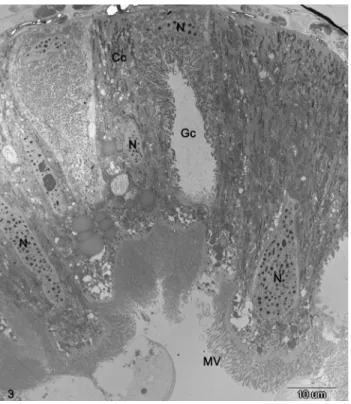
The midgut epithelium of cotton bollworm larvae consists of three types of cells: columnar and goblet cells, which make up most of the midgut epithelium, and the basal regenerative cells (Figure 3). Small PIB (< 1mm)
were observed in some of the columnar cells 1 or 2 days af-ter treatment with HaCPV. The PIBs sizes were 0.5 to 3.5 mm. The diameter of virions measures from 41 to 51 nm, with a mean of 46 nm.
On day 3 after infection, infected columnar cells were packed with PIB that virtually fill the whole cell (Figure 4). At this stage of infection the microvilli, which cover the apical ends of columnar cells, were not affected. However, at more advanced stages of infection and just prior to cell rupture, the apical microvilli deteriorated, revealing the bare and distended apices of infected cells (Figure 5). The distended cells eventually ruptured through their apices, re-leasing PIB into the gut lumen (Figure 6).
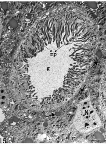
On day 4 after infection, deterioration of mitochon-dria and rough endoplasmic reticulum were observed (not shown). The mitochondria became swollen and eventually disintegrated. There is little or no rough endoplasmic retic-ulum within infected cells (not shown). At this stage, the nucleus in many of the infected columnar cells was still present (not shown). Examinations of sectioned tissues with the electron microscopes showed that infections oc-curred in the cells of the fat body. Because adjacent cells usually differed in the degree and stage of infection, the vi-rus in cells in earlier stages of infection varied in size; those in terminally infected cells were uniformly large, an indica-tion that individual virus grow during the infecindica-tion. On day 5 or 6 after treatment, PIB were observed in some goblet cells (Figure 7). The process of infection of goblet cells is similar to that described for columnar cells. Infected goblet cells degenerate to such an extent that only a few of the original microvillus-like cytoplasmic projections and cell organelles were presented, as compared with control (Fig-ures 7, 8).
Discussion
The midgut epithelium is mainly involved in the ab-sorption of nutrients and other useful substances and their transport. In addition, it has an important role in the re-moval of unwanted and harmful substances from the body. Figure 2- Gut of HaCPV-infected larvae ofHelicoverpa armigera.
Figure 3- Surface of midgut epithelium of a normal cotton bollworm larva, showing distinct nucleus (N), microvilli (Mv), columnar cells (Cc), and goblet cell (Gc).
The histopathology of HaCPV in the midgut of the cotton bollworm was similar to that of reported for the corn ear-worm (Bong and Sikorowski, 1991) and the silkear-worm (Iwashita, 1971; Kawase and Miyajima, 1971) infected with cypoviruses, especially regarding to changes in the co-lumnar cells. Small PIB were found in some midgut colum-nar cells of the cotton bollworm as early as one day after treatment. Penetration of the Cytoplasmic polyhedrosis vi-rus particles into the cotton bollworm midgut incidented much earlier. In the silkworm, penetration of the virus par-ticles into midgut cells occurs within 10 min of inoculation (Kobayashi, 1971) and small PIB are observed in the midgut cells 48 h later (Kawase and Miyajima, 1971). Ap-parently, formation of PIB is more rapid in the cotton bollworm than that in the silkworm.
The PIB in cotton bollworm are in general smaller (0.5 to 3.5mm) than those reported for silkworm and other lepidopterous insects (Aizawa, 1971; Cunninghamand longworth, 1968), the PIB as large as 5mm occur in larvae of the monarch butterfly,Danaus plexippus(L.) (Arnottet al., 1968). The virion (46 nm) in the cotton bollworm is
smaller than the general size range (60-80 nm) reported for CPV (Fenner, 1976). The virion measures 69 nm in the silk-worm (Aizawa, 1971)and 67 nm in monarch butterfly lar-vae (Arnottet al., 1968).
Kobayashi (Kobayashi, 1971) showed that the viro-genic stroma is the developmental focus of viral synthesis where formation of PIB takes place. Formation of HaCPV PIB in the cotton bollworm is similar to that in the silkworm that infected with CPV. Empty capsids described by Arnott et al.(1968) in larvae of the monarch butterfly which in-fected with CPV are not observed in the cotton bollworm. In infected columnar cells, the microvilli, which play a vital role as the absorptive lining of the gut lumen, are severely affected. Whereas, microvilli of infected columnar cells are partially or completely absent and they contain large num-ber of PIB. This differs from that reported for the corn ear-worm,Helicoverpa zea(Boddie) that infected with CPV (Bong and Sikorowski, 1991).
Most digestive and adsorptive functions of the insect occur in the midgut. So, its damage by HaCPV would ad-versely affect the normal growth and development of cot-ton bollworm. Marzbanet al.(2009) found that exposure of H. armigeralarvae to HaCPV have negative impact on its Figure 5- HaCPV-Infected midgut lining of Helicoverpa armigera,
showing the bare apices (arrow) of heavily infected columnar cells imme-diately prior to rupture. Note the large number of polyhedra inclusion bod-ies (P).
growth and development which resulted in reducing pupa-tion and pupal weight and an extended developmental pe-riod. Also, viral synthesis requires energy. The large num-ber of PIB within infected cells indicates a great expenditure of the insect’s metabolic energy. Marzban (2012) observed that HaCPV-infected cotton bollworm re-duced significantly not only body weight of larvae, but also glycogen, soluble protein, and total lipid content. This is in-dicative of extensive diversion of normal metabolism for synthesis of viral materials adversely affecting normal functions of the insect.
As infection advances, columnar cells becomes filled with PIB, and so distended that they rupture through their apices releasing PIB into the gut lumen.
Infected columnar cells of the cotton bollworm exhibit additional changes. The mitochondria and rough endo-plasmic reticulum degenerate 3 or 4 days after the onset of infection. Kobayashi (Kobayashi, 1971) reported similar ob-servations for rough endoplasmic reticulum in CPV-infected silkworm midgut, but found that mitochondria remained normal except for several enlarged ones near the virogenic stroma. In advanced stages of infection, the nuclei of in-fected cells are obscured by the large number of PIB.
Goblet cells are infected by HaCPV 5 or 6 days after treatment and undergo serious changes leading them to de-terioration. Goblet cells are responsible for potassium ion transport from heamolymph to the gut lumen (Aizawa, 1971). These histopathological data is part of researches evaluating the potential of HaCPV as a biocontrol agent in the integrated pest management of the cotton bollworm.
Acknowledgments
The authors are grateful to Dr. M. motazeri of Iranian Research Institute of Plant Protection and two anonymous reviewers for their critical review and comments on the manuscript. We are indebted to Dr Jiang Zhong (Fudan University) who most generously provided cytoplasmic polyhedrosis virus ofH. armigerafor this study; also ex-tend our appreciation to Dr Wang Mo (Hua Zhong agricul-tural University) for providing H. armigera pupae. This research was funded by “973” Projects (2006CB100204) and the State Key Research Programs of Science and Tech-nology Ministry (2006BAD08A07-05) of PR China.
References
Aizawa K (1971) Structure of polyhedra and viral particles of cy-toplasmic polyhedrosis.In: Aruga, H., Tanada, Y. (eds).The Figure 7- HaCPV-infected goblet cell ofHelicoverpa armigera, showing
numerous PIB (P). Mitochondria and rough endoplasmic reticulum are ab-sent in the cell, and degenerated microvilli (arrow). Goblet chamber (g).
cytoplasmic polyhedrosis virus of the silkworm. Tokyo, p. 23-59.
Anderson E, Harvey WR (1966) Active transport by the cecropia midgut. Fine structure of the midgut epithelium. J Cell Biol 31:134-160.
Arnott HJ, Smith KM, Fullilove SL (1968) Ultrastructure of a cy-toplasmic polyhedrosis virus affecting the monarch butter-fly, Danaus plexippus. Development of virus and normal polyhedra in the larva. J Ultra Mol Struct R 24:479-507. Bong CFJ, Sikorowski PP (1991) Histopathology of Cytoplasmic
polyhedrosis virus (Reoviridae) infection in corn earworm Helicoverpa zea (Boddie) larvae (Insecta: Lepidoptera: Noctuidae). Can J Zool 69:2121-2127.
Bot J (1966) Rearing Helicoverpa armigera (Hubner) and Prodenia lituraF. on an artificial diet. J Agric Sci 9:538-539.
Cunningham JC, longworth JF (1968) The identification of some cytoplasmic polyhedrosis viruses. J Invertebr Pathol 11:196-202.
Dwyer G, Dushoff J, Yee SH (2004) The combined effects of pathogens and predators on insect outbreaks. Nature 430:341-345.
Fenner F (1976) The classification and nomenclature of virus. Intervirology7:1-115.
Flower NE, Filshie BK (1976) Goblet cell membrane differentia-tion in the midgut of a lepidopteran larva. J Cell Sci 20:357-375.
Gunning R, Dang H, Christiansen I (2001) Play your part in resis-tance testing. Australian Cotton Grower 22:18.
Iwashita Y (1971) Histopathology of cytoplasmic polyhedrosis virus of the silkworm.In: Aruga, H., Tanada, Y. (eds).The cytoplasmic polyhedrosis virus of the silkworm. Tokyo, p. 23-59.
Katagiri K (1981) Pest control by cytoplasmic polyhedrosis vi-ruses, pp.433-440.In: H. D. Burges (ed.).Microbial control of pests and plant diseases 1970-1980. Academic Press, New York.
Katagiri K, Iwata Z (1976) Control ofDendrolimus spectabilis with a mixture of cytoplasmic polyhedrosis virus and Bacil-lus thuringiensis.Appl Entomol Zool 11:363-364.
Katagiri K, Iwata Z, Kushida T, Fukuizumi Y, Ishizuka H (1977) Effects of application ofBt, HACPV and a mixture ofBtand HACPV on the survival rates in populations of the pine cat-erpillar, Dendrolimus spectabilis. J Jpn Fore Soc 59:442-448.
Katagiri K, Iwata Z, Ochi K, Kobayashi F (1978) Aerial applica-tion of a mixture of CPV andBacillus thuringiensisfor the control of the pine caterpillar, Dendrolimus spectabilis. J Jpn Fore Soc 60:94-99.
Kawase S, Miyajima S (1971) Multiplication of cytoplasmic poly-hedrosis virus.In: Aruga, H., Tanada, Y.(eds).The cytoplas-mic polyhedrosis virus of the silkworm. Tokyo, p. 23-59. Kobayashi M (1971) Cycle of cytoplasmic polyhedrosis virus as
observed with the electron microscope. In: Aruga, H., Tanada, Y.(eds).The cytoplasmic polyhedrosis virus of the silkworm. Tokyo, p. 23-59.
Li G, Wu KM, Gould F, Feng H, He Y, Guo Y (2004) Frequency ofBtresistance genes inHelicoverpa armigerapopulations from the Yellow River cotton-farming region of China. Entomol Exp Appl 112:135.
Li Y, Tan L, Chen W, Zhang J, Hu Y (2006) Identification and ge-nome characterization of Heliothis armigera cypovirus types 5 and 14 andHeliothis assultacypovirus type 14. J Gen Virol 87:87-394.
Martignoni ME, Iwai PJ (1981) A catalogue of viral disease of in-sects, mites, and ticks.In: Burges, H.D.(ed).Microbial con-trol of pests and plant diseases. academic press, New York, p.897-911.
Marzban R, He Q, Liu XX, Zhang QW (2009) Effects ofBacillus thuringiensistoxin Cry1Ac and Cytoplasmic polyhedrosis virus ofHelicoverpa armigera(Hübner) (HaCPV) on Cot-ton bollworm (Lepidoptera: Noctuidae). J Invertebr Pathol 101:71-76.
Marzban R (2012) Midgut pH Profile and Energy Differences in Lipid, Protein and Glycogen Metabolism of Bacillus thuringiensis Cry1Ac Toxin and Cypovirus-infected Helicoverpa armigera(Hübner) (Lepidoptera: Noctuidae). J Entomol Res Soc 14:45-53.
Mertens PPC, Pedley S, Crook NE, Rubinstein R, Payne CC (1999) A comparison of the genomic dsRNA segments of six cypovirus isolates by cross-hybridization of their dsRNA genome segments. Arch Virol 144:561-566.
Mertens PPC, Rao S, Zhou H (2004)Cypovirus. In: Fauquet, C.M., Mayo, M.A., Maniloff, J., Desselberger, U., Ball, L.A.(eds). Virus taxonomy: eighth report of the Interna-tional Committee on Taxonomy of Viruses. Elsevier/Aca-demic Press, London, p. 522-533.
Payne CC, Rivers CF (1976) Aprovisional classification of cyto-plasmic polyhedrosis virus based on the size of the RNA ge-nome segments. J Gen Virol 33:71-81.
Shen J, Zhou W, Wu Y, Lin X, Zhu X (1998) Early resistance of Helicoverpa armigera (Hubner) to Bacillus thuringiensis and its relation to the effect of transgenic cotton lines ex-pressing Bt toxin on the insect. Acta Entomol Sin 41:8-14. Sikorowski PP, Thampson AC (1979) Effects of cytoplasmic
polyhedrosis virus on diapausing Heliothis virescens. J Invertebr Pathol 33:66-70.
Wiygul G, Sikorowski PP (1991) Oxygen uptake in larval bollworm (Heliothis zea) infected with iridescent virus. J Invertebr Pathol 58:252-256.
Wiygul G, Sikorowski PP (1978) Oxygen uptake in tobacco budworm larvae(Heliothis virescens) infected with cyto-plasmic polyhedrosis virus. J Invertebr Pathol 32:95-191. Ying SL (1986) A decade of successful control of pine caterpillar,
Dendrolimus punctatus walker (lepidoptera: Lasiocampidae), by microbial agents. Forest Ecol Manag 15:69-74.
Zhang H, Zhang J,Yu X,Lu X, Zhang Q, Jakana J, Chen DH, Zhang X, Zhou ZH (1999) Visualization of proteRNA in-teractions in cytoplasmic polyhedrosis virus. J Virol 73:1624-1629.


