A Regulated Response to
Mitochondrial Dysfunction Modulates
Longevity and Rates of Living
David Zeferino d’Azevedo Cristina
Dissertação apresentada para obtenção do grau de doutor em Genética Molecular pelo Instituto de Tecnologia Química e Biológica da Universidade
Acknowledgements
First, I would like to thank Cynthia Kenyon for taking me into her lab and allowing me to pursue my scientific passions while zealously looking over my scientific progress. Through Cynthia, I learned that, on the road to becoming a great scientist, a little enthusiasm goes a long way.
I would like to thank Jorge Carneiro for supporting me as my national advisor. I would also like to thank Jose Leal and Monica Dias for being a part of my thesis committee.
I thank the UCSF community for allowing me to enjoy such a wonderfully creative and dynamic working environment.
I thank Fundação para a Ciência e a Tecnologia and the Fundo Social Europeu and the POCI2010 for generous financial support (SFRH/BD/9603/2002).
I thank the Fundação Calouste Gulbenkian and the Programa
Gulbenkian de Doutoramento em Biomedicina for the first year of my PhD and for allowing me to be a part of such a amazing community.
I would like to thank all the wonderful and talented Kenyon lab members, past and present, that taught me so much and made lab work so enjoyable. In particular, I would like to thank Malene Hansen for her friendship and advice, which helped shape me as a scientist and as a person. I would also like to thank Lev Oscherovich, Marta Gaglia, and Laura Mitic for great scientific insight and for being such tremendously interesting people and good friends.
I would like to thank Paulo Almeida and Tania Vinagre for proofreading this thesis.
Abstract
Sumário
Table of contents
Acknowledgements i
Abstract iii
Sumário v
Table of contents vii
List of figures x
List of tables xi
CHAPTER I -INTRODUCTION page 1
1.1 - Aging in Biology – “What is aging?” page 2
1.2 - Theories of Aging – “Why do we age?” page 2
1.3 - Cellular Causes of Aging – “How do we age?” page 5 1.3.1 - Oxidants and Aging page 5 1.3.2 - Mitochondria and Aging page 9 1.3.3 - Other potential causes of aging page 17 1.4 - Modulation of Aging – “Can we age more gracefully?” page 21
1.4.1 - Dietary Restriction (DR) page 23 1.4.2 -Insulin/IGF-1 (IIS) dependent regulation of aging page 25
1.4.3 -Long-lived mitochondrial mutants page 28 1.5 - C. elegans as a model organism for studying lifespan
regulation – Why old worms? Page 35
Chapter II - Timing of Action of Mitochondrial Mutations page 38
2.1 - Summary page 38
2.2 - Background page 38
2.3 - Results page 39
2.4 - Discussion page 41
Chapter III - A Regulated Longevity Response to Mitochondrial
Respiration in C. elegans page 43
3.1 - Summary page 43
3.2 - Background page 44
3.3 - Results page 47
3.3.1 - clk-1 mutants exhibit a conserved retrograde response page 47 3.3.2 - fstr-1/2 and aqp-1 contribute to the increased longevity of
clk-1mutants page 53
3.3.3 - fstr-1/2 knockdown increases the behavioral rates of clk-1
Mutants page 56
3.3.4 - fstr-1/2 is necessary for gene expression changes observed
in clk-1mutants page 59
3.3.5 - fstr-1 may act in the intestine and/or nervous system to slow
down clk-1mutants page 62 3.3.6 - A similar retrograde response in animals with reduced
respiration page 64
3.3.7 - cdr-2 RNAi suppresses the increased longevity of isp-1
mutants page 67
3.3.8 - fstr-1/2’s regulatory function is specific to clk-1 mutants page 68
3.4 - Discussion page 70
3.4.1 - A C. elegans retrograde response page 71 3.4.2 - Long-term reductions in respiration are not necessary
to maintain expression of the retrograde response page 75 3.4.3 - The retrograde response is probably required for the
longevity of C. elegans mitochondrial mutants. page 76 3.4.4 - The molecular function of FSTR-1/2 page 78 3.4.5 - Different paths to a similar phenotype page 79
3.5 - Methods page 81
3.6 - Acknowledgements page 85
Chapter IV - Final Considerations on Mitochondria and Aging page 82
List of Figures
Figure 1 – Mitochondrial structure page 10
Figure 2 – Overview of aerobic respiration. page 12 Figure 3 – Electron transport chain (ETC) page 14 Figure 4 - Mitochondrial knock down after the L3
postembryonic stage is insufficient to increase longevity page 40 Figure 5 - Mitochondrial DNA quantification page 53 Figure 6 - fstr-1/2 and aqp-1 contribute to the increased
longevity of clk-1mutants page 55
Figure 7 - fstr-1/2 RNAi speeds up clk-1(-) animals. page 58 Figure 8 - fstr-1/2 RNAi inhibits expression of the clk-1
retrograde response page 61
Figure 9 - fstr-1 is expressed in the intestine and three
neurons in a clk-1(-)background. page 63
Figure 10 - cdr-2 RNAi suppresses the increased longevity
of isp-1 mutants. page 68 Figure 11 - fstr-1/2 RNAi did not shorten the lifespan of isp-1
mutants page 69
Fig. 12. fstr-1/2 RNAi does not affect gpd-2 expression in an
isp-1 mutant page 70
Figure 13 - C. elegans retrograde response. page 74 Figure 14 - The short lifespan of mev-1 mutants is increased
by respiratory-chain RNAi. page 75
Figure 15 - The glyoxylate cycle gene gei-7 is partially
necessary for cyc-1 RNAi to increase longevity. page 78 Fig. 16 - C. elegans mitochondrial mutants activate similar
List of Tables
Table 1 - Up-regulated GO categories in wormand yeast
mitochondrial mutants page 50
Table 2. The most significant differentially-expressed genes
Chapter I
Introduction
“To get back my youth I would do anything in the world, except take
exercise, get up early, or be respectable.” – Oscar Wilde
Aging is a widespread process with deep-rooted implications in our lives and societies. Humanity has struggled to understand this mystery and its implications for millennia, and even presently, many questions remain. It has captured the imagination of many philosophers and artists. Great literary works were created either embracing the inevitability of aging or fantasizing about eternal youth. The idea of prolonged life is a common mythological theme in many cultures, and an intrinsic part of our collective imagination. More so, the search for long life is not a recent trend, in fact, Juan Ponce de Leon, an influential Spanish explorer, in 1513 found himself dissatisfied with his riches and set out on a quest for the fountain of youth, discovering Florida in the process (Oviedo 1535; Gómara 1551). Already many years before this “conquistador’s” adventure, in the 1300s, alchemy attempted to, besides the noble goal of turning household items into gold, create a liquid “panacea” that would heal all illnesses and allow eternal life (Burland 1989).
economic growth since the main consumer base consists of younger people (Krulwich 2006). Also, as the fraction of elderly individuals increases, it is important to understand the underlying physiological changes of aging and age-related disease, developing appropriate medical procedures in the process.
More relevantly to this work, the aging process represents a major unanswered biological question of tremendous scientific interest and potential. A complex task that requires an interdisciplinary approach, its study generates a better understanding of both fundamental biological processes and age-related disease. It is a compelling question that fuels the imagination of scientists and non-scientists alike.
1.1 - Aging in Biology –
“What is aging?”
There is no simple definition since there are several phenotypes associated with aging, some of which are shared between a wide range of species, some of which are unique. However, aging can be defined broadly as the age-associated decline of physiological and neurological processes leading to a decrease in overall function of an organism.
It is important to note that bacteria and certain mammalian cells in culture have the ability to divide almost indefinitely, so, clearly, the potential to keep cellular stability through time exists. Yet, despite of this, most organisms lose function, age and eventually die. So the question remains, why do organisms age?
1.2 - Theories of Aging –
“Why do we age?”
resources. Along these lines, a commonly heard argument for aging as an evolutionarily selected process is that it increases generation turnover, thus facilitating adaptation to changing environments. This theory is no longer believed to be accurate, mostly due to three evolutionary arguments: Firstly, in the wild, animals tend to die early from extrinsic causes (predation, climate, disease), thus making the effects of aging as a population control mechanism negligible (Medawar, 1952). Secondly, since the majority of the animals die young, natural selection cannot act on phenotypes that occur later in life. Thirdly, because senescence is mainly post-reproductive, mutations that affect this life period are less likely to be selected for, since they should have minimal effects on fitness. Hence, it is hard to imagine how a mechanism with no function other than to regulate longevity may have been selected for. This does not mean, however, that genes cannot affect longevity, but that aging per se was not evolutionarily selected for.
Along these lines, we have a more widely accepted theory of aging, the mutation accumulation theory of aging. This theory states that because natural selection cannot act later in life, populations will tend to randomly accumulate mutations that will have deleterious effects in older organisms. The mutations that act the latest are exposed to lower selective pressures, which allows an accumulation of progressively worse phenotypes as animals age (Kirkwood 2002). This theory predicts that over the span of many generations there will be a severe decrease in overall function in older organisms, in other words, aging.
argued that a consequence of this reasoning is that any mutation extending longevity should decrease fitness of younger animals.
This has prompted the disposable soma theory of aging, stating that animals have limited resources to tap over the course of their lives. These resources are optimized for maintenance of the germline in liu of longevity, hence any condition that delays aging should negatively affect fertility (Kirkwood and Holliday 1979). In a simplified way, why spend valuable resources on protective enzymes that will allow the animal to live to 30 years when those resources would be better spent on reproduction, which ends at 15 years of age? This theory predicts that, any process that increases longevity leads to decreased fertility (Kirkwood and Holliday 1979). However, experimentally, this “trade-off” is not always present, thus suggesting that this theory is incomplete. Either the organism’s resources are not limited or there is room for further optimization of their allocation that would allow increases in longevity without a reproductive “trade-off”.
Currently, most evolutionary biologists tend to defend a mixed model involving the “mutation accumulation” and “antagonistic pleiotropy” theories. It is interesting to note that, although aging is a widespread phenomenon with shared characteristics throughout different species, evolutionary biologists predict it not to be genetically regulated (Kirkwood 2002). However, work done in the last 15 years has uncovered numerous lifespan-increasing mutations and has started to suggest that this view may be an oversimplification.
and lifespan remained elusive. With the discovery of cellular oxidants (see section 1.3.1) however, this theory gained momentum as these provided the perfect connection between metabolic rate and longevity. Increased metabolic rates led to more oxidant-related damage which, in turn, decreased longevity. Although it is an attractive hypothesis due to its simple and intuitive nature, presently most scientists regard it as incomplete, mostly because the correlation between metabolic rate and lifespan is inconsistent across species. This is particularly true for birds and primates, which tend to live longer than predicted by their metabolic rates (Finkel and Holbrook 2000). Analysis of free radical formation in these species led to the finding that they have decreased production of toxic oxidants, suggesting that this could be the cause of their increased longevity. For a complete answer to our questions, theoretical models of aging need to be accompanied by experimental data. This experimental approach allows us to address another outstanding question in the field of aging research: How do we age? What are the causes of aging?
1.3 - Cellular Causes of Aging –
“How do we age?”
Until now, we have discussed a non-specific decrease in function of living organisms, but on a more mechanistic note, exactly what causes the decrease in function observed with aging? If it is non-specific damage, why does it consistently affect a subset of processes?
1.3.1 - Oxidants and Aging
Presently, the most widely accepted culprits for age-associated decline in cellular and organismal function are reactive oxygen species (ROS), according to the free radical theory of aging (Harman 1956).
with several organic molecules (nucleotides, proteins or lipids) and, in doing so, can cause considerable damage, impairing normal cellular function (Finkel and Holbrook 2000; Kirkinezos and Moraes 2001).
In the mid 1950s Denham Harnham introduced his free-radical theory of aging by speculating that cells generated endogenous oxygen radicals, which, in turn, resulted in a pattern of age-related cumulative damage.
Although, at first, the concept of endogenous oxidants was highly controversial, it gained momentum with the discovery of the first superoxide dismutase (SOD) enzyme (McCord and Fridovich 1969). Superoxide dismutases belong to a large family of important enzymes that detoxify ROS. SODs, in particular, are responsible for converting the superoxide anion (.O2-) into the more stable hydrogen peroxide (H2O2). H2O2, in turn, can be broken down into innocuous water (H2O) and oxygen (O2) by the enzymes catalase (Loew 1900) and glutathione peroxidase (Finkel and Holbrook 2000). However, if left unchecked, both superoxide and hydrogen peroxide molecules can lead to the production of the highly reactive hydroxyl radical (O2- ), which is thought to be responsible for most cellular oxidative damage (Beckman and Ames 1998). Besides the aforementioned detoxifying enzymes, several other non-enzymatic, low molecular mass molecules can be found in cells that share their role in scavenging ROS. These include ascorbate, pyruvate, flavonoids, carotenoids and perhaps most importantly, glutathione (Finkel and Holbrook 2000).
single- and double-strand breaks in the nucleotide backbone, or cross-linking to other molecules. These altered nucleotides can eventually lead to mutation, DNA rearrangements or problems during transcription (Beckman and Ames 1998). Oxidation of proteins by oxygen radicals leads to formation of carbonyls, protein-protein cross-linking, peptide fragmentation and inactivation of proteins with iron-sulfur clusters (Beckman and Ames 1998). Overall, any of these interactions between ROS and biomolecules promote cellular dysfunction.
superoxide dismutase leads to an increased lifespan in yeast (Harris, Bachler et al. 2005) and overexpression of SOD and catalase increase longevity of D. melanogaster (Orr and Sohal 1994; Sun and Tower 1999). Interestingly, flies can benefit from an increase in longevity when SOD is overexpressed only in the neurons arguing that this could be a rate-limiting tissue (Parkes, Elia et al. 1998), or there could be non-autonomous regulation originating in the neurons. Also, exposure to superoxide/catalase mimetics can extend C. elegans longevity up to 44% (Melov, Ravenscroft et al. 2000), although that work has been recently been put into question (Keaney, Matthijssens et al. 2004), interestingly, the latter authors saw a decrease in oxidative damage, but observed no effects on lifespan. Many long-lived C. elegans mutants show increased stress resistance which correlates with increased expression of sod-3, the nematode’s SOD (Honda and Honda 1999; Feng, Bussiere et al. 2001; Henderson, Bonafe et al. 2006). In addition, certain short-lived C. elegans mitochondrial mutants, which show hallmarks of accelerated aging, have increased ROS-related damage (Ishii, Fujii et al. 1998; Kayser, Sedensky et al. 2004). However, the correlation between accelerated aging and increased ROS has been broken in several instances, which leads us to an important question:
Is oxidative stress resistance an absolute requirement for longevity? Is it, moreover, the sought-after mechanism of lifespan regulation? Experimental evidence suggests that the answer to these questions may not be a simple one. For instance, chico mutants in Drosophila are long-lived, but exhibit normal levels of resistance to oxidative stress (Clancy, Gems et al. 2001). In addition, mice knockout mutants for SOD1, SOD2,
long-lived mutants (Murphy et al. 2003, Lee, Kennedy et al. 2003). Thus, further research is required to determine the exact relationship between oxidative stress resistance and the regulation of longevity.
It is well established that during oxidative stress, accumulation of ROS leads to cellular damage. However, under physiological circumstances ROS have several “legitimate” cellular functions: they act as essential signaling molecules involved in wound healing (Sen 2003), recruitment of leukocytes during the adaptive immune response (Guzik, Korbut et al. 2003), regulation of aconitase activity (Gardner and Fridovich 1991; Gardner, Raineri et al. 1995; Irani, Xia et al. 1997) and regulation of the Ras pathway (Irani, Xia et al. 1997; Shibata, Branicky et al. 2003), mostly in the form of nitric oxide (NO·) and superoxide (.O2-). These newly-found roles of ROS in cellular signaling may have significant implications for their relationship to the aging process. Since the data suggests that there is an increase in free radicals in older organisms, it is tempting to speculate that many ROS-signaling dependent cellular processes will be altered with age, leading to a decrease in overall function of the organisms (Beckman and Ames 1998). This is an attractive hypothesis because potentially, smaller, more physiological amounts of “illegitimate” ROS could cause significant cellular miss-regulation and lead to aging.
1.3.2 - Mitochondria and Aging
themselves. In this way, mitochondria may be predicted to have a role in aging as the cell’s main sources of ROS.
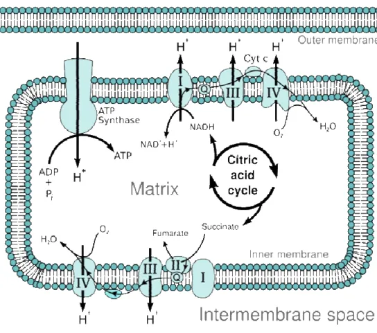
Mitochondria are complex cellular organelles consisting of two lipid membranes (outer and inner) with unique properties that separate two distinct compartments: the intermembrane space and the matrix (space inside the inner membrane) (Fig. 1).
Figure 1 – Mitochondrial structure. Mitochondria have two distinct membranes (inner membrane and outer membrane) which separate two distinct compartments: matrix and inter membrane space. Mitochondria have their own genetic material, the nucleoid, which is circular and encodes their own set of translational machinery (ribosomes). These ribosomes are similar to bacterial ribosomes and distinct from eukaryotic ribosomes. This image was adapted from a wikicommons image.
2001) from an endosymbiotic proteobacteria that survived an endocytosis event. This pre-mitochondrial bacterium or proto-mitochondrion provided an evolutionary advantage to the host cell by performing aerobic respiration in cells that would have otherwise relied on less-efficient pathways of glycolysis and fermentation for energy (Sagan 1993). Although the majority of mitochondrial proteins are encoded in the nucleus, mitochondria contain their own genetic material (mtDNA) and translational machinery, reminiscent in many aspects of their prokaryotic evolutionary origins (Emelyanov 2003; O'Brien 2003). It is thought that over the millions of years since the appearance of eukaryotic organisms, much of the mitochondrial genetic material was transferred to the nucleus (Alberts et al., 2002). mtDNA molecules are usually circular, small (≈ 16 kb), attached to the inner mitochondrial membrane and code for ≈37 mitochondrial proteins essential for aerobic respiration (this number varies between species) (Alberts et al., 2002). This means that, although most mitochondrial proteins are encoded in nuclear DNA, cells with deficiencies in mtDNA are unable to perform mitochondrial respiration (see yeast “petites”, section 1.4.3).
Mitochondria serve several functions in eukaryotes: through unique metabolic reactions, they are the cell’s main source of energy, in the form of adenosine-triphosphate (ATP); they are essential in sequestering potentially toxic Ca2+ ions (Chan 2006); they serve an important role in apoptosis (Hengartner 2000); and they are necessary for steroid and heme synthesis (Chan 2006). Nevertheless, the most intensively studied and arguably the most important of mitochondrial functions is energy generation.
Figure 2 – Overview of aerobic respiration. ETC – electron transport chain; OAA – oxaloacetate; α-KG – α-ketoglutarate; Pyr. Decarb. – pyruvate decarboxilation. NADH produced during glycolysis may also be used by the ETC to produce ATP, however its transport into the mitochondria requires energy expenditure.
form of one GTP molecule that can be readily converted to ATP. The Krebs cycle is not only a central process in energy generation, but also produces many biochemical intermediates essential to other metabolic pathways such as glutamate, a key intermediate in amino acid metabolism that is generated from α-ketoglutarate (Butow and Avadhani 2004). Besides pyruvate decarboxylation, which yields acetyl-CoA, several other catabolic pathways provide intermediates for the Krebs cycle, especially when these intermediates are scarce. These are called anaplerotic pathways (Butow and Avadhani 2004). There are several different anaplerotic pathways, which can “feed” into different steps of the Krebs cycle (see chapter 3.4.1).
Figure 3 – Electron transport chain (ETC). Thin interconnected arrows depict flow of electrons. Q - electron carrier ubiquinone. Cyt c – electron carrier cytochrome c. H+ - proton. Pi – phosphate. This image is adapted from a
wikicommons image.
which is used by the highly conserved ATP synthase (sometimes called complex V) to generate ATP.
we can predict that the increase in ROS seen with age can also lead to increased metabolic dysregulation, with grave and pleiotropic cellular consequences.
be proven conclusively. It is still unclear whether the increase in frequency of ∆mtDNA and mtDNA point mutations is causally related to increased oxidative damage, or is merely a biomarker of aging.
Further emphasizing the connection between mitochondria and aging, work done in C. elegans and S. cerevisiae has uncovered several mutants with impaired mitochondrial function that have altered lifespans. Some of these mutants have shortened lifespans (Ishii, Fujii et al. 1998; Kayser, Sedensky et al. 2004), whereas others, interestingly, live considerably longer than their wild-type counterparts (Wong, Boutis et al. 1995; Kirchman, Kim et al. 1999; Epstein, Waddle et al. 2001; Feng, Bussiere et al. 2001; Traven, Wong et al. 2001; Dillin, Hsu et al. 2002; Lee, Lee et al. 2003). These fascinating long-lived mitochondrial mutants and the regulation of their phenotypes will be the focus of this PhD thesis (for a more detailed discussion of these mutants, please see section 1.4.3).
1.3.3 - Other potential causes of aging
This section presents an overview of other cellular processes potentially involved in aging that are beyond the scope of this thesis.
information. But of course, even telomeres would eventually “wear down”, so in order to re-elongate them to their original length, an enzyme called telomerase is required (Blackburn 1990). Telomeres also prevent chromosome fusions that can lead to cancer (DePinho 2000). As a result, when telomeres become too short, possibly in order to avoid becoming cancerous, cells either stop dividing and enter a senescent state, or self-destruct through apoptosis (DePinho 2000). Because of this, cells have a limited number of divisions they can go through, linked to telomeric length, the Hayflick limit (Hayflick 1965). In this sense, telomeres can act as biological “clocks”. This idea generated much interest in the aging research community, but, despite this, the effect of telomeres and senescence on aging remains controversial (Campisi 2005). On the one hand, many lines of evidence connect ROS damage to increased senescence and telomere shortening (Chen and Ames 1994; von Zglinicki, Saretzki et al. 1995; Oexle and Zwirner 1997; Lee, Fenster et al. 1999; Passos, Saretzki et al. 2007), providing a potential role for telomeres in aging. On the other hand, experiments attempting to establish a causal link between telomere length, senescence and aging have come up with unclear results (Gonzalez-Suarez, Geserick et al. 2005). Overall, the relationship between cancer, aging and telomeres is complex and needs further study.
cell’s ability to cope with protein misfolding stress is altered with age. Still, the argument could be made that there is a constant accumulation of misfolded proteins irrespective of the organism’s age, and that some critical mass must be obtained before disease symptoms occur, thus separating disease from aging. Interestingly, recent studies contradict this hypothesis. In C. elegans, time of onset of aggregation can be delayed by approaches that increase longevity, such as inhibiting insulin pathway activity (Morley, Brignull et al. 2002; Hsu, Murphy et al. 2003). This is a strong argument for a connection between protein aggregation and aging. There are two possible explanations for this observation: On one hand, some of the factors contributing for aging could also be causing protein aggregation, it is tempting to speculate that as organisms age, there is a decrease in quality of protein folding mechanisms. On the other hand, protein aggregation itself could be causal to aging, as it is already clear that increases in misfolded proteins impair organismal function (Hsu, Murphy et al. 2003). In all likelihood both of these are accurate to some extent.
and Johnson 1997). So, what evidence is there supporting somatic mutations as a potential cause of aging? A compelling line of evidence is the high correlation between DNA repair ability and lifespan (Hart and Setlow 1974; Cortopassi and Wang 1996). This is indicative that increased DNA repair may play a role in increasing longevity. However, it has been demonstrated that effective DNA repair is necessary but not sufficient to increase longevity (Hart and Setlow 1974; Cortopassi and Wang 1996). The role of somatic mutations and DNA repair mechanisms in aging remains controversial. Clearly, organisms need an effective DNA repair system to avoid cancer and realize their longevity potential. The question is whether this DNA repair system and somatic mutations play a role in normal aging.
1.4 - Modulation of Aging
– “Can we age more gracefully?”
mutants relative to wild-type (Klass 1983) many more gerontogenes (genes affecting longevity) have been identified in worms (Kenyon, Chang et al. 1993; Lee, Lee et al. 2003; Hamilton, Dong et al. 2005; Hansen, Hsu et al. 2005). Despite the large number of genes, they can generally be broadly grouped into one of three distinct pathways influencing longevity: reduced food intake (dietary restriction or DR), reduced insulin signaling or other conditions that activate the DAF-16/FOXO transcription factor, or impaired mitochondrial function (which is the main focus of this thesis). These three pathways have been shown to be conserved in C. elegans and rodents, suggesting the presence of an evolutionarily conserved mechanism for regulation of lifespan.
response? In order to address this question we need to understand each of these pathways in more detail. Of the three pathways I will provide greater background on the mitochondrial inhibition pathway of aging, since it is the main point of this thesis.
1.4.1 - Dietary Restriction (DR)
Over 70 years ago Mcay et al. reported that decreasing food intake increased lifespan in rats (McCay, Crowell et al. 1989). Since then, this finding has been reproduced in several typical model organisms such as: mice (Mair and Dillin 2008), nematodes (C. elegans) (Klass 1977; Houthoofd, Braeckman et al. 2003), budding yeast (S. cerevisiae) (Jiang, Jaruga et al. 2000), fruit flies (D. melanogaster) (Chapman and Partridge 1996) and in some more atypical model organisms such as water striders, grasshoppers, water fleas, fish, hamsters, dogs and spiders (Mair and Dillin 2008). Studies on rhesus monkeys (Hansen and Bodkin 1993; Kemnitz, Weindruch et al. 1993) and squirrel monkeys (Ingram, Cutler et al. 1990) are ongoing and results are promising if still preliminary. In spite of the amount of time elapsed since its discovery and its impressive evolutionary conservation, the mechanism by which DR acts remains elusive. For many years, the field of DR research was lacking molecular studies, probably because most studies were done in rodents, which are not as amenable to genetic manipulation as some of the invertebrate model organisms.
An argument for DR’s evolutionary conservation is that dietary-restricted animals share multiple phenotypes across different species in addition to the increased longevity. Mainly, as mentioned before, a decrease in overall reproductive potential and a delay in onset of reproduction have been observed in fruit flies (Mair and Dillin 2008), worms (Klass 1977; Bishop and Guarente 2007; Bishop and Guarente 2007) and rats (Mair and Dillin 2008). Also, DR in model organisms has been shown to delay onset of age-related diseases such as: autoimmune disease, osteoporosis, cataracts, neurodegenerative diseases, diabetes and several forms of cancer (for a more detailed review see chapter V of Masoro, 2002). This means that dietary-restricted animals do not live longer as frail aged individuals, but are instead maintained in a healthier state until later in life.
DR is defined as a significant decrease in caloric intake that does not constitute starvation, usually 30-40% lower than fully fed. DR has been modeled in many different organisms and in many different ways: from decreasing all food availability (Klass 1977; Fabrizio and Longo 2003), to specifically altering the animal’s diet (Zimmerman, Malloy et al. 2003; Miller, Buehner et al. 2005), to mutations that decrease food intake (Lakowski and Hekimi 1998), (for a detailed listing of all, please see (Mair and Dillin 2008)). As a consequence of all these different methodologies, results can vary significantly between researchers, which has considerably slowed the field. The complexity and variability of the response to CR, when administered under different conditions, was unexpected. It will be very interesting to learn its basis at the molecular level.
that overexpression of Sir2p in yeast (Lin, Defossez et al. 2000), worms (Tissenbaum and Guarente 2001) and flies (Rogina and Helfand 2004) extends longevity, which clearly implicates this conserved family of proteins (sirtuins) in aging. However, new lines of evidence have put the initial yeast findings into question (Kaeberlein, Kirkland et al. 2004; Kaeberlein and Powers 2007), and this, in turn, has significantly weakened the connection between DR and sirtuins. Consequently, at this point, the connection between sirtuins and DR remains unproven. Another potential modulator of lifespan through DR is the target of rapamycin (TOR) pathway (Mair and Dillin 2008). The TOR nutrient sensing pathway has been implicated in longevity in several model organisms (Schieke and Finkel 2006), and is the best candidate for a conserved regulator of DR, found so far. Recently, two transcription factors skn-1, which is induced in response to oxidative stress (Bishop and Guarente 2007) and pha-4, a forkhead transcription factor (Panowski, Wolff et al. 2007) have been identified as necessary for DR-mediated increased longevity in C. elegans. These are promising molecules which may allow genetic probing of DR regulatory pathways. However, it remains to be shown whether their effects are conserved across different species. Interestingly, recent data has pointed to a role of autophagy in DR-mediated increased longevity (Jia and Levine 2007; Hansen, Chandra et al. 2008), this is suggestive that recycling of cellular components has an important role in DR and longevity in C. elegans
1.4.2
-
Insulin/IGF-1 (IIS) dependent regulation of aging
The best characterized pathway regulating longevity is the Insulin/IGF-1 (IIS) dependent regulation of aging. This pathway was originally identified in
mice (Bluher, Kahn et al. 2003; Holzenberger, Dupont et al. 2003), making this an important therapeutic target for longevity modulation in humans (Bartke 2008; Suh, Atzmon et al. 2008).
IIS is an important nutrient-sensing pathway that coordinates processes such as development, fat metabolism, growth and reproductive maturation. As such, this pathway is a good candidate for regulating longevity in response to energy availability, much like what was proposed earlier for DR. A possible model is that less energy availability leads animals to post-pone their reproductive maturation in order to increase likelihood of viable progeny (Wolff and Dillin 2006). This is consistent with the function of IIS pathway in worms, in that it regulates a hibernation-like state (the dauer diapause) that can occur during development if resources are scarce (Riddle, Swanson et al. 1981). Animals in this dauer stage are very resistant to thermal (Lithgow, White et al. 1995) and oxidative stresses (Honda and Honda 1999) and are considerably long-lived. It is all-together possible that the long lived C. elegans IIS mutants are activating and benefiting from the same protective mechanisms that allow dauers to live long, but without the hibernation phenotypes. However, the time during the animals’ life when the IIS pathway controls reproduction is different from the time when it affects aging (Dillin, Crawford et al. 2002). Also, mutants with different alleles have normal reproduction profiles. These are a strong arguments against a causal connection between rebroduction and aging .
Microrray studies of daf-2(-), daf-16(-) and daf-2(-); daf-16(-) mutants showed that the DAF-16 forkhead transcription factor modulates expression of several downstream genes affecting longevity (Murphy, et al. 2003). Interestingly, individual knock downs of each these genes in long-lived daf-2 mutants were insufficient to completely suppress longevity. This suggests that the total lifespan observed in long-lived daf-2 mutants is a result of small cumulative effects from multiple genes downstream of daf-16. Therefore, based on this finding, even though aging is regulated by a few key pathways, many downstream genes affect longevity.
Mutations in daf-2 lead to significant metabolic remodeling of the organisms as can be ascertained from gene expression studies (Murphy, et al. 2003). One interesting change is an increase in expression of enzymes necessary for the glyoxylate cycle (Murphy, et al. 2003). The glyoxylate cycle is an anaplerotic pathway that feeds intermediates into the Krebs cycle, thus allowing production of energy from stored fats. Another characteristic of daf-2 mutants is an increase in stress resistance (Larsen 1993; Vanfleteren 1993; Adachi, Fujiwara et al. 1998), which is accompanied by an increase in expression of several stress protective genes under control of daf-16 (Vanfleteren and De Vreese 1995; Honda and Honda 1999; Murphy, et al. 2003; Lin, Hsin et al. 2001). It is interesting to note that, there is a high (albeit imperfect) correlation between longevity and stress resistance in most long-lived mutants. This holds true for mutants that live long through DR, IIS inhibition or mitochondrial impairment.
In C. elegans germline removal also affects longevity through the daf-16
pathways. On the other hand, if these animals lack their entire gonad they do not exhibit an extended lifespan except when combined with some classes of daf-2 mutants, raising the possibility of partial mediation of this extension by the IIS (Hsin and Kenyon 1999). These findings support a connection between longevity and reproduction, although the nature of this connection is still unclear.
There is also significant evidence that sensory neurons affect longevity (Apfeld and Kenyon 1999; Alcedo and Kenyon 2004) in an IIS partially-dependent fashion. When sensory neuron function is impaired, worms live longer (Apfeld and Kenyon 1999). Certain subsets of sensory neurons can affect longevity in opposite ways. Different gustatory neurons can both extend and shorten longevity in a IIS dependent fashion (Alcedo and Kenyon 2004). Olfactory neurons, on the other hand, can affect lifespan in a distinct pathway that is connected to the reproductive system (Alcedo and Kenyon 2004) and not dependent on insulin signaling.
1.4.3
-
Long-lived mitochondrial mutants*
*(For a summarized version of this section please see section 3.2)
In section 1.3.2 (Mitochondria and Aging), we showed several lines of evidence linking mitochondria to longevity. One of the most compelling arguments is that in budding yeast, nematodes and mice, mutations that affect normal mitochondrial function can lead to increased longevity. This observation in itself is somewhat paradoxical. Since mitochondria and mitochondrial respiration are vital processes in cellular metabolism, it is unexpected that from their impairment organisms could reap longevity benefits.
binding domain is essential for this, but the mechanism remains unclear (Liu, Sekito et al. 2003). One of the initial markers for the retrograde response was increased expression of the gene CIT2, which encodes a citrate synthase (Liao, Small et al. 1991), this suggested that there might be some form of metabolic response to mitochondrial impairment.
inhibited and the Krebs cycle cannot be completed. In summary, inhibition of mitochondrial respiration and electron flow through the ETC, leads to interruption of the Krebs cycle and accumulation of succinate. This inability of the Krebs cycle to be completed, in turn, leads to a decrease in α -ketoglutarate availability and to a depletion of cellular glutamate (Butow and Avadhani 2004). Hence, it is likely that cells are increasing expression of Krebs cycle enzymes and anaplerotic pathways in order to keep the cycle flowing, if only partially to allow for production of glutamate.
These two microarray analyses also showed very significant up-regulation of glyoxylate cycle enzymes. Glyoxylate cycle is an important anaplerotic pathway. It bypasses the succinate-to-fumarate step of the Krebs cycle through the formation of glyoxylate, and eventually leads to the formation of pyruvate, which can be converted into alpha-ketoglutarate. This metabolic process occurs in the peroxisomes (Fig. 13). Another class of gene that was seen to have increased expression in these long-lived yeast “petites” was the stress response class(Epstein, Waddle et al. 2001; Traven, Wong et al. 2001). These stress response genes encoded mitochondrial chaperones (Traven, Wong et al. 2001), heat shock proteins (Epstein, Waddle et al. 2001; Traven, Wong et al. 2001) and stress response transcription factors (Epstein, Waddle et al. 2001; Traven, Wong et al. 2001). It is interesting to note that there was one inconsistenciy between the two studies, namely Traven et al. found increased expression of numerous genes encoding mitochondrial proteins, strongly suggesting that the “petites” were trying to compensate for the deficient mitochondrial respiration. On the other hand, Epstein et al. saw no change in expression levels of these genes. This can potentially be explained by differences in the strains used for each study (Butow and Avadhani 2004).
unclear whether its role in longevity is specific to yeast, or whether retrograde responses can be important processes in regulating longevity of other, multicellular, organisms. This question is addressed in chapter III of this thesis.
seems independent of DR or the IIS pathways for controlling longevity, which sets them up as a bona fide independent pathway for longevity regulation. Importantly, clk-1’s role in longevity is likely conserved, since heterozygous mice with reduced levels of Mclk-1 live longer than controls (Stepanyan, Hughes et al. 2006).
Another long-lived C. elegans mitochondrial mutant, isp-1, was identified in a mutagenesis screen and was found to have several phenotypes in common with clk-1(-) animals (Feng, Bussiere et al. 2001). These isp-1(-) animals harbor a point mutation in an iron-sulfur protein located in complex III of the ETC (Feng, Bussiere et al. 2001). isp-1
mutants exhibit extraordinarily decreased rates of living and delayed development to adulthood (Feng, Bussiere et al. 2001). In many respects these mutants have similar, although more dramatic, phenotypes than clk-1. However, they differ in a significant trait; isp-1 mutants have a significant decrease in mitochondrial respiration as measured by oxygen consumption (Feng, Bussiere et al. 2001). Also, as a potential mechanism for their increased longevity these mutants are resistant to oxidative damage and increase expression of antioxidant enzymes (Feng, Bussiere et al. 2001).
significant role for mitochondrial respiration in lifespan (Dillin, Hsu et al. 2002; Lee, Lee et al. 2003). These studies showed that knocking down several different components of the ETC significantly increased longevity and decreased rates of living (Dillin, Hsu et al. 2002; Lee, Lee et al. 2003). These phenotypes are similar to the ones observed in clk-1 and isp-1
mutants. These long-lived mitochondrial RNAi knockdowns were also smaller than wild type animals and had decreased ATP production (Dillin, Hsu et al. 2002).
Importantly, when looking at long-lived mitochondrial mutants in C. elegans, even though they share several phenotypes, there is an important distinction between the ones that do not affect respiration and energy production, clk-1 mutants; and the ones that do, isp-1 mutants and RNAi-induced mitochondrial knockdowns. This has prompted a long-standing question in the field, which is: How similar are these long-lived mitochondrial mutants to each other? Since they share so many phenotypes the simplest explanation is that they share a common mechanism in regulating their increased longevities. If so, however, why do they have different phenotypes? These questions are addressed in chapter III.
lifespan (Dillin, Hsu et al. 2002). In addition, lifespan extension does not correlate with resistance to oxidative stressors (Lee, Lee et al. 2003;Rea, Ventura et al. 2007).
clk-1 mutations, on the other hand, have very little effect on respiration, hence it is unlikely they are reducing ROS in this way. Overexpression of
clk-1 shortens lifespan and increases movement rates in C. elegans (Felkai, Ewbank et al. 1999) suggesting that whatever the mechanism, its influence on longevity and rates of living is rate-limiting in the animal. However the exact mechanism remains elusive.
In chapter III we present experimental evidence for a likely mechanism by which these mitochondrial mutants have increased longevity and introduce a potential regulatory pathway governing their mutant phenotypes.
1.5 -
C. elegans
as a model organism for studying
lifespan regulation –
Why old worms?
The nematode C. elegans as a model organism has pioneered the genetic study of aging (Kenyon, Chang et al. 1993; Duhon and Johnson 1995; Klass 1977). This small nematode has been used in elucidating several different biologic phenomena ranging from apoptosis (Ellis and Horvitz 1986; Hengartner, Ellis et al. 1992), RNA interference (Fire, Xu et al. 1998; Timmons, Court et al. 2001), behavioral neuroscience (Bargmann, Thomas et al. 1990) and, more recently, aging. Its genetic tractability, small size (I mm in length), and transparency of its outer cuticle (which allows visualization of all its 959 cells), have made it an extremely used and useful model organism.
these have been shown to be evolutionarily conserved in higher organisms. Importantly, although the study of the genetic regulation of aging is in its relatively early stages, results to date indicate that it is very likely that most mechanisms regulating longevity in C. elegans are evolutionarily conserved.
A relatively short lifespan (the mean lifespan of wild type C. elegans is approximately two weeks at 20˚C) and the small size of the animals facilitate population studies of longevity. This, in turn, has enabled swift progress in identifying and characterizing a number of genes involved in lifespan regulation.
Some of these genes have proved to be components of known regulatory pathways (such as the IIS pathway, see section 1.4.2), while the role of others is yet to be clarified. In addition, recent technological advances, such as the fully sequenced C. elegans genome (1998) and the RNA interference technique (RNAi) (Fire, Xu et al. 1998; Timmons, Court et al. 2001), are now allowing the rapid identification of additional longevity genes.
wrinkled and pale as the animal ages. Thus, age-specific tissue deterioration gives the animal a characteristic macro- and microscopic appearance similar to that of aging humans.
Chapter II
Timing of Action of Mitochondrial
Mutations
2.1 – Summary
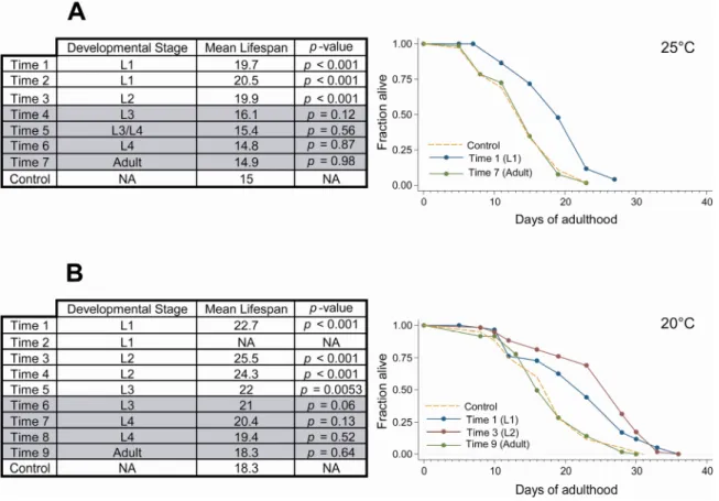
Mitochondrial deficiencies can only increase longevity if caused during development. In this section, I identified the developmental time before which mitochondrial function must be decreased in order to affect lifespan. I found it to be during the L3/L4 stages of post-embryonic development. This suggests that the “decision” to live long in response to mitochondrial dysfunction occurs late in development, just before the final transition to adulthood.
2.2 – Background
As mentioned in section 1.4.3, mitochondrial knock down using RNAi significantly extends longevity, but only if performed during development. Inhibiting mitochondrial function during adulthood has no effect on lifespan (Dillin, Hsu et al. 2002; Rea, Ventura et al. 2007). This supports the notion that the phenotypes observed in these long-lived mitochondrial mutants are somehow regulated. If the increase in lifespan was simply due to a passive process (such as decreased ROS production) one would expect to see some longevity benefit, if only partial, from an adult-only treatment. Bearing this in mind, I was interested in understanding the nature of this developmental regulatory mechanism.
been extensively documented. Namely, with regards to mitochondria there is a several-fold increase in mitochondrial number during the L3 and L4 stages, related to sexual maturation (Tsang and Lemire 2003).
I was interested in finding the important stage for the “decision” regulating longevity in long-lived mitochondrial RNAi-treated animals.
2.3 – Results
In order to identify the “decision” stage for mitochondrial RNAi mutants, I knocked down mitochondrial function at different stages during post-embryonic development and measured longevity of each population (Fig. 4). The knockdowns were achieved using an RNAi clone specific for cyc-1,
which is a component of complex III of the ETC (Dillin, Hsu et al. 2002). The experiment was done at two different temperatures (25°C and 20°C) with similar results. We found that knocking down mitochondrial function after the L3 stage was insufficient to significantly extend longevity (note that we are using a p < 0.01 cut-off for significant differences from control). If we take into account that RNAi by feeding takes ≈ 8-12 hours to decrease RNA levels (Dillin, Hsu et al. 2002; Rea, Ventura et al. 2007), we can estimate the important stage for mitochondrial-mediated longevity to be late L3 or L4. This is consistent with the observation that when animals were exposed to
2.4 – Discussion
Knocking down mitochondrial function during adulthood has no effect in longevity. These experiments show that the decisive stage in the long-life response to mitochondrial dysfunction is L3/L4. This is consistent with previous work showing a big proliferation of mtDNA during this stage of the animals life (Tsang and Lemire 2003), which in turn coincides with reproductive maturation. This energy intensive developmental stage is an optimal candidate for sensing the ability of cells to produce energy, since it is the time where mitochondrial deficiencies would have the most obvious consequences.
Similar findings were published recently (Rea, Ventura et al. 2007), but interestingly where these authors report an “all-or-nothing” effect I saw a gradual decrease in longevity as mitochondrial function was inhibited closer to adulthood. It is interesting to note that when the experiment was done at 20°C, populations in which the RNAi treatment was started as L2s actually lived longer than animals treated from hatching. The reason for this is unclear, however it is possible that starting the treatment too early has detrimental effects. It is also possible that this result was merely experimental variation, since it was not repeated by Rea et. al (Rea, Ventura et al. 2007).
2.5 – Methods
Strains
The strain used in this study was: N2-Bristol (WT)
RNAi feeding at different developmental times
Ahringer RNAi library and confirmed by sequencing. This RNAi clone was picked based on experiments from Dillin et al., 2002. Populations were synchronized as L1s overnight through larval arrest. Animals were moved from a master population exposed to control RNAi, to plates containing bacteria expressing cyc-1 dsRNA at different times through development. Timepoints were previously determined to reflect all different stages of post-embryonic development. Each population’s developmental stage was determined by visual examination prior to transfer to RNAi treatment.
Survival measurements
Chapter III
A Regulated Longevity Response to
Mitochondrial Respiration in
C. elegans
*
*adapted from Cristina, D., Cary, M., Lunceford, A., Clarke, C., Kenyon, C., (2008). A regulated longevity response to mitochondrial dysfunction. PLoS Genetics. (Submitted)
3.1 - Summary
Mitochondrial respiration generates energy in the form of ATP. When respiration is inhibited in C. elegans, rates of behavior and growth are slowed and lifespan is extended. We find that inhibiting respiration increases the expression of genes predicted to protect and metabolically remodel the animal. This pattern of gene expression is reminiscent of the expression profile of long-lived respiration-defective yeast and mammalian cells, the “retrograde response”, suggesting ancient evolutionary conservation. As in yeast, genes switched on in C. elegans mitochondrial mutants extend lifespan, suggesting an underlying evolutionary conservation of mechanism. Mutations in clk-1, which inhibit the synthesis of ubiquinone, do not reduce ATP levels, nevertheless they produce gene expression, longevity, and behavioral phenotypes similar to those produced by inhibiting components of the respiratory chain. We find that knocking down the activities of two similar genes, fsrt-1 and fstr-2, partially suppresses the phenotypes of clk-1 mutants and inhibits the clk-1(-)
phenotypes produced by inhibiting the gene isp-1, which encodes a component of the respiratory chain. Our findings suggest that different types of mitochondrial perturbations activate distinct pathways that converge on similar downstream processes to slow behavioral rates and extend lifespan.
3.2 - Background
The link between mitochondria and aging is fascinating and complex. Mitochondria generate most of the cell’s energy as well as its reactive oxygen species (ROS), and mitochondrial dysfunction can cause disease and accelerate aging. Paradoxically, mitochondrial dysfunction can also increase longevity. Yeast petite mutants, which lack mitochondrial DNA and do not carry out respiration, have an increased replicative lifespan (Kirchman, Kim et al. 1999). In C. elegans, two types of mutations that affect mitochondrial function also increase lifespan. The first type reduces respiration. One such mutant, isp-1(qm150), was identified in an EMS screen for mutants with delayed development and defecation rates. These animals harbor a mutation in an iron-sulfur protein in complex III of the electron transport chain and have reduced rates of oxygen consumption (Feng, Bussiere et al. 2001). In addition, two independent RNA interference (RNAi) longevity screens found that knock-down of genes encoding components of the respiratory chain or ATP synthase decreased ATP production and rates of respiration, reduced behavioral rates and increased lifespan (Dillin, Hsu et al. 2002; Lee, Lee et al. 2003). Mitochondrial RNAi-treated animals are smaller than isp-1 mutants (Dillin, Hsu et al. 2002) (Hekimi and Guarente 2003), implying either a more severe reduction in respiration or, conceivably, a qualitatively different response.
The second type of mitochondrial mutant is exemplified by clk-1
(Lakowski and Hekimi 1996). clk-1 mutants lack a mitochondrial hydroxylase necessary for synthesis of ubiquinone, a prenylated benzoquinone lipid required for shuttling electrons from complexes I and II to complex III during respiration (Ewbank, Barnes et al. 1997). In yeast, the
clk-1 homologue COQ7 is necessary for respiration, and coq7 mutants are unable to grow on non-fermentable carbon sources. In contrast, C. elegans clk-1 mutants are not only viable, but they have close to normal levels of respiration and ATP (Braeckman, Houthoofd et al. 1999; Felkai, Ewbank et al. 1999). clk-1 mutants compensate for the lack of endogenous ubiquinone, Q9 (the subscript refers to the number of isoprene units) with bacterial Q8, provided in their diet (Jonassen, Larsen et al. 2001; Larsen and Clarke 2002). In the absence of clk-1, the animals accumulate the Q9 precursor demethoxyubiquinone (DMQ9). There is some debate over what role DMQ9 plays in the clk-1 phenotypes, but the data suggest that they are likely due to absence of Q9 (Larsen and Clarke 2002; Branicky, Nguyen et al. 2006; Padilla, Jonassen et al. 2004). A role for clk-1 in lifespan determination may be conserved evolutionarily, since mice with reduced levels of Mclk-1 are also long lived (Liu, Jiang et al. 2005).
paraquat (Lee, Lee et al. 2003). Likewise, little is known about the mechanism by which clk-1 mutations, which have very small effects on respiration, extend lifespan. Overexpression of clk-1 shortens lifespan and increases movement rates in C. elegans (Felkai, Ewbank et al. 1999) suggesting that whatever the mechanism, its influence on longevity and rates of living is rate-limiting in the animal.
In this study, we have asked how mitochondrial mutations affecting respiration and ubiquinone biosynthesis might extend the lifespan of C. elegans. It seems possible that these mutations extend lifespan in a similar way, since their effects on growth and behavioral rates are so similar. In yeast, loss of mitochondrial DNA triggers a robust transcriptional response. This change in gene expression is known as a “retrograde response”, so named because it implies a reversal in the normal direction of information flow between the mitochondria and nucleus (Liao and Butow 1993; Kirchman, Kim et al. 1999; Parikh, Morgan et al. 1987). The genes expressed during the retrograde response lead to a metabolic remodeling of the cell, heat-shock resistance, and increased mitochondrial biogenesis (Epstein, Waddle et al. 2001; Traven, Wong et al. 2001; Butow and Avadhani 2004; Miceli and Jazwinski 2005). The retrograde response has been shown to be required for the increased longevity of these so-called yeast “petites”. Thus, lifespan extension in these yeast cells is actively regulated, and not simply a passive consequence of decreased respiration. Yeast are not the only cells that exhibit a retrograde response to loss of respiration. A similar response has been observed in cultured mammalian cells when mitochondrial DNA is depleted using ethidium bromide (Miceli and Jazwinski 2005).
Consistent with this possibility, C. elegans isp-1 mutants have increased levels of expression of at least one protective gene, the superoxide dismutase sod-3 (Feng, Bussiere et al. 2001). In this study, we carried out microarray analysis of C. elegans mitochondrial mutants to better understand the molecular basis of their longevity.
3.3 - Results
3.3.1 -
clk-1
mutants exhibit a conserved retrograde response
9074 and 30170 encompass genes involved in amino acid metabolism; GO categories 6825 and 5375 include genes involved in Cu transport; GO category 6629 includes genes involved in lipid metabolism; GO category 46040 includes genes involved in nucleotide metabolism; and GO categories 4499 and 16758 include genes involved in xenobiotic response and maintenance of cellular redox state.
When we looked at the level of individual genes, we observed up-regulation of genes encoding enzymes required for glycolysis, such as GPD-2, GPD-3, (gliceraldehyde 3-phosphate dehydrogenase), T05D4.1 (Aldolase A homologue), and LDH-1 (lactate dehydrogenase). GEI-7, which is an enzyme necessary for the glyoxylate cycle in worms (see section 3.4.1), was also up-regulated. We also observed increased expression of an isocitrate dehydrogenase C30G12.2, likely involved in the Krebs cycle, and other alcohol dehydrogenases (dhs-29, dhs-3) that could potentially act in anaplerotic pathways. We found increased expression of proteins involved in oxidative phosphorylation such as asg-2 (subunit of ATP synthase complex) and F17A9.4 (NADH oxidoreductase). Also, several genes coding for enzymes involved in amino acid and nucleotide metabolism showed increased expression in clk-1 mutants. There was also a significant increase in enzymes involved in cellular detoxification, including UDP-glycosyl transferases (UGT-53, UGT-13, UGT-43, UGT-6, UGT-39), gluthathione S-transferases (GST-4, GST-13, GST-36), superoxide dismutase (SOD-3), flavin-containing monooxygenases (FMO-1, FMO-3) and other gene classes potentially involved in xenobiotic metabolism (cytochrome P450s, alcohol dehydrogenases and ABC transporters).
software to the most differentially expressed genes reported in those studies and compared the results to those of clk-1 mutants. Among the top ranking GO categories (p < 0.1), we observed a remarkable degree of similarity (p < 0.001) between the clk-1 and yeast petite BiNGO results (Table 1).
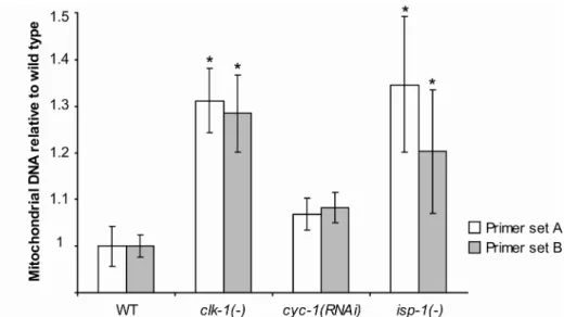
Figure 5 - Mitochondrial DNA quantification. Percent increase in total mitochondrial DNA relative to wild type, as measured by qPCR. Data are shown for two different mitochondrial primer pairs across five independent biological repeats. (*) indicates significance of p < 0.05 after Bonferroni multiple comparison correction. Error bars are ± SEM. clk-1(-) primer set A: m (mean) = 1.31 ± 0.07, primer set B: m = 1.29 ± 0.08; cyc-1(RNAi) primer set A: m = 1.07 ± 0.03, primer set B: m = 1.08 ± 0.03; isp-1(-) primer set A: m = 1.35 ± 0.14, primer set B: m = 1.21 ± 0.13.
3.3.2 -
fstr-1/2
and
aqp-1
contribute to the increased longevity of
clk-1
mutants
To identify genes necessary for the increased longevity of clk-1(-)
measured lifespan. Out of the initial list of 75, we collected lifespan data for 63 genes over two independent trials. We established a significance cut-off of p < 0.05 and selected RNAi clones that decreased clk-1 longevity significantly in both trials or were statistically significant in one experiment and showed a decrease of at least 5% in the other (a 5% decrease in overall lifespan corresponds to an ~25% decrease in the lifespan extension produced by clk-1 mutation). We retested the positives in clk-1(-) and wild-type animals for effects on longevity. Out of 63 RNAi clones tested, only two decreased clk-1 mutant longevity in all three trials (Fig. 6). Neither of these clones significantly shortened wild-type lifespan, suggesting that they may play a role specifically in clk-1 mutant lifespan. One of these clones corresponded to aqp-1, which encodes a glycerol channel (Huang, Lamitina et al. 2007). aqp-1 RNAi decreased the lifespan extension that would normally be produced by clk-1 mutations from 26% to 7%, and did not affect wild-type longevity in two separate experiments. Interestingly, aqp-1 (also called dod-4) has already been shown to contribute to the long lifespan of
daf-2 insulin/IGF-1-receptor mutants (Murphy et al. 2003). The other RNAi clone, corresponding to a gene we call fstr-1 (for “faster”, also known as gfi-1) decreased the lifespan extension produced by clk-1 mutations from 26% to 3% (close to total suppression on this trial) while not affecting wild-type lifespan in two separate experiments. The effects of aqp-1 RNAi and fstr-1
RNAi on the longevity of clk-1 mutants were tested three times, with consistent results, although the extent of suppression varied between experiments. We examined the genome for the possibility that fstr-1 RNAi might cross-inhibit another gene, and found that the RNAi clone was likely to knock down a close homolog (with 98% inferred DNA sequence similarity) located next to fstr-1 that did not exhibit clk-1-dependent regulation in our microarray analysis. We call this gene fstr-2, and henceforth we refer to their combined functions, as inferred from RNAi, as
Figure 6 - fstr-1/2 and aqp-1 contribute to the increased longevity of clk-1
mutants A. aqp-1 RNAi significantly decreased the lifespan extension produced by
13.8 days. In two other trials, fstr-1/2 RNAi reduced the lifespan increase produced by clk-1 mutations from 33% to 27% (not statistically significant) and from 17% to 0%.
3.3.3 -
fstr-1/2
knockdown increases the behavioral rates of
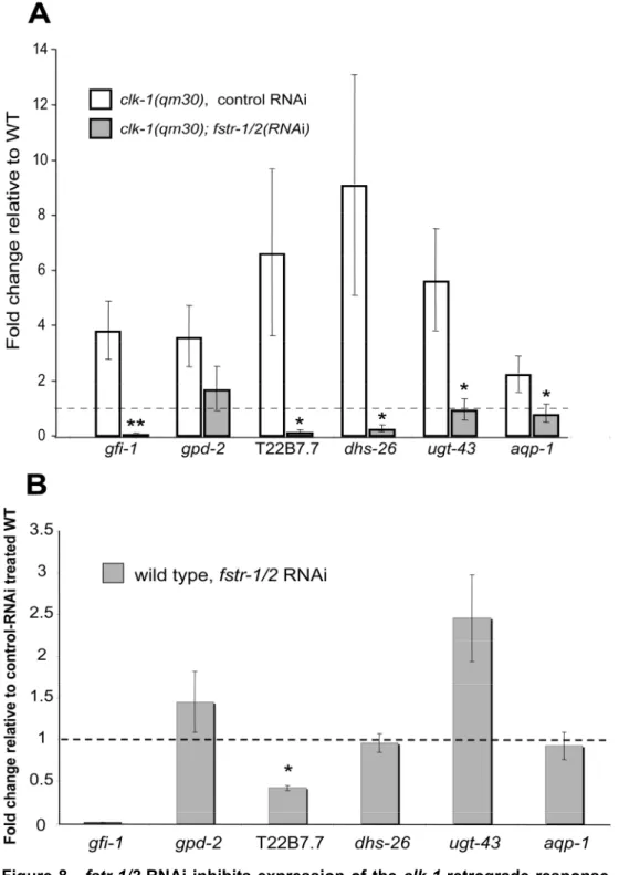
clk-1
mutants
In addition to increased longevity, the most striking phenotypes of mitochondrial mutants are their decreased behavioral rates. In principle, these rates could decrease as a direct consequence of impaired mitochondrial function. Alternatively, it is possible that their slowed behavioral rates reflect a regulated response to mitochondrial perturbation; for example, to conserve energy. To look for genes that slow the behaviors of clk-1 mutants, we inhibited the top 100 up-regulated genes from microarrays of L4 and pre-fertile adults using RNAi and measured time it took for L1 larvae to develop to adulthood. We found that knockdown of fstr-1/2 in clk-1 mutants consistently increased the rate of growth to adulthood (Fig. 7A). This phenotype was most striking when the animals were examined 75-80 hours after hatching. At this time, no control animals had reached adulthood, whereas 95-100% of the fstr-1/2 RNAi treated animals were adults. fstr-1/2 RNAi treatment also increased the behavioral rates of
clk-1(-) animals, as measured by thrashing and pumping (Fig 7B and 7C). These effects were not observed in wild type; in fact, in wild type, knock-down of these genes had the opposite effect, slowing development and decreasing rates of thrashing and pumping.
mutant pattern of ubiquinone species in clk-1 mutants subjected to fstr-1/2
Figure 7 - fstr-1/2 RNAi speeds up clk-1(-) animals.A. Time to adulthood. clk-1
every experiment. WT subjected to control RNAi: 47.4 ± 1.6 hours; WT subjected to
fstr-1/2 RNAi: 53.2 ± 1.7 hours; clk-1(-) mutants subjected to control RNAi: 82.2 ± 1.2 hours; clk-1(-) mutants subjected to fstr-1/2 RNAi: 72 ± 1.7 hours. B. Boxplots illustrating thrashing rates measured on day 3 of adulthood. fstr-1/2 RNAi treatment significantly reduced the average thrashing rate of wild-type animals and significantly increased the average thrashing rate of clk-1(-) mutants. WT subjected to control RNAi: 115.6 ± 3.1 thrashes/min; WT subjected to fstr-1/2 RNAi: 97.5 ± 4.3 thrashes/min; clk-1(-) mutants subjected to control RNAi: 61.2 ± 5 thrashes/min;
clk-1(-) mutants subjected to fstr-1/2 RNAi: 78.9 ± 5.7 thrashes/min. C. Boxplots illustrating pumping rates. fstr-1/2 RNAi decreased the average pumping rate of wild type and increased the pumping rate of clk-1(-) mutants. WT subjected to control RNAi: 273.3 ± 41.1 pumps/min; WT subjected to fstr-1/2 RNAi: 231.8 ± 24.7 pumps/min; clk-1(-) mutants subjected to control RNAi: 188.5 ± 41 pumps/min; clk-1(-) mutants subjected to fstr-1/2 RNAi: 238.9 ± 36.4 pumps/min. In figures 3A, 3B and 3C error bars depict SEM; * depicts a significance of p < 0.05 when compared to controls, ** depicts significance of p < 0.008 when compared to controls, ¥¥ depicts significance of p < 0.008. P < 0.008 is the cut-off set by the Bonferroni correction for multiple comparisons. D. HPLC analysis of quinone content. The chromatograms show a representative run of three independent experiments for each of the different conditions (WT subjected to control RNAi, WT subjected to
fstr-1/2 RNAi, clk-1(-) mutants subjected to control RNAi and clk-1(-) mutants subjected to fstr-1/2 RNAi). The Q9 peak is absent from clk-1(-) animals and
instead the intermediate DMQ9 is detected. fstr-1/2 RNAi had no effect in Q9 levels
in wild-type or clk-1(-) mutants, in clk-1 mutants Q9 levels remained below detection
threshold.