Investigations on DNA damage and frequency of micronuclei
in occupational exposure to electromagnetic fields (EMFs)
emitted from video display terminals (VDTs)
NK Lakshmi
1, R Tiwari
1, SC Bhargava
2and YR Ahuja
3 1UGC Research Unit, Bhavan’s New Science College, Hyderabad, Andhra Pradesh, India.
2
Department of Electrical and Electronic Engineering, Sree Nidhi
Institute of Science and Technology, Hyderabad, Andhra Pradesh, India.
3
Genetics Unit, Vasavi Medical and Research Centre, Hyderabad, Andhra Pradesh, India.
Abstract
The potential effect of electromagnetic fields (EMFs) emitted from video display terminals (VDTs) to elicit biological response is a major concern for the public. The software professionals are subjected to cumulative EMFs in their oc-cupational environments. This study was undertaken to evaluate DNA damage and incidences of micronuclei in such professionals. To the best of our knowledge, the present study is the first attempt to carry out cytogenetic investiga-tions on assessing bioeffects in personal computer users. The study subjects (n = 138) included software profession-als using VDTs for more than 2 years with age, gender, socioeconomic status matched controls (n = 151). DNA damage and frequency of micronuclei were evaluated using alkaline comet assay and cytochalasin blocked micronucleus assay respectively. Overall DNA damage and incidence of micronuclei showed no significant differ-ences between the exposed and control subjects. With exposure characteristics, such as total duration (years) and frequency of use (minutes/day) sub-groups were assessed for such parameters. Although cumulative frequency of use showed no significant changes in the DNA integrity of the classified sub-groups, the long-term users (> 10 years) showed higher induction of DNA damage and increased frequency of micronuclei and micro nucleated cells.
Key words:electromagnetic fields (EMFs), video display terminals (VDTs), comet assay, DNA damage, micronuclei. Received: March 25, 2009; Accepted: July 22, 2009.
Introduction
Electromagnetic technologies like personal comput-ers and televisions have brought social and economic bene-fits to large sections of society. At the same time, their biological effects are raising concern due to the electro-magnetic radiation emitted from video display terminals (VDTs). The video display units comprising cathode ray tube have a large number of applications in all spheres of life like communication and broadcasting, space research and medicine. There is a widespread apprehension that ex-cessive exposure to these electromagnetic fields (EMFs) may hamper fundamental biological processes in the hu-man body.
The potential effects of EMFs on human health vary widely depending on the frequency and intensity of the fields. In spite of years of research, there is still ongoing discussion whether radiofrequency (RF)-EMFs and extre-mely low frequency (ELF) -EMFs could induce any
physi-ologically relevant effects (Krewskiet al.2007). The stud-ies envisaging the possible health effects of EMF exposure at such field ranges have mainly focused on biological end-points such as DNA damage (Lai and Singh 1996; Hooket al.2004; Sunet al.2006; Yaoet al.2008), increase in free radicals (Ticeet al.2002; Bolandet al.2002; Ferreiraet al. 2006; Simkoet al.2006), induction of heat shock proteins (Lantowet al., 2006; Sanchezet al., 2007; Valbonesiet al. 2008) and cellular alterations (Kimet al.2008; Schwarzet al.2008).
Relatively less attention has been paid to health haz-ards from exposure to radiation in the intermediate EMFs, including the radiation emitted from personal computer cathode ray tube monitors, in the frequencies of 20 kHz. The workers are subjected to cumulative EMFs in their oc-cupational environments comprising EMFs of 50 Hz powerline frequencies as well as 15-25 kHz RF-EMFs. Epi-demiological studies have suggested that occupational ex-posure to VDTs is associated with increased risk of various health effects, particularly reproductive disorders, depres-sion and cancer. However, the experimental and epidemio-logical data from the intermediate frequency (IF) range are Send correspondence to Tiwari Ravindra. UGC Research Unit,
Bha-van’s New Science College, Narayanguda, Hyderabad, 500029 Andhra Pradesh, India. E-mail: [email protected].
sparse. Therefore, assessment of acute health risks in the IF range is currently based on known hazards at lower fre-quencies and higher frefre-quencies.
The conflicting results have raised attention for fur-ther research on bioeffects of EMF fields taking into ac-count exposure levels and duration. Apparently very few studies have documented genotoxicity in personal com-puter users. The present investigation reports DNA damage and chromosomal damage in peripheral blood lymphocytes of the exposed populations by alkaline comet assay or Sin-gle Cell Gel Electrophoresis (SCGE) and cytochalasin blocked micronuclei test (CBMN). To the best of our knowledge, there is no report from India on the genotoxic potential of occupational exposure to VDTs. Hence, this study was carried out to investigate the effect of occupa-tional EMFs exposure on DNA damage and frequency of micronuclei in peripheral blood leukocytes of the VDT us-ers. Analysis of the data was carried for all the exposed sub-jects pooled together as well as in sub-groups based on the duration and intensity effect of exposure.
Subjects and Methods
Participants
The study included 138 subjects occupationally ex-posed to video display terminals for more than two years. The exposed subjects were screened along with 151 age, sex and diet matched controls with similar socioeconomic status. The exposed subjects were software professionals from software companies and consultancies in Hyderabad, India. In the detailed questionnaire, duration of exposure (years), frequency of exposure in hours/day were noted. Age, diet, gender, recent infection, smoking, drinking alco-hol and exercise were also recorded for both exposed and unexposed populations.
Sampling
After taking informed consent, 2 mL peripheral blood was collected from each participant by venepuncture into heparinised disposable syringe and placed in ice to prevent exogenous damage. The sample was processed in the labo-ratory within an hour of collection for assessing DNA dam-age and micronucleus frequency.
Chemicals
The sources of chemicals were as follows: Agarose [low melting point (20 °C) and regular melting point (35 °C)], sodium lauryl sarcosinate, Triton X-100, silver ni-trate (all from Sigma-USA); tungstosilicic acid(Koch-Light Laboratories, UK); sodium chloride, sodium hydrox-ide, potassium chlorhydrox-ide, TRIS, EDTA, potassium dihy-drogen phosphate and sodium phosphate dibasic (all from Glaxo, Mumbai, India); zinc sulphate and ammonium ni-trate (Fisher, Madras, India); thiobarbituaric acid, butylated hydroxyl toluene, sulphosalicyclic acid and N-1-napthyl
ethylene diamine dihydrochloride, potassium chloride, me-thanol, acetic acid (all from SD Fine Chemicals, Mumbai, India); RPMI-1640 media - Himedia, Phytohaemagglutinin – Gibco, Penicillin, Streptomycin - Himedia. Phytohae-magglutinin (PHA), Cytochalasin B (all from Sigma, USA), DMSO - (Merck, Germany).
Alkaline comet assay
Alkaline comet assay or single cell gel electrophore-sis (SCGE) after Singhet al.(1998) was used to study DNA damage.
On a clean, dry, plain slide 100mL of 0.75% normal melting agarose (NMA) prepared in phosphate buffered sa-line (PBS) was layered. These precoated slides were dried at 37 °C. On top of this layer, 30mL of whole blood, mixed with 70mL of 0.5% low melting agarose (LMA) prepared in PBS was layered. The third layer consisted of 100mL of LMA. The slides were incubated in cold lysis buffer (2.5 M NaCl, 100 mM Na2EDTA, 10 mM Tris; 1% sodium lauryl
sarcosinate; 1% Triton X-100 and 10% DMSO added fresh) at 4 °C overnight.
The slides were removed from the lysing solution and placed side by side in a horizontal electrophoretic unit. The slides were completely immersed in freshly prepared alka-line electrophoretic buffer (1 mM Na2EDTA and 300 mM
NaOH; pH 13) for 30 min to facilitate the DNA unwinding and expression of alkali labile sites. After alkali treatment, the electrophoresis was carried out for 30 min at 300 mA and 0.67 V/cm. The slides were carefully lifted from the buffer and gently washed with neutralizing buffer (0.4 M Tris buffer, pH 7.5). The slides were then washed with dis-tilled water and air dried.
The air dried slides were immersed in the fixing solu-tion (15% w/v trichloroacetic acid, 5% w/v zinc sulphate and 5% w/v glycerol) for 10 min and washed gently with double distilled water several times. For staining, 32 mL of staining solution A (5% w/v Na2CO3) was mixed with
68 mL of staining solution B (0.02% w/v NH4NO3, 0.02%
w/v AgNO3, 0.1% w/v tungstosilicic acid and 0.05% v/v
formaldehyde) and poured over the slides so as to cover the slides uniformly. This step was repeated until with a fresh mixture of staining solution a grayish colour developed on the slides. To stop staining, the slides were immersed in stopping solution (1% acetic acid) for 5 min, washed with double distilled water and air dried.
For visualization of DNA damage, a bright field, transmission light microscope (Leitz) was used at 400x magnification. Comet tail length was measured, using an ocular micrometer fitted in the eyepiece, in 200 cells per slide (in duplicate). Mean comet tail length, which is an es-timate of DNA damage, was calculated for each sample.
Micronuclei (MN) were observed in cytokinesis-blocked cells using cytochalasin B (Cyt-B) following the method suggested by Fenech and Morley (1985).
About 0.2 mL of PHA was added to 5 mL of RPMI 1640 medium using 1 mL syringe. 15 drops of blood was added to each vial. Samples were initiated in duplicates. The culture vials were incubated for 72 h at 37 °C and shaken for proper mixing. Cyt-B (6mg/mL) was added at 44thhour after initiation of culture and incubated further for another 28 h at 37 °C and then cultures were harvested.
The cultures were centrifuged at 1000 rpm for 5-10 min. The supernatant was discarded and 5 mL of prewarmed hypotonic solution (0.56%) was added to the pellet drop by drop slowly on by vortexing and incubated for 10 min for 37 °C. Then the vials were centrifuged for a minute and supernatant was discarded. Cells were fixed in 5 mL of fixative (3:1 methanol: acetic acid) followed by two more changes of fixative.
The slides were prepared in triplicate by gently drop-ping the cell suspension onto the precleaned slides and flame dried. Slides were stained with 2% Giemsa for 10 min, rinsed and air dried.
To determine the MN yield, nearly 1000 binucleated (BN) cells were scored for each experimental condition un-der magnifications of 400x and finally 1000x from 2 coded slides/culture. Identification of MN was according to the criteria summarized by Countryman and Heddle (1976). Routinely on average ~2000 BN cells were scored for the presence of MN in each subject and mean values of the re-sults were calculated.
Statistical analysis
The slides were coded during processing and decoded at the time of statistical analysis. For statistical evaluation, observations on each parameter for each group were pooled and mean±SD was calculated. Student’s t-test (paired and unpaired comparisons) and one way ANOVA were per-formed to evaluate various differences. Multiple regression analysis was done to study the effects of confounding
fac-tors and correlation analysis was carried out to adjudge the sensitivity of parameters used. Statistical software SPSS 15 was used to carry out statistical analysis.
Results
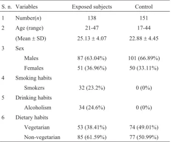
The general characteristics of the study group and controls are shown in Table 1. The mean (±SD) duration of VDT use was 7 (±4.8) years with a mean (±SD) cumulative frequency of 389 (±147) minutes per day.
The results of basal DNA damage assessed by alka-line comet assay in terms of mean comet tail length±SD are summarized in Table 2. Independentttest showed no sig-nificant difference in the mean comet tail length values of exposed and controls. The results of CBMN assay on binu-cleated cells, percentage micronuclei (%MN) and percent-age micronucleated cells (%MNC) of the exposed groups are shown in Table 3. Overall, there was no significant dif-ference in the frequency of micronuclei between the ex-posed subjects and the controls (Table 3). However, results Table 1- General characteristics of the study group and controls.
S. n. Variables Exposed subjects Control
1 Number(n) 138 151
2 Age (range) 21-47 17-44
(Mean±SD) 25.13±4.07 22.88±4.45
3 Sex
Males 87 (63.04%) 101 (66.89%)
Females 51 (36.96%) 50 (33.11%)
4 Smoking habits
Smokers 32 (23.2%) 0 (0%)
5 Drinking habits
Alcoholism 34 (24.6%) 0 (0%)
6 Dietary habits
Vegetarian 53 (38.41%) 74 (49.01%)
Non-vegetarian 85 (61.59%) 77 (50.99%)
Table 2- Mean±SD values of DNA damage in exposed and control subjects.
Parameters Exposed subjects Control t value p value
N Mean±SD N Mean±SD
Comet tail length (arbitrary units) 138 3.76±1.38 151 3.69±1.13 1.895 0.061NS
NS
: Non-significant at 5%.
Table 3- Mean±SE values of frequency of micronuclei in exposed and control subjects.
Groups Sample size Number of BN cells Total number of micronuclei Mean±SE of MN cells (%) Mean±SE of MN (%)
IF-EMF 34 35053 413 1.16±0.48NS 1.39±0.63NS
Control 60 64026 661 1.04±0.52NS 1.24
±0.67NS
NS
from one-way ANOVA revealed significant differences in DNA damage and incidence of micronuclei among sub-groups based on duration of exposure (years) of exposed subjects (Table 4). The significant mean comet tail length and incidences of micronuclei was observed in sub-group having duration of exposure more than 10 years. The two sub-groups based on frequency of exposure (< 420 min/day and > 420 min/day) had no significant difference in damage levels (Table 5).
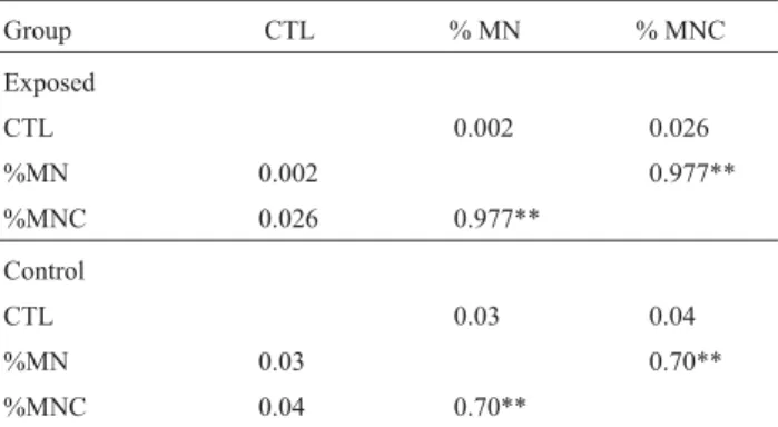
Pearson’s coefficient of correlation was carried out be-tween different parameters to assess the extent of relation-ship between the endpoints of comet assay and CBMN assay (Table 6). The mean comet tail length and frequency of micronuclei showed no significant correlation in the exposed as well the control subjects. Of the various confounding fac-tors studied, no significant effect could be seen in multiple regression analysis with respect to gender, age and habits.
Discussion
Non-panel video display screens of computer moni-tors produce significant EMFs despite improvements in
technology over the last decade or so. In India presently there is a boom in Information Technology (IT) and Busi-ness Process Outsourcing centers. The IT sector witBusi-nessed considerable activity since 2004 including a ramping up of operations by major multinational corporations. Apart from the powerline frequencies (ELF-EMFs) the software em-ployees are exposed to EMFs emitted from personal com-puters.
The workers are subjected to cumulative EMFs in their occupational environments. To the best of our knowl-edge, the present study is the first attempt to carry out mul-tiple markers on assessing bioeffects in subjects occupa-tionally exposed to Cathode Ray Tube (CRT) from personal computers. The aim of our study was to investi-gate genotoxicity in workers occupationally exposed to CRT VDTs through the induced DNA damage and micro-nuclei in leukocytes and lymphocytes respectively.
Overall, the results on DNA damage and micronuclei frequency showed no significant differences. With expo-sure characteristics, such as total duration (years) and fre-quency of use (minutes/day), sub-groups were also as-sessed for such parameters. The long-term users (> 10 years) showed higher induction of DNA damage and in-creased frequency of micronuclei and micronucleated cells. The cumulative frequency of use showed no significant changes in the DNA integrity between the classified sub-groups.
The subjective symptoms reported by the PC usage include predominantly headache, followed by sleepless-ness and neck pain. Duration and intensity was not signifi-cant predicting factors for the reported symptoms. Very fewin vivostudies have directly evaluated the cytogenetic damage in computer workers. Carbonariet al.(2005) indi-cated significant cytogenetic damage by micronuclei assay in computer workers, reinforcing the data obtained in our study. Estecio and Silva (2002) also evaluated possible nu-clear alterations in microcomputer’s workers and found that exposed individuals had two times more chromosomal aberrations in cultured lymphocytes than control individu-als. The rationale behind such findings have been proposed to be the genotoxic influences of EMFs may be through in-Table 4- DNA damage and frequency of micronuclei in relation to
dura-tion of exposure to VDTs.
Observation Group based on duration of exposure (years)
t value p value
1-6 > 7
CTL N 73 65 21.96 < 0.001
M 3.15±0.24 4.09±0.26
%MNC N 20 14 28.39 < 0.001
M 1.00±0.19 2.73±0.15
%MN N 20 14 5.80 < 0.001
M 1.19±0.24 1.63±0.18
CTL-comet tail length, M micronuclei, MNC- micronucleated cells, N-sample size, M-mean±SE.
Table 5- DNA damage and incidences of micronuclei in relation to
fre-quency of exposure (minutes/day) to VDTs.
Observation Group based on frequency of exposure (minutes/day)
t value
p value
A 180-420
B 420-720
CTL N 64 74 1.259 0.210
Mean±SE 3.5991±0.16 3.8914±0.17
%MNC N 20 14 1.177 0.249
Mean±SE 1.2382±0.11 1.0474±0.12
%MN N 20 14 1.026 0.317
Mean±SE 1.5085±0.14 1.2526±0.16
CTL-comet tail length, M micronuclei, MNC- micronucleated cells, N-sample size, M-mean±SE.
Table 6- Pearson’s correlation analysis in exposed and control subjects.
Group CTL % MN % MNC
Exposed
CTL 0.002 0.026
%MN 0.002 0.977**
%MNC 0.026 0.977**
Control
CTL 0.03 0.04
%MN 0.03 0.70**
%MNC 0.04 0.70**
creased free radical activity or acceleration of electron transfer in different enzymes and proteins.
In the present study, no thermal effects of EMFs could be seen. However, Gangi and Johansson (2000) de-tected an increased incidence of skin and central nervous system (CNS) alterations among microcomputer workers. Andersson (1996) reported that female workers exposed to VDU presented obstetric complications, besides skin, ocu-lar and CNS diseases.
Thus we conclude that this epidemiological study on occupational EMF exposure was significant to provide pre-liminary evidence on health risk along with the pertinent reliable parameters carried out as biomarkers. The long-termed effect also implicated the need for close monitoring of health hazards associated in long-term VDT users.
Acknowledgments
The authors acknowledge the financial assistance from Union Grants Commission India.
References
Andersson B (1996) A cognitive-behavioral treatment of patients suffering from “electric hypersensitivity”. Subjective ef-fects and reactions in a double-blind provocation study. J Occup Environ Med 38:752-758.
Boland A, Delapierre D, Mossay D, Dresse A and Seutin V (2002) Effect of intermittent and continuous exposure to electro-magnetic fields on cultured hippocampal cells. Bioelectro-magnetics 23:97-105.
Carbonari K, Goncalves L, Roth D, Moreira P, Fernández R and Martino-Roth MG (2005) Increased micronucleated cell fre-quency related to exposure to radiation emitted by computer cathode ray tube video display monitors. Genet Mol Biol 28:469-474.
Countryman PI and Heddle JA (1976) The production of micro-nuclei from chromosome aberrations in irradiated cultures of human lymphocytes. Mutat Res 41:321-332.
Estecio MRH and Silva AE (2002) Chromosome abnormalities caused by computer video display monitor’s radiation. Rev Saude Publica 36:330-336.
Fenech M and Morley AA (1985) Measurement of micronuclei in lymphocytes. Mutat Res 147:29-36.
Ferreira AR, Knakievicz T, Pasquali MA, Gelain DP, Dal-Pizzol F, Fernández CE, de Salles AA, Ferreira HB and Moreira JC (2006) Ultra high frequency electromagnetic field irradia-tion during pregnancy leads to an increase in erythrocytes micronuclei incidence in rat offspring. Life Sci 80:43-50.
Gangi S and Johansson O (2000) A theoretical model based upon mast cells and histamine to explain the recently proclaimed sensitivity to electric and/or magnetic fields in humans. Med Hypoth 54:663-671.
Hook GJ, Zhang P and Lagroye I (2004) Measurement of DNA damage and apoptosis in Molt-4 cells afterin vitroexposure to radiofrequency radiation. Radiat Res 161:193-200. Kim TH, Huang TQ, Jang JJ, Kim MH, Kim HJ, Lee JS, Pack JK,
Seo JS and Park WY (2008) Local exposure of 849 MHz and 1763 MHz radiofrequency radiation to mouse heads does not induce cell death or cell proliferation in brain. Exp Mol Med 40:294-303.
Krewski D, Glickman BW, Habash RW, Habbick B, Lotz WG, Mandeville R, Prato FS, Salem T and Weaver DF (2007) Re-cent advances in research on radiofrequency fields and health: 2001-2003. J Toxicol Environ Health 10:287-318. Lai H and Singh NP (1996) Single- and double-strand DNA
breaks in rat brain cells after acute exposure to radiofrequen-cy electromagnetic radiation. Int J Radiat Biol 69:513-521. Lantow M, Lupke M, Frahm J, Mattsson MO, Kuster N and
Simko M (2006) ROS release and Hsp 70 expression after exposure to 1800 MHz radiofrequency electromagnetic fields in primary human monocytes and lymphocytes. Radiat Environ Biophys 45:55-62.
Sanchez S, Haro E, Ruffié G, Veyret B and Lagroye I (2007)In vi-tro study of the stress response of human skin cells to GSM-1800 mobile phone signals compared to UVB radia-tion and heat shock. Radiat Res 167:572-580.
Schwarz C, Kratochvil E and Pilger A (2008) Radiofrequency electromagnetic fields (UMTS, 1,950 MHz) induce geno-toxic effectsin vitroin human fibroblasts but not in lympho-cytes. Int Arch Occup Environ Health 81:755-767. Simko M, Hartwig C, Lantow M, Lupke M, Mattsson MO,
Rahman Q and Rollwitz J (2006) Hsp 70 expression and free radical release after exposure to non-thermal radio-frequen-cy electromagnetic fields and ultrafine particles in human Mono Mac 6 cells. Toxicol Lett 161:73-82.
Singh NT, McCoy NT and Tice RR (1998) A simple technique for quantification of low levels of DNA in individual cells. Exp Cell Res 517:184-191.
Sun LX, Yao K and He JL (2006) Effect of acute exposure to mi-crowave from mobile phone on DNA damage and repair of cultured human lens epithelial cellsin vitro. Zhonghua Lao Dong Wei Sheng Zhi Ye Bing Za Zhi 24:465-467. Tice RR, Hook GG and Donner M (2002) Genotoxicity of
radio-frequency signals. I. Investigation of DNA damage and micronuclei induction in cultured human blood cells. Bio-electromagnetics 23:113-126.
Valbonesi P, Franzellitti S, Piano A, Contin A, Biondi C and Fabbri E (2008) Evaluation of HSP70 expression and DNA damage in cells of a human trophoblast cell line exposed to 1.8 GHz amplitude-modulated radiofrequency fields. Radiat Res 169:270-279.
Yao K, Wu W, Wang K, Ni S, Ye P, Yu Y, Ye J and Sun L (2008) Electromagnetic noise inhibits radiofrequency radiation-in-duced DNA damage and reactive oxygen species increase in human lens epithelial cells. Mol Vis 14:964-969.
Associate Editor: Catarina S. Takahashi