Increased micronucleated cell frequency related to exposure to radiation
emitted by computer cathode ray tube video display monitors
Karina Carbonari
1, Luciane Gonçalves
1, Daniela Roth
2, Patrick Moreira
3, Ricardo Fernández
3and Maria da Graça Martino-Roth
1,31
Universidade Federal de Pelotas, Departamento de Zoologia e Genética, Pelotas, RS, Brazil.
2Monash University, Department of Pharmaceutical Biology and Pharmacology, Melbourne,
Victoria, Australia.
3
Universidade Católica de Pelotas, Escola de Medicina, Pelotas, RS, Brazil.
Abstract
It is well recognized that electromagnetic fields can affect the biological functions of living organisms at both cellular and molecular level. The potential damaging effects of electromagnetic fields and very low frequency and extremely low frequency radiation emitted by computer cathode ray tube video display monitors (VDMs) has become a concern within the scientific community. We studied the effects of occupational exposure to VDMs in 10 males and 10 females occupationally exposed to VDMs and 20 unexposed control subjects matched for age and sex. Genetic damage was assessed by examining the frequency of micronuclei in exfoliated buccal cells and the frequency of other nuclear abnormalities such as binucleated and broken egg cells. Although there were no differences regarding binucleated cells between exposed and control individuals our analysis revealed a significantly higher frequency of micronuclei (p < 0.001) and broken egg cells (p < 0.05) in individuals exposed to VDMs as compared to unexposed. We also found that the differences between individuals exposed to VDMs were significantly related to the sex of the individuals and that there was an increase in skin, central nervous system and ocular disease in the exposed individuals. These preliminary results indicate that microcomputer workers exposed to VDMs are at risk of significant cytogenetic damage and should periodically undergo biological monitoring.
Key words:micronucleus test, mutagenicity, electromagnetic fields. Received: October 25, 2004; Accepted: February 22, 2005.
Introduction
Constant advances in the electronics industry and the increasing use of electrical appliances have resulted in con-sumers being increasingly exposed to the effects of ex-tremely low frequency electromagnetic fields (ELF-EMF). Concerns have been raised (Ivancsitset al, 2002) about the possible health hazards of chronic exposure to extremely low frequency (ELF) and very low frequency (VLF) elec-tromagnetic radiation from various appliances including computer cathode ray tube (CRT) video display monitors (VDMs). Over the past few decades several studies (Wertheimer, Leeper, 1979; Li and Theriault, 1997) have demonstrated that exposure to ELF-EMF may be related to an increased risk of certain types of cancer, including leu-kemia, central nervous system cancer, and lymphoma, al-though other studies (Verkasaloet al., 1993; Schreibneret al., 1993) have failed to find such association.
Chromosomal mutation is a key event in carcinogenesis (Fenech, 2000) and the study of DNA dam-age at the chromosome level is an essential part of genetic toxicology. Micronuclei (MN) are acentric fragments or complete chromosomes which fail to attach to the mitotic spindle during cytokinesis and are normally excluded from the nuclei (Maluf and Erdtmann, 2000). The quantification of micronuclei by the micronucleus test (MT) is an impor-tant non-invasive cytogenetic method which is a good indi-cator of chromosome mutation (Majeret al., 2001) and has been extensively used for monitoring populations exposed to known mutagens and carcinogens. The major advantages of the micronucleus test over other techniques are that it can be applied to interphase cells and does not require cell cul-ture or the preparation metaphase cells, a further advantage being that because of the low cost of this test it is suitable for the large-scale screening of populations (Titenko-Hollandet al, 1994).
The major aim of our study was to use the presence of investigate a possible link between genotoxic effects and www.sbg.org.br
Sendo correspondence to Maria da Graça Martino-Roth. Rua Vis-conde de São Gabriel 131, Bairro Areal, 96077-260 Pelotas, RS, Brazil. E-mail: [email protected].
microcomputer users occupationally exposed to CRT VDMs through the quantification of cells with micro-nucleus (MNC), binucleated (BNC) and broken egg (BEC) cells.
Material and Methods
Subjects
The study was carried out on 10 male and 10 female microcomputer users who were occupationally exposed to CRT VDMs and 20 control subjects matched for age and sex. At least five years of working was the criteria used for sample selection of the exposed individuals. To obtain nec-essary data on lifestyles and personal factors (age, working period, diets, etc.), all subjects were interviewed, according to the protocol published by the International Commission for Protection against Environmental Mutagens and Car-cinogens (Carrano, 1988). The subjects were either Cauca-sian or mixed race Brazilians of African and CaucaCauca-sian descent living in the city of Pelotas in the southern Brazil-ian state of Rio Grande do Sul. The exposed group worked principally as copy typists, word processor operators or computer programmers while the unexposed subjects were selected from the general population of the same city.
The effects of occupational exposure to VDMs were investigated by the micronucleus test applied in exfoliated buccal cells from the exposed and unexposed individuals.
Cytological preparations
The buccal mucosa of each individual tested was washed with distilled water and then swabbed with a wooden tongue depressor. Two coded slides were prepared for each individual by smearing the exfoliated buccal cells onto pre-cleaned slides. The smears were then air-dried, fixed with methanol, stained by the Feulgen reaction and counterstained with fast-green as described by Stich and Rosin (1982) and examined using bright-field microscopy. For each individual 2,000 cells were assessed for genetic damage by observing the frequency of micronucleated, binucleated and broken egg cells in exfoliated buccal cells and the frequency of. The number of cells assessed was se-lected to ensure an untruncated spontaneous dispersion of micronucleated, binucleated and broken egg cells.
Statistical analysis
Independent variables such as age, working period, ocular, skin, and central nervous system (CNS) diseases, theoretically expected to be associated with the occurrence of MN (dependent variables), were classified as qualitative (no parametrical). These non-parametric factors were ana-lyzed between controls and exposed individuals by Mann-Whitney test, while the Student t-test was used for age and time comparisons. The frequencies of MNC, BNC and BEC between exposed and control groups were compared through Mann-Whitney U test. The association of
inde-pendent variables and the occurrence of MNC, BNC and BEC was analyzed by Mann-Whitney U test, and further evaluation, if necessary, performed by non-parametric Spearman rank correlation coefficient. The hypothesis was considered to be 5%.
Results
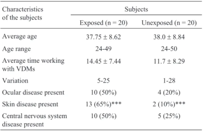
The main characteristics of the exposed and control subjects (average of age, working time, sex and ocular, skin and CNS diseases) are summarized in Table 1. None of the groups studied showed significant difference in age, period of work, ocular and CNS diseases, except for skin diseases found in a higher incidence (65%) in the exposed individu-als (p < 0.001).
Table 2 shows the average numbers of MNC, BNC and BEC of both exposed and control individuals included in this investigation. Assessment of MN frequencies in ex-foliated buccal cells revealed a significantly higher fre-quency of micronuclei (p < 0.001) in individuals exposed to VDMS (6.20±4.44) compared to unexposed (2.05±2.16). In the same way, we observed a significant difference (p < 0.05) in BEC frequency, between exposed workers (2.80±2.50) and controls (1.40±2.30). However, no sta-tistically significant differences regarding BNC frequency between exposed (3.40 ± 3.89) and control individuals
were observed (2.15±2.54).
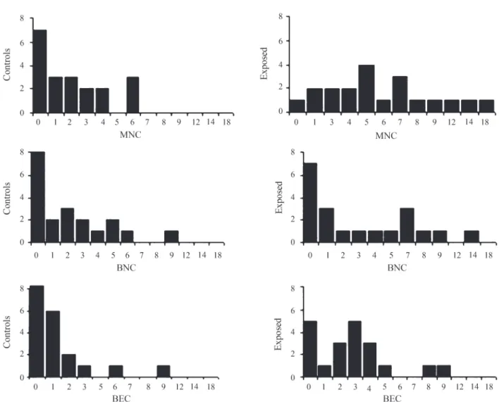
The number of cells (per 2,000 cells) with micronucleus, binucleated, and broken egg cells of each in-dividual is showed in Figure 1. Within the control group, 35% showed no cells with micronucleus, and 40% of them showed only one to three MNC, resulting in mode zero. Six MNC was the maximum number found in non-exposed in-dividuals. In contrast, in the exposed group, we found a variable distribution of cells with micronucleus, ranging from zero to 18, resulting in mode five. Similarly, the distri-bution of BEC was different between control and exposed
Table 1- General characteristics of 10 male and 10 female workers occupationally exposed to cathode ray tube computer video display monitors (VDMs) as compared to unexposed control individuals matched for age and sex.
Characteristics of the subjects
Subjects
Exposed (n = 20) Unexposed (n = 20)
Average age 37.75±8.62 38.0±8.84
Age range 24-49 24-50
Average time working
with VDMs 14.45±7.44 11.7±8.29
Variation 5-25 1-28
Ocular disease present 10 (50%) 4 (20%)
Skin disease present 13 (65%)*** 2 (10%)***
Central nervous system disease present
10 (50%) 5 (25%)
groups. While 90% of the controls showed zero to three BEC, resulting in multiple modes (zero and three), only 70% of the exposed individuals showed the same range of BEC. In contrast, the distribution of BNC found was simi-lar between both groups, resulting in mode zero. Zero to three was the maximum number of BNC found in more than 50% of exposed and unexposed individuals.
Comparisons between independent variables age (within the following intervals:£35 and > 35 years), work-ing time (£10 and > 0 years) and the frequencies of MNC, BEC and BNC was investigated among exposed and con-trols. Our results suggest that there is no association be-tween the age, working time, of subjects, and the number of MN and nuclear abnormalities (Table 3).
Among the exposed females the MNC, BEC and BNC frequency in the above cited intervals, did not differ (n = 5; 8.00±4.64, and n = 5; 8.80±5.36). Among the
ex-posed males, alike the females, in the same intervals, pre-sented a small increase, however, not significant (n = 3; 2.00 ± 2.65, and n = 7; 4.86 ± 2.73).
Exposed individuals presented an increased fre-quency of ocular (50%), skin (65%), and CNS (50%) dis-eases, however association between these diseases and the frequency of MNC, BNC and BEC was only obtained for binucleated cells among exposed individuals with ocular and CNS diseases (p < 0.05) (Table3).
The frequency of MNC, BNC and BEC between males and females was also compared, and, in both exposed
and control groups, the frequencies of MNC and nuclear abnormalities were significantly higher in females. Among the exposed group, females showed at least twice more MNC, BNC and BEC than males (p < 0.05), (Table 3). The Spearman correlation coefficient between MNC and sex was positive (p = 0.029).
Among the females and males, the exposed group showed more MNC than the control group (p < 0.01) (Table 4).
Based on the p-values estimated by Spearman rank coefficients, in our analysis sex was the main factor affect-ing MNC frequency in exfoliated cells.
Discussion
For many years scientists and engineers believed that electromagnetic fields of low frequency could not cause al-terations in human cells. This hypothesis was based on the idea that EMF could not generate a sufficient amount of heat to increase the body temperature, not causing damage at the dna level (Lechter, 1991). However, data available in
Table 2- Cytological observations made on 10 males and 10 females occupationally exposed to cathode ray tube computer video display monitors (VDMs) as compared to unexposed control individuals matched for age and sex. We assessed 2,000 cells for each individual.
Cytological observation
Subjects
Exposed (n = 20) Unexposed (n = 20)
Micronucleated cells
Mean 6.20** 2.05**
Standard deviation ±4.44 ±2.16
Minimum number 0 0
Maximum number 18 6
Binucleated cells
Mean 3.40 2.15
Standard deviation ±3.89 ±2.54
Minimum number 0 0
Maximum number 13 9
Broken egg cells
Mean 2.80* 1.40*
Standard deviation ±2.50 ±2.30
Minimum number 0 0
Maximum number 9 9
*Significant at p < 0.05 by the Mann-Whitney U test. **High significant at p < 0.001 by the Mann-Whitney test.
Table 3- Cytological observations made on 10 males and 10 females occupationally exposed to cathode ray tube computer video display monitors (VDMs). We assessed 2,000 cells for each individual.
Cytological observations
Exposed individuals
Micronucleated cells (Mean±SD)
Binucleated cells (Mean±SD)
Broken egg cells (Mean±SD)
Age (years)
£35 (n = 8) 5.75±4.89 3.63±3.62 2.13±1.46
>35 (n = 12) 6.50±4.32 3.25±1.46 3.25±2.99
Number of years working with VDMs
£10 (n = 7) 4.57±3.84 3.57±3.91 2.00±3.23
>10 (n = 13) 7.08±4.63 3.31±4.03 3.23±2.86
Ocular disease
Present (n = 9) 7.67±4.18 5.78±3.83* 3.89±3.14
Absent (n = 11) 5.00±4.47 1.45±2.72* 1.91±1.45
Skin disease
Present (n = 13) 6.23±4.83 2.85±2.48 3.69±3.99
Absent (n = 7) 6.14±3.98 2.71±2.75 2.86±3.93
Central nervous system disease
Present (n = 10) 6.60±4.30 5.10±4.15* 3.80±2.97
Absent (n = 10) 5.80±4.78 1.70±2.87* 1.80±1.48
Sex
Females (n = 10) 8.40±4.77* 6.30±3.56*** 4.40±2.32**
Males (n = 10) 4.00±2.91* 0.50±0.71*** 1.20±1.48**
the literature suggest that exposure to elf-emf directly causes genetic changes in biological systems (McCanet al., 1993, 1998; Murphyet al., 1993; Simkoet al., 1998a, b;
Repacholi and Greenebaum, 1999; Walleczeket al., 1999). Furthermore, some studies suggest the hypothesis that EMF is not involved on the onset of the tumoral growth, but may be acting as a promoter or co-promoter in this process (Goodman and Shirley-Henderson, 1990).
The evidence that EMF could cause genotoxic effects in human cells can reinforce the relation between exposure to EMF and increased incidence of cancer in exposed indi-viduals. Gangi and Johansson (1997) detected an increased incidence of skin and CNS alterations among microcom-puter’s workers. In their study, the workers reported to feel skin - and mucosa - related symptoms, such as pain, itch, heat sensation, erythema, papules, and pustules. The CNS symptoms were,e.g.dizziness, tiredness, and headache, the same symptoms caused by sunburn. Kisner and Fedeman (1998) reported that female workers exposed to VDM pre-sented obstetric complications, besides skin, ocular, and CNS diseases. However, the authors conclude that the data reviewed were inconsistent or methodically flawed, and be-lieved that continued research had to be carried out to fur-ther define and elucidate the risk of EMF produced by
Figure 1- Distribution of number of cells with micronuclei (MNC), binucleated (BNC) and broken egg (BEG) cells (2,000 cells per individual).
Table 4- Cytological observations made on 10 males and 10 females occupationally exposed to cathode ray tube computer video display monitors (VDMs) as compared to unexposed control individuals matched for age and sex. We assessed 2,000 cells for each individual.
Cytological observations
Individuals Micronucleated
cells (Mean±SD)
Binucleated cells (Mean±SD)
Broken egg cells (Mean±SD)
Females
Exposed (n = 10) 8.40±4.74* 6.30±3.56 4.40±2.32**
Unexposed (n = 10) 3.60±1.90* 4.10±2.23 1.90±1.66**
Males
Exposed (n = 10) 4.00±2.91** 0.50±0.71 1.20±1.48
Unexposed (n = 10) 0.50±0.97** 0.20±0.42 1.0±2.83
video display terminals. In our study we also detected skin, ocular and CNS diseases among approximately 50% of the exposed group, maybe as a result of continual exposure to EMF emitted by microcomputer monitor. However, no as-sociation was found between these diseases and frequen-cies of MNC and BEC, and only frequency of BNC was related with ocular and CNS diseases within the exposed individuals
Significantly our results obtained from the investiga-tion of micronucleus in exfoliated cells indicate that ex-posed individuals have a significant increased number of MNC. Exposed individuals showed three times more cells with micronucleus than control individuals. In the same way, the frequency of BEC among exposed individuals was two times higher than in the control group. Estécio and Silva (2002) also evaluated possible nuclear alterations in microcomputer’s workers and found that exposed individu-als had two times more chromosomal aberrations in cul-tured lymphocytes than control individuals, reinforcing the data obtained in our study.
Similarly, Ivancsitset al. (2002) showed that inter-mittent exposure to a 50Hz magnetic field causes a repro-ducible increase in DNA strand breaks in cultured human diploid fibroblasts. They evaluated the genotoxic effect of cultured fibroblasts from skin biopsy of healthy donors that were continuously exposed to ELF-EMF, through the comet assay, and obtained significant results. Although these findings suggest that the exposure to EMF increase the frequency of chromatic and/or chromosomal breaks, it is not clear yet the exactly molecular mechanism affected by this type of radiation.
Several hypotheses like induction of electric currents, increased free radical activity or acceleration of electron transfer in different enzymes and proteins have been pro-posed, but all of these hypotheses have been mostly specu-lative. Recent observations have shown that DNA can transfer electrons within its base pairs. These studies sug-gest that EMF may initiate transcription by generating re-pulsive forces causing chain separation at specific DNA sequences. As in the state of transcription the DNA presents itself as a vulnerable target to genotoxic influences, the on/off switching at intermittent ELF-EMF exposure may lead to DNA disruption, causing DNA strand breaks (Ivancsitset al., 2002).
According to Estécio and Silva (2002) the chromatic aberrations could be a result of malfunction of enzymes that duplicate and repair DNA, leading to alterations on the DNA strand recently polymerized. Despite of the molecu-lar mechanism the EMF leads to chromosomal destabilization. Thus, individuals who have this destabilization could present cells with high frequency of mutations, chromosomal aberrations, micronucleus, and increased risk of cancer growth than individuals with stable genome.
Several reports have described an association be-tween chronological aging and aneuploidy. The chromo-somes most frequently lost are the X chromosome in females and Y chromosome in males. The autosomes may be lost at the same frequency as the sex chromosomes, but these events should lead to death as they contain genes re-quired for cell survival. As lagging chromosomes and frag-ments can be incorporated into a MN and the increase in aneuploidy occur according to the age, this factor should be correlated with the increase in MN formation (Burkvicet al., 2001). In contrast, our results suggest that there is no as-sociation between the individual’s age and the frequency of MNC, BNC, and BEC among microcomputer’s workers. However, when this factor was analyzed among male workers, it was observed a slight increase of MNC and nu-clear abnormalities in older workers, although this result was not statistically significant.
In the same way, analysis of MNC, BNC and BEC frequencies after different working times showed a small, not significant increase among the exposed group on the longest working time (< 10 years).
Particularly evident is the increment observed in women with respect to men. In females it was observed sig-nificant higher frequencies of MNC, BNC, and BEC than in males, and the Spearman correlation coefficients between MNC and sex were positive (p = 0,029). In agreement with our data, some reports in this area have been shown similar results. Burkvic et al., 2001 demonstrated that there are 22% more sexual chromosomes lost in females than in males, confirming that females cells are more sensitive to action of genotoxic agents. Both the preferential involve-ment of sex chromosomes in somatic aneuploidy in women and the relation between malsegregation of X chromo-somes and age are well documented. However, the molecu-lar mechanisms underlying instability of sex chromosomes in females are not fully elucidated yet. Studies have demon-strated the high susceptibility of sex chromosomes to be lost at mitosis, as revealed by the high incidence of micronucleated X-positive lymphocytes in female donors. Further investigation by in situ hybridization of cytokinesis-bloked lymphocytes using a X chromosome’s centromere probe, demonstrated that both, chromosomal loss and non-disjunction, contribute to the malsegregation of sex chromosomes of the females (Zijnoet al., 1996).
Different physiological parameters, such as men-strual cycle, estrogen levels, and oral contraceptives may affect the frequency of sister chromatid exchange (SCE) (Ghosh and Ghosh, 1988; Joseph-Lerneret al., 1993), but in our investigation we did not take in consideration neither estrogen levels nor oral contraceptive assumption.
sex of the individuals. Finally, extensive studies and stan-dardized tests to evaluate biological damage at different levels are recommended to public agencies concerned with environmental quality and public health. Genotoxic evalua-tion is a necessary measure to ensure environmental quality and occupational health, as is the workers orientation to in-crease awareness and dein-crease the risk for serious disease caused by computer cathode ray tube video display moni-tors radiation.
Acknowledgements
The authors are very grateful to Abílio Novack Alves, technical assistant of the Laboratório de Genética of the UCPel, Álvaro Moreira Martins and Vilma Ruas da Silva, technical assistants of the Departamento de Zoologia e Genética of the UFPel, and the 40 workers who spontane-ously took part in this study. This research was supported by the Universidade Católica de Pelotas and the Univer-sidade Federal de Pelotas.
References
Bukvic N, Gentile M, Susca F, Fanelli M, Serio G, Buonadonna L, Capurso A and Guanti G (2001) Sex chromosome loss, micronuclei, sister chromatid exchange and aging: A study including 16 centenarians. Mutat Res 498:159-167. Carrano AV (1988) Considerations for population monitoring
us-ing cytogenetic techniques. ICPEMC – International Com-mission For Protection Against Environmental Mutagens And Carcinogens. Publication n. 14. Mutat Res 204:379-406.
Estécio MRH and Silva AE (2002) Chromosome abnormalities caused by computer video display monitor’s radiation. Rev Saúde Pública 36:330-336.
Fenech M (2000) Thein vitromicronucleus technique. Fundam
Molec Mechan Mut 455:81-95.
Gangi S and Johansson O (1997) Skin changes in “screen dermati-tis” versus classical UV - And ionizing irradiation-related damage - Similarities and differences. Exp Dermatol 6:283-91.
Ghosh R and Ghosh PK (1988) Sister chromatid exchanges in the lymphocytes of control women, pregnant women and women laking oral crontraceptives effects of cell culture temperature. Mutat Res 12:179-183.
Goodman R and Shirley-Henderson A (1990) Exposure of cells to extremely low-frequency electromagnetic fields: Relation-ship to malignancy? Cancer Cells 2:355-9.
Ivancsits S, Diem E, Pilger A, Rüiger HW and Jahn O (2002) In-duction of DNA strand breaks by intermittent exposure to extremely-low-frequency electromagnetic fields in human diploid fibroblasts. Muta Res 519:1-13.
Joseph-Lerner N, Fejgin M, Ben-Nun I, Leguin C and Arniel A (1993) The correlation between the frequency of sister chromatid exchange and human reproductive hormones. Mutat Res 300:247-252.
Kisner RS and Federman DG (1998) Video display terminals: Risk of electromagnetic radiation. Southern Med J 91:12-16. Lechter GS (1991) A radiação eletromagnética. PC Mag Brás
12:44-54.
Li CY and Theriault RS (1997) Residential exposure to 60 Hz magnetic fields and adult cancers in Taiwan. Epidemiol 8:25-30.
Majer BJ, Laky B, Knasmüller S and Kassie F (2001) Use of the micronucleus assay with exfoliated epithelial cells as a biomarker for monitoring individuals at elevated risk of ge-netic damage and in chemoprevention trials. Mutat Res 489:147-172.
Maluf SW and Erdtmann B (2000) Follow-up study of the genetic damage in lymphocytes of pharmacists and nurses handling antineoplastic drugs evaluated by cytokinesis-block micronuclei analysis and single cell gel electrophoresis as-say. Mutat Res 471:21-27.
McCann J, Dietrich F, Rafferty C and Martin AO (1993) A critical review of genotoxic potential of electric and magnetic fields. Mutat Res 297:61-95.
McCann J, Dietrich F and Rafferty C (1998) The genotoxic poten-tial of electric and magnetic fields: An update. Mutat Res 411:45-86.
Murphy JC, Kaden DA, Warren J and Sivak A (1993) Power fre-quency electric and magnetic fields: A review of genetic toxicology. Mutat Res 296:221-240.
Repacholi MH and Greenebaum B (1999) Interaction of static and extremely low frequency electric and magnetic fields with living systems: Health effects and research needs. Bioelectromagnetics 20:133-160.
Schreibner GH, Swaen GMH, Meijers JMM, Slangen JJM and Sturmans F (1993) Cancer mortality and residence near electricity transmission equipament: A retrospective cohort study. Int J Epidemiol 22:9-15.
Simko M, Kriehuber R and Lange S (1998a) Micronucleus forma-tion in human amnion cells after exposure to 50 Hz MF ap-plied horizontally and vertically. Mutat Res 418:101-111. Simko M, Kriehuber R, Weiss DG and Luben RA (1998b) Effects
of 50 Hz EMF exposure on micronucleus formation and apoptosis in transformed and nontrasformed human cell lines. Bioelectromagnetics 19:85-91.
Stich HF and Rosin MP (1982) Micronuclei in exfoliated human cells as a tool for studies in cancer risk and cancer interven-tion. Cancer Let 22:241-253.
Titenko-Holland N, Moore LE and Smith MT (1994) Measure-ment and characterization of micronuclei in exfoliated hu-man cells by fluorescence in situ hybridization with a centromeric probe. Mutat Res 312:39-50.
Verkasalo PK, Pukkala E, Hongisto MY, Valjus JE, Järvinen PJ, Heikkilâ KV and Koskenvuo M (1993) Risk of cancer in Finnish children living close to power lines. Br Med J 307:895-899.
Walleczek J, Shiu EC and Hahn GM (1999) Increase in radia-tion-induced HPRT gene mutation frequency after nonthermal exposure to non-ionizing 60 Hz electromagnetic fields. Radiat Res 151:489-497.
Wertheimer N and Leeper E (1979) Electrical wiring configura-tions and childhood cancer. Am J Epidemiol 109:273-284. Zijno A, Leopardi P, Marcon F and Crebelli R (1996) Sex
chro-mosome loss and non-disjunction in women: Analysis of chromosomal segregation in binucleated lymphocytes. Chromosoma 104:461-467.