Ide ntificatio n o f the m ain ge ne rato r
so urce o f lo ngitudinal m uscle
co ntractio n in the e arthwo rm
ve ntral ne rve co rd
Departamento de Fisiologia, Universidade Federal do Paraná, Curitiba, PR, Brasil
Y.C. Chang, Z. Assmé, E.C.L. Caffaro and A.B. Bartoszeck
Abstract
The main generator source of a longitudinal muscle contraction was identified as an M (mechanical-stimulus-sensitive) circuit composed of a presynaptic M-1 neuron and a postsynaptic M-2 neuron in the ventral nerve cord of the earthworm, Amynthas hawayanus, by simul-taneous intracellular response recording and Lucifer Yellow-CH in-jection with two microelectrodes. Five-peaked responses were evoked in both neurons by a mechanical, but not by an electrical, stimulus to the mechanoreceptor in the shaft of a seta at the opposite side of an epidermis-muscle-nerve-cord preparation. This response was corre-lated to 84% of the amplitude, 73% of the rising rate and 81% of the duration of a longitudinal muscle contraction recorded by a mechano-electrical transducer after eliminating the other possible generator sources by partitioning the epidermis-muscle piece of this preparation. The pre- and postsynaptic relationship between these two neurons was determined by alternately stimulating and recording with two micro-electrodes. Images of the Lucifer Yellow-CH-filled M-1 and M-2 neurons showed that both of them are composed of bundles of longitudinal processes situated on the side of the nerve cord opposite to stimulation. The M-1 neuron has an afferent process (A1)in the first nerve at the stimulated side of this preparation and the M-2 neuron has two efferent processes (E1 and E3) in the first and third nerves at the recording side where their effector muscle cell was identified by a third microelectrode.
Co rre spo nde nce
Y.C. Chang
Departamento de Fisiologia Setor de Ciências Biológicas, UFPR Caixa Postal 8621
80011-970 Curitiba, PR Brasil
Fax: + 55-41-266-2042 E-mail: changyc@ cce.ufpr.br Part of a Doctoral thesis presented by Z. Assmé to the Departamento de Fisiologia, UFPR, Curitiba, PR, Brasil.
Received January 20, 1997 Accepted July 27, 1998
Ke y wo rds
•Earthworm muscle contraction
•Main generator source •M-1 and M-2 neurons •Mechanical stimulus
Intro ductio n
This series of three papers (the present paper and Refs. 1 and 2) describes a study of the interaction mechanism between substrates in the central and peripheral nervous systems of one familiar yet seldom studied animal, the South American earthworm, Amynthas hawayanus. The mode of this interaction was suggested in a preliminary report (3)
mechanism. A generator source, possibly located in the central nervous system, evokes this muscle contraction. A modulator which may be in the peripheral nervous system determines the magnitude of this contrac-tion. If this modulation is facilitatory, then the muscle contraction magnitude must be proportional to the amount of peripheral neu-ral substrate left in the preparation. Earth-worms have a well-developed peripheral nervous system (4) with several functions proposed (5-7). Our first objective was to identify the neural substrate of a main gen-erator source of muscle contraction and an-other study (1) was conducted to identify the neural substrate of a modulator with a facili-tatory effect on this main generator source. These two neural substrates may either be the known neurons identified by us (8,9) or by others (10,11), or must be identified as new neurons in these two articles. We com-pared the interaction between these two neu-ral substrates (2) to the conditioning para-digm of the other animals (12-14) and to that of the intact earthworm (15,16). This com-parison is expected to contribute to the knowl-edge of learning and memory mechanism in general.
Mate rial and Me tho ds
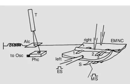
Mature South American earthworms iden-tified as Amynthas hawayanus and raised in a terrarium outside the laboratory were used in this study. Only the middle body between the 25th and 70th segments was used for their repeated neuronal organization (17) important for reproducible results. Dissec-tion of the EMNC (Figure 1), electrophysi-ological equipment and experimental proce-dures were described in previous articles (8,9).
Stimulatio n and re co rding
Stimuli were square pulses delivered from a CURITIBA-l stimulator (manufactured by
Extracel-lular electrical responses were recorded from the segmental nerves (N; Figure 2) by suc-tion electrodes on a Tektronix 5113 oscillo-scope (Beaverton, OR). Intracellular electri-cal responses were recorded from neurons and muscle cells by microelectrodes (AM, 6010, Everett, USA) filled with 7% Lucifer Yellow-CH on another trace of this oscillo-scope. A high-impedance amplifier bridge (Model 8500, Dagan Corp., Minneapolis, MN) enabled each of these microelectrodes to be used for recording, stimulation and dye-injection simultaneously. These electrodes were mounted on separate micro-manipulators (Model CS-56-3, Cus-tom Instruments, Whippany, USA) to facili-tate the search for neurons and muscle cells by independent scanning over the entire sur-face of this preparation. Mechanical re-sponses, i.e., muscle contractions (C; Figure 2), were recorded with a mechano-electrical transducer. Although such transducers are available commercially, or can be constructed on the basis of literature data (e.g., 18), a low-damping device was designed to pre-serve the rising and falling rates of a contrac-tion as much as possible. The device con-sisted of a photographic photocell (Metrawatt, AG, Nürenberg, Germany) (Phc; Figure 1) with a small light spot shining on it from a battery-operated (for reducing 60-cycles interference with the records) toy torch (T; Figure 1). The amount of light received by this photocell was controlled by the move-ment of an aluminum foil cover (Alc; Figure 1) pulled by the muscle contraction. One end of this aluminum foil cover was connected to the anterior end of the right side of this preparation and the other end to a small spring. By adjusting this spring, the move-ment of this aluminum foil cover could be made linearly proportional to the amount of light it allowed to pass to the photocell in a range of 0.1 to 5.0 g of force on the Tektronix 5113 oscilloscope trace (Osc; Figure 1). Notice that the mechanical friction of this setup was low because the two connecting
lines at both ends of this aluminum foil cover were suspended in the air without contacting any solid object. Both the velocity (in straight lines) and acceleration (in curves) of these rising and falling rates (in g/s) of a muscle contraction were preserved as much as pos-sible by this design. In addition to the rising and falling rates, the amplitude (in grams) and the duration (in seconds) at 2/3 ampli-tude of muscle contraction were also re-corded and measured for comparison.
Iso latio n o f the main ge ne rato r so urce o f
muscle co ntractio n
Four systems are known to be capable of generating contraction in an earthworm muscle cell. They are the central nervous system (2,5-7,11), the peripheral nervous system (2,7), the muscle system which trans-mits impulses through their nexi (5,7,19) and the spontaneous activation in a single muscle cell membrane (20). The experiments in this section were carried out to determine which of them is the “main” generator source
T
Alc to Osc
Phc left
right EM NC
1 2
3 S
M S ES
which generates the largest percentage of the muscle contraction and to isolate it by differ-ential sectioning (Figure 2). Contractions evoked by peripheral electrical (left column; Figure 2) and mechanical (right column; Fig-ure 2) stimuli were used to identify these sources. Two kinds of differential sectioning were done in these experiments. Sectioning the afferent (Figure 2-3), or the efferent (Fig-ure 2-4), segmental nerves was performed to interrupt the transmission routes through the central nervous system. Sectioning, or parti-tioning, the epidermis-muscle piece (Figure 2-5) was performed to interrupt the trans-mission routes through the peripheral ner-vous system, the muscular system and the single muscle cell. While isolating the cen-tral circuits, it must be kept in mind that all segmental nerves contain afferent and effer-ent fibers. In order to isolate the affereffer-ent from the efferent fibers, one must sacrifice the reflex arcs with ipsilateral afferent and efferent routes and to preserve only the arc with the contralateral routes. This was done by stimulating only the left side of this prepa-ration and recording only from the right side as shown in Figures 1 and 2. All responses recorded from the left side nerves, Nl, were then considered as purely afferent and all responses recorded from the right side nerves, Nr, were purely efferent in this arrangement (21) although the opposite would be equally valid because the body plane is bilaterally symmetrical in this animal.
Ide ntifying the ne uro n circuit o f the main
ge ne rato r so urce
The pre- and postsynaptic relationship between two neurons in the mechanical-stimulus-sensitive circuit (abbreviated as the M-circuit) in the nerve cord was determined by alternately stimulating and recording with two microelectrodes (Figure 3). There may be three situations of transmission between two neurons: “no transmission”, where exci-tation of either one of them did not evoke a C
C Nr Nl E M
C
N
E
M
C
NI
Nr E
M
C
N
E
M
N
C
Nr NI
C
N
E M
C NI Nr E
M
C
N E
M
2-1
2-2
2-3
2-4
2-5
E M
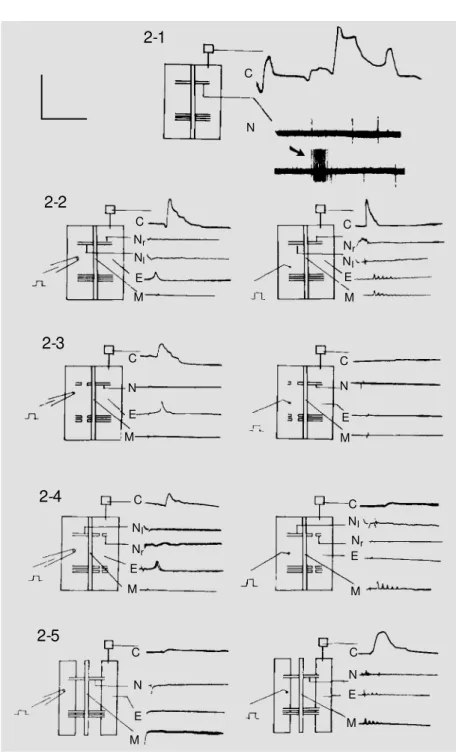
Figure 2 - Neuron and muscle cell activities.
2-1, Spontaneous activities of an intact EM NC preparation w ithout stimulation.
2-2, Responses of an intact EM NC preparation evoked by electrical (left column) and mechanical (right column) stimuli.
2-3, Responses of an EM NC preparation evoked by electrical and mechanical stimuli after section of all afferent (left) segmental nerves.
2-4, Responses of an EM NC preparation evoked by electrical and mechanical stimuli after section of all efferent (right) segmental nerves.
2-5, Responses of an EM NC preparation evoked by electrical and mechanical stimuli after section, or partition, of the epidermis-muscle piece.
C, Contraction; N, segmental nerves; Nl, left segmental nerve; Nr, right segmental nerve; E,
response in the other, “rectifying, or ortho-dromic, transmission”, where excitation in a presynaptic M-1 neuron evoked a response in a postsynaptic M-2 (Figure 3-1, right) neuron but excitation in an M-2 neuron did not evoke a response back in the M-1 neuron (Figure 3-1, left), and “non-rectifying trans-mission” where excitation in either one of them evoked a response in the other. Only the second situation, or “rectifying transmis-sion”, was further studied in this article. The third situation, or “non-rectifying transmis-sion”, may also be authentic between some of these neurons but was not studied because of the difficulty in further confirmation. The diffusion of Lucifer Yellow-CH (22) injected into one of these neurons was also tested for its rectifying or non-rectifying properties. Another characteristic which may also help distinguish the M-1 and M-2 neurons is their possible different connection to other neu-rons outside their own circuit. Their differ-ent connections to higher cdiffer-enter interseg-mental neurons were tested by stimulating the anterior end of the nerve cord and record-ing its response from these two neurons (Fig-ure 3-2).
Re sults
Spo ntane o us activitie s
Spontaneous activities are the electrical discharges from the segmental nerves (N; Figure 2-1) and muscle contractions (C; Figure 2-1) not correlated to the deliv-ered stimulus. These two activities were found not to be correlated, i.e., they do not seem to evoke a response from each other. Both of them have irregular frequencies, amplitudes, rising and falling rates and durations. Some of these spontaneous con-tractions had no measurable duration be-cause they did not return to baseline (C; Figure 2-1). The spontaneous electrical dis-charges from the segmental nerves had ir-regular intervals but maintained an average
frequency of 0.74 ± 0.21 Hz in 50 prepara-tions measured for 30 min each (top line (N); Figure 2-1). There were also high fre-quency bursts without correlation with any noticeable events (arrow, bottom line; Figure 2-1). The resting potentials of both muscle cells and neurons decreased and in-creased unpredictably between 30 and 80 mV when recorded continuously for longer than 1 min. Spontaneous discharges of these cells might or might not accompany these resting potential fluctuations. These sponta-neous activities, although part of behavior, were not studied in the present investigation. Only responses evoked by the delivered stimuli were studied in the following sections.
E
3-1
M -2
M -1
M -2
M -1
3-2
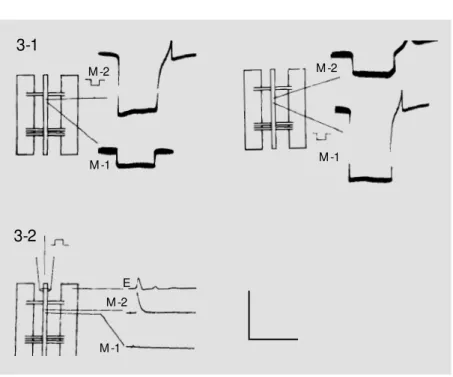
Figure 3 - Synaptic connection betw een the main generator neurons.
3-1, Rectifying synaptic transmission demonstrated by alternate intracellular stimulation w ith hyperpolarizing 7.0-nA, 50-ms pulses. Notice that a postsynaptic (M -2) response evoked by this stimulus is not transmitted to the presynaptic (M -1) neuron in the left figure w hile a presynaptic (M -1) response evoked by this stimulus is transmitted to the postsynap-tic (M -2) neuron in the right figure.
3-2, Different connections to higher intersegmental centers examined by extracellular depolarizing pulse to the anterior end of the nerve cord. Only postsynaptic (M -2) neuron has connection to the higher center. Presynaptic (M -1) neuron does not respond to this stimulus.
E, Effector muscle response. Calibration: 50 mV and 50 ms for both figures. M -2
Effe cto r re spo nse s
Effector responses are the muscle con-tractions (C; Figure 2-2) and electrical re-sponses recorded from the muscle cells (E; Figure 2-2) and from the nerves (Nr, Nl; Figure 2-2) on one side (right) of this prepa-ration evoked by a mechanical, or electrical, stimulus on the other side (left). The con-tractions evoked by either electrical (C, left column; Figure 2-2), or mechanical (C, right column; Figure 2-2) stimuli in an intact EMNC preparation were similar. Three pa-rameters were measured from 50 muscle contractions in 23 preparations, i.e., ampli-tude (2.15 ± 0.19 g), rising rate (8.72 ± 0.16 g/s) and duration at 2/3 amplitude (0.31 ± 0.11 s).
The electrical responses evoked in the effector muscle cell by the mechanical and electrical stimuli were different. Those evoked by the electrical stimuli were single-peaked (E, right column) but those evoked by the mechanical stimuli were five-peaked (E, left column).
Main ge ne rato r so urce
Although both electrical (left column; Figure 2-2) and mechanical (right column; Figure 2-2) stimuli evoked muscle contrac-tions, C, on the opposite side of this prepara-tion, the electrical stimulus did not evoke an afferent response in the left segmental nerves, N1, or in the neurons in the nerve cord, but the mechanical stimulus evoked both affer-ent, N1, and efferent, Nr, nerve responses and five-peaked responses, M, in many neu-rons in the nerve cord.
Section of all afferent (left) segmental nerves (Figure 2-3) did not change the effec-tor muscle cell electrical response, E, and contraction, C, evoked by the electrical stimu-lus (left column) but eliminated the efferent nerve response, Nr, central neuron response, M, and muscle cell electrical, E, and chanical, C, responses evoked by the
me-chanical stimulus (right column). These two experiments (Figure 2-2 and 2-3) showed that the responses evoked by the electrical stimulus were not transmitted through the neurons in the nerve cord to reach the effec-tor muscle cell. Only the response evoked by the mechanical stimulus was transmitted through the neurons in the nerve cord to reach the effector muscle cell.
Section of all efferent nerves (right) did not attenuate much of the muscle contrac-tion, C, evoked by the electrical stimulus (left column; Figure 2-4) but reduced signif-icantly the amplitude evoked by mechanical stimuli (right column; Figure 2-4) to 38%, or 0.81 ± 0.14 g, and the rising rate to 25%, or 2.21 ± 0.33 g/s, and increased its duration at 2/3 amplitude to 225%, or 0.71 ± 0.21 s in 50 contractions. Section of the suspected trans-mission route of the electrical stimulus-evoked response by partitioning the epider-mis-muscle piece along its midline while leaving all segmental nerves intact (Figure 2-5) almost eliminated, but never completely, the muscle contraction, C, evoked by electri-cal stimulus (left column) but only reduced non-significantly the amplitude to 84%, or 1.85 ± 0.13 g, and the rising rate to 73%, or 6.35 ± 0.46 g/s, and increased the duration at 2/3 amplitude to 123%, or 0.38 ± 0.10 s, equivalent to a reduction of 81%, in 50 con-tractions evoked by the mechanical stimulus (right column). The last three percentages were believed to be the percentages of a muscle contraction evoked by the mechani-cal stimulus alone without the nonsignifi-cant contribution from the electrical stimu-lus.
Synaptic co nne ctio n be twe e n the main
ge ne rato r ne uro ns
response transmitted from the stimulated neuron. As these neurons hardly responded to an intracellular depolarizing electrical stimulus, a hyperpolarizing (square pulse symbol in Figure 3-1 and 3-2) 50-ms pulse slightly above the threshold value (7.0 nA) was always used to evoke an off-response in the form of a single-peaked action potential in one of these neurons (M-2; Figure 3-1). If no response was evoked in the next neuron (M-1; Figure 3-1, left) except for the spread-ing square pulse, stimulation and recordspread-ing were switched around in these two micro-electrodes (Figure 3-1, right). If the first neuron did not respond to the response in the second, this neuron pair was discarded as “no transmission”, i.e., they do not form a circuit. Only when the postsynaptic neuron (M-2) did not evoke a response in the presyn-aptic neuron (M-1; Figure 3-1, left) but the presynaptic neuron evoked a response in the postsynaptic neuron (M-2; Figure 3-1, right) was this pair accepted as forming a rectify-ing two-neuron circuit. Thirty-one of the identified 42 pairs (62 neurons among 84) satisfied this criterion. Non-rectifying pairs which responded in both ways were also not used in this study because they could not be further distinguished as to whether they were composed of only one, or two, neurons. On the other hand, diffusion of Lucifer Yellow-CH seemed to be always non-rectifying from one neuron to the next (22) in this circuit but never to a third neuron. The last test con-firmed that this generator source was formed by a two-neuron circuit.
When the ventral nerve cord anterior to the identified neuron pair was stimulated extracellularly with a suction electrode (Fig-ure 3-2), none of these 31 identified M-1 neurons responded but 26 M-2 neurons in 13 pairs responded. This additional characteris-tic showed that only the postsynapcharacteris-tic, but not the presynaptic, neuron was commanded by a higher intersegmental center and was also used for their identification. Thus, only the last 13 pairs were physiologically
identi-fied as the main generator source.
Mo rpho lo gy o f the main ge ne rato r so urce
R
WM
E1
A1
V
M -1 M -2
E3
Figure 4 - M orphology of themain generator source. Fluores-cence microscope photograph and camera-lucida draw ing of Lucifer Yellow -CH-filled images. R, Radial dimensional view ; WM , w hole-m ount ing preparat ion; A1, afferent process in the first nerve of the stimulated (left) side of this preparation; E1, efferent process in the first nerve of the recording (right ) side of t his preparation; E3, efferent process in the third nerve of the record-ing (right) side of this prepara-tion; V, varicosities in the central region of these identified neu-rons; M -1, presynaptic neuron; M -2, postsynaptic neuron. Tw o longit udinal bundles of pro-cesses representing these tw o neurons are also visible in the upper middle region of V. AP, Ant eropost erior view of t he same preparation at a level near V. Some of the branches extend dorsoventrally and are not de-tectable in the R view above. Calibration: 100 µm for all fig-ures.
E1
A1
E3
V
D iscussio n
Only 18 neurons identified in nine prepa-rations (one pair in each) were accepted as the main generator source in the present study although a total of 84 neurons in 42 preparations satisfied the primary criterion that their five-peaked responses were evoked by a threshold mechanical stimulus to the setal mechanoreceptor and their responses were correlated to the electrical responses and contractions of the contralateral muscle cell. These nine neuron pairs were selected because of their physiological and morpho-logical consistency. Circuits with variations were rejected in this procedure. Most of these variations were related to their synap-tic transmission mechanism. Only rectifying pre-(M-1) and post-(M-2) synaptic neurons in an M-circuit were accepted. It was sus-pected that many of them had non-rectifying transmission. They were rejected because their pre- and postsynaptic relationship could not be determined and because one could not distinguish morphologically if they were com-posed of a single neuron or two. On the other hand, all these neurons were non-rectifying to Lucifer Yellow-CH diffusion (22). Those having rectifying diffusion were rejected because only one neuron was morphologi-cally visible. Therefore, this high rejection rate was the result of a predetermined bias of
the experimenters. Actual two-neuron M-circuits for generating 70-80% of a contrac-tion in the contralateral effector muscle cell must be more numerous than the nine pairs reported here. Slight physiological or mor-phological variation may actually be the rule rather than the exception. This predetermined bias of selection was nevertheless upheld in this article because a precise quantitative measurement was a necessity for comparing the muscle contraction magnitude modifica-tions in the two subsequent studies (1,2). But this predetermined bias in their isolation and identification imposed a limit on the compa-rability of the results of this article to the stimulus-response paradigm in intact prepa-rations tested by experimental psychological methods (15,16), where not only all segmen-tal neuronal and muscular circuits but also intersegmental circuits from the highest to the lowest centers are included. In addition, one must also not overlook the interpretation of one single stimulus, such as that of a chemical, into a mixture of mechanical and thermal stimuli, by the conscious subject in experimental psychology. This limit in com-parability may be mitigated by adding more factors in future experiments, such as a study of the Q (chemical-stimulus-sensitive) cir-cuit (23), but cannot be addressed in the preliminary experiments reported in the pres-ent article.
Re fe re nce s
1. Chang YC, Assmé Z & Bartoszeck AB (1998). Facilitation of the main generator source of earthw orm muscle contraction by a peripheral neuron. Brazilian Journal
of M edical and Biological Research, 31:
1295-1302.
2. Chang YC, Caffaro ECL, Assm é Z & Bartoszeck AB (1998). Persistent attenua-tion and enhancement of the earthw orm main muscle contraction generator re-sponse induced by repeated stimulation of a peripheral neuron. Brazilian Journal of
M edical and Biological Research, 31:
1303-1311.
3. Chang YC & Assmé Z (1989). Neurônios no circuito reflexo periférico da minhoca,
Amynthas haw ayanus. IV Reunião Anual
da Federação de Sociedades de Biologia
Experimental, Caxambu, M G, Brasil, 21
(Abstract).
4. Zyeng DH (1930). Distribution of inter-muscular nerve cells in the earthw orm.
Tohoku Imperial University Science Re-ports, Series 4, 5: 449-466.
5. Prosser CL (1934). The nervous system of the earthw orm. Quarterly Review of
Biology, 9: 181-200.
6. Prosser CL (1946). The physiology of
ner-vous systems of invertebrate animals.
Physiological Review s, 26: 337-382.
7. Gardner CR (1976). Neuronal control of locomotion in earthw orm. Biological Re-view s, 51: 25-52.
8. Chang YC & Assmé Z (1989). Identifica-tion of neurons involved in the earth-w orm, Amynthas haw ayanus, reflex ac-t iviac-t y. Com parat ive Biochem ist ry and
Physiology, Section A, 92: 171-179.
9. Chang YC, M archioro M & Assm é Z (1991). Tw o kinds of peripheral afferent neurons in the earthw orm reflex arc.
Physiolo-gy,Section A, 100: 563-569.
10. Günther J (1972). Giant motor neurons in the earthw orm. Comparative
Biochemis-try and Physiology, Section A, 42:
967-973.
11. Drew es CD (1984). Escape reflexes in earthw orm and other annelids. In: Eaton RC (Editor), Neural M echanism in Startle
Behavior. Plenum Press, New York,
43-91.
12. Carew TJ, Walters ET & Kandel ER (1981). Classical conditioning in a simple w ith-draw al reflex in Aplysia californica.
Jour-nal of Neuroscience, 1: 1426-1437.
13. Lukow iak K & Sahley C (1981). The in vitro classical conditioning of the gill w ith-draw al reflex of Aplysia californica. Sci-ence, 212: 1516-1518.
14. Sastry BR, Goh JW & Auyeung A (1986). Associative induction of posttetanic and long-term potentiations in CA1 neurons of rat hippocampus. Science, 232: 988-990.
15. Ratner SC & M iller KR (1959). Classical conditioning in earthw orms, Lumbricus
terrestris. Journal of Comparative and
Physiological Psychology, 52: 102-105.
16. Abram son CI & Buckbee DA (1995). Pseudoconditioning in earthw orms (
Lum-bricus terrestris): Support for
nonassocia-tive explanation of classical conditioning phenomena through an olfactory para-digm. Journal of Comparative Psychology, 109: 390-393.
17. Smith PH & M ittenthal JE (1980). Inter-segmental variations of afferent pathw ays to giant interneurons of the earthw orm,
Lumbricus terrestris L. Comparative
Bio-chemistry and Physiology, Section A, 140:
351-363.
18. Drew es CD & Pax RA (1971). M echanical response of body w all muscle of the earthw orm, Lumbricus terrestris, to seg-mental nerve stimulation. Canadian
Jour-nal of Zoology, 49: 1527-1534.
19. Belestin BE (1982). M embrane physiolo-gy of excitable cells in Annelids. In: Podesta RB (Editor), M embrane
Physiolo-gy of Invertebrates. M arcel Dekker, New
York, 199-260.
20. Chang YC (1969). M embrane potential of muscle cells from the earthw orm,
Phere-tima haw ayana.American Journal of
Phys-iology, 216: 1258-1265.
21. Assmé Z (1983). Controle reflexo dos músculos somáticos de Amynthas haw
a-yanus (Rosa, 1891) (M egascolecidae,
Oligochaeta). M aster’s thesis, Universida-de FeUniversida-deral do Paraná, Curitiba, PR, Brasil. 22. Brink PR & Ramanan SV (1985). A model for the diffusion of fluorescent probes in the septate giant axon of earthw orm.
Bio-physical Journal, 48: 299-309.
23. Chang YC, Assmé Z & Bartoszeck AB (1996). Distribuição de frequências da resposta aferente evocada pelo estímulo quím ico perif érico na m inhoca. IV. Neurônios centrais que respondem aos est ím ulos quím icos na m inhoca. XI
Reunião Anual da Federação de
Socieda-des de Biologia Experimental, Caxambu,