Facilitatio n o f the m ain ge ne rato r
so urce o f e arthwo rm m uscle
co ntractio n by a pe riphe ral ne uro n
Departamento de Fisiologia, Universidade Federal do Paraná, Curitiba, PR, Brasil
Y.C. Chang, Z. Assmé and A.B. Bartoszeck
Abstract
A constant facilitation of responses evoked in the earthworm muscle contraction generator neurons by responses evoked in the neurons of its peripheral nervous system was demonstrated. It is based on the proposal that these two responses are bifurcations of an afferent response evoked by the same peripheral mechanical stimulus but converging again on this central neuron. A single-peaked generator response without facilitation was demonstrated by sectioning the afferent route of the peripheral facilitatory modulatory response, or conditioning response (CR). The multipeaked response could be restored by restimulating the sectioned modulatory neuron with an intracellular substitutive conditioning stimulus (SCS). These multi-peaked responses were proposed to be the result of reverberating the original single peaked unconditioned response (UR) through a parallel (P) neuronal circuit which receives the facilitation of the peripheral modulatory neuron. This peripheral modulatory neuron was named “Peri-Kästchen” (PK) neuron because it has about 20 peripheral processes distributed on the surface of a Kästchen of longitudinal muscle cells on the body wall of this preparation as revealed by the Lucifer Yellow-CH-filling method.
Co rre spo nde nce
Y.C. Chang
Departamento de Fisiologia Setor de Ciências Biológicas, UFPR Caixa Postal 8621
80011-970 Curitiba, PR Brasil
Fax: + 55-41-266-2042 E-mail: changyc@ cce.ufpr.br
Part of a Doctoral thesis presented by Z. Assmé to the Departamento de Fisiologia, UFPR, Curitiba, PR, Brasil.
Received January 20, 1997 Accepted July 27, 1998
Ke y wo rds
•Peri-Kästchen neuron •Facilitation of main
generator response •Single and multipeaked
responses
Intro ductio n
After the main generator source of effec-tor muscle contraction of the earthworm,
Amynthas hawayanus, was identified as an M (mechanical-stimulus-sensitive) circuit composed of two neurons (1), the purpose of this second paper was to identify the mech-anism by which the responses of this main generator source is influenced by another circuit in its epidermis-muscle-nerve-cord preparation, resulting in the modified effec-tor muscle contraction magnitude (2).
Neu-ronal circuits which modify the magnitude of body wall muscle contraction are known. One of them has been identified as the intra-nerve-trunk (INT) neurons (3). The mech-anism of influence from one of these INT neurons to the M-neurons will be identified in the present article. This mechanism will be compared to the influence of the condi-tioning response (CR) on the unconditioned response (UR) using an associative condi-tioning paradigm reported in the third article (4) in this series.
must be eliminated by differential sectioning (Figure 2) and a substitutive conditioning response (SCR; Figure 1) must be evoked in this neuron by a manipulable substitutive conditioning stimulus (SCS; Figure 1).
Mate rial and Me tho ds
The same epidermis-muscle-nerve-cord preparation (EMNC; Figure 1) of the earth-worm, Amynthas hawayanus, illustrated in Figure 2-5 in the first study (1) in this series, with a partitioned epidermis-muscle piece connected to the ventral nerve cord through three pairs of intact segmental nerves, was used in the present study. A mechanical stimulus (Figure 1) was delivered to the setal mechanoreceptor (S; Figure 1) at the parti-tioned left (stimulated) epidermis-muscle piece. Four microelectrodes with Lucifer Yellow-CH were used to record the responses evoked by this mechanical stimulus in the M-1 and M-2 neurons on the right (record-ing) side of the nerve cord (2), in the effector muscle cell (E;Figures 1 and 7) and in one of the INT neurons in the left third nerve (3). The postsynaptic response from the M-2 neuron was used as the control response or UR in the present study.
As a first step, it must be demonstrated that only an unconditioned stimulus (US) but not the conditioning stimulus (CS) evokes a UR in the M-2 neuron. This was demon-strated by alternately sectioning the left first (Figure 2-1) and third (Figure 2-2) segmental nerves while the mechanical stimulus was delivered. After eliminating the original CS by sectioning the left third nerve, a substitu-tive conditioning stimulus SCS (Figure 1) consisting of a 50-ms electrical pulse was delivered to the modulatory neuron (PK; Figure 3) through the microelectrode in the proximal (cordal) stump of this sectioned left third nerve while leaving the left first nerve intact for delivering the unconditioned stimulus. The effect of this electrical SCS on the modulatory and generator neurons was Figure 1 - Proposed neuronal circuit and their responses. EM NC,
Epidermis-muscle-nerve-cord preparation w ith the anterior end at the top of the figure. The tw o lateral columns are the partitioned epidermis-muscle pieces. The central column is the ventral nerve cord. Three pairs of intact segmental nerves, w ith numbers, connect the tw o epidermis-muscle pieces to the ventral nerve cord. Solid lines indicate identified neurons and muscle cells. Broken lines indicate still unidentified neurons and routes. Sample responses from these neurons and muscle cells are show n on the right side of the figure. M S, M echanical stimulus to the setal mechanoreceptor, S, in the left epidermis-muscle piece; UR, uncondi-tioned response; CR, conditioning response; C, muscle contraction. The square symbol is the mechano-electrical transducer. M -1, Presynaptic mechanical-stimulus-sensitive neuron; M -2, postsynaptic mechanical-stimulus-sensitive neuron; P, parallel neuronal circuit; E, effector muscle cell; PK, Peri-Kästchen neuron; SCS, substitutive conditioning stimulus; SCR, substitutive conditioning response. Conventional symbols of circles and lines for cat spinal neurons are used to represent neurons and muscle cells in this figure. Their realistic morphology is show n in Figures 6, 7 and 8 of this article. Calibration: for C, 5 g and 5 s; for M -1, M -2, E and SCR, 50 mV and 50 ms; for SCS, 50 nA and 50 ms.
M-circuit and one of the INT neurons (here designated as “Peri-Kästchen” (PK) neuron, see explanation below) was proposed in a diagram (Figure 1) based on the fact (1,3) that responses in both circuits were evoked by the same mechanical stimulus (MS; Fig-ure 1). The afferent pathway from the same mechanoreceptor (S; Figure 1) was proposed to bifurcate into the UR (Figure 1) and the CR (Figure 1). This proposal of bifurcated afferent pathways was used as a guide for the identification of this mechanism in the pres-ent article and for comparison of mecha-nisms in the third article (4). However, if the UR and CR are assumed to be simultaneous, they cannot be independently manipulated in experiments such as varying the interstimulus interval (ISI) between them. The natural CR in the INT (or PK) neuron
C
SCS
E
PK 3
2
S CR M S
M -1
M -2
E
SCR
M -1
M -2 1
P EM NC
tested by different combinations of polarity, duration and amplitude of this pulse without the unconditioned mechanical stimulus (Fig-ure 3). Restoration of this modulatory facili-tation to the unconditioned response was tested by delivering both US and SCS in different ISI to this preparation with a sec-tioned left third nerve (Figure 4). Summa-tion of the CS and SCS effects on the UR was tested by delivering both stimuli to the intact modulatory neuron without sectioning the left third nerve (Figure 5).
Lucifer Yellow-CH was injected into the generator neurons, modulatory neuron and the effector muscle cell while stimulating and recording. The morphology of the iden-tified modulatory neuron was photographed and camera-lucida drawn in three dimen-sions (3) (Figure 6). This neuron was tenta-tively named the Peri-Kästchen (PK) neuron by its morphology (see Results). Its anatomi-cal position relative to the generator M-neu-rons and to the effector muscle cell was photographed in the radial dimension (PK, Figure 7). Its morphological relation to other intramural, or intermuscular, neurons in this preparation was demonstrated (Figure 8) by replicating the histological method used in references 5-7 based on hematoxylin-eosin staining and paraffin embedding.
Re sults
Characte ristics o f the m ain ge ne rato r so urce
re spo nse
After sectioning the left first nerve (Fig-ure 2-1) which contains the afferent process (A-l) of the main generator source (1), the five-peaked responses were no longer re-corded from the M-1 and M-2 neurons al-though the afferent response evoked by the mechanical stimulus was continuously re-corded from the PK neuron in the intact third nerve. This experiment showed that the UR was evoked only by the US but not by the bifurcated branch of the CS in the PK
neu-ron. On the other hand, section of the left third nerve while leaving the left first nerve intact (Figure 2-2) eliminated the four subse-quent peaks while leaving the first peak in-tact in the responses recorded from the M-1 and M-2 neurons. This experiment suggests that only the first peak was the response evoked in the generator source by the US alone while the four subsequent peaks were the result of facilitation by the modulatory PK neuron. This suggestion will be further tested in the following sections.
Effe ct o f SCS o n the ge ne rato r and
mo dulato ry ne uro ns
The simultaneous occurrence of CS and US was inconvenient for further study. The natural CS was always eliminated by sec-tioning of the left third nerve and a more controllable SCS (Figure 3) was always de-livered to the PK neuron. The effect of this SCS on both the generator (M-2; Figure 3) and modulatory (PK; Figure 3) neurons with-out the mechanical US was tested in 44 preparations. Consistent results were
ob-Figure 2 - Elimination of UR peaks.
2-1, Elimination of all peaks by sectioning the left first nerve. 2-2, Elimination of four subse-quent peaks by sectioning the left third nerve.
C, M uscle contraction recorded by transducer (square); N, nerve response recorded by suction electrodes; M -1, M -2, E and PK recorded by microelectrodes. Same abbreviations and calibra-tions as in the legend to Figure 1.
2-1
2-2
C N
M -1
PK E
C N
M -1
M -2
tained in 23 preparations. A series of on-responses was evoked in the generator neu-rons (M-2; Figure 3-2 and 3-3) when a delarizing SCS reached 5.0 nA. An action po-tential was evoked in the modulatory neuron (PK; Figure 3-3) when it reached 7.0 nA while the on-response in the generator neu-rons continued. When seen in high speed oscilloscope tracings (Figure 3-4 and 3-5), this series of generator neuron on-responses had a time course similar to that of the excitatory postsynaptic potentials (EPSP) (M-2; Figure 3-5). No effector muscle re-sponse or contraction was evoked by the action potential in the modulatory neuron or by the EPSP in the generator neuron. Hyper-polarizing SCS evoked no action potential in the modulatory neuron (PK; Figure 3-6) but evoked a series of off-responses, also
similar to EPSP, in the generator neurons (M-2; Figure 3-6) when the SCS reached 5.0 nA.
Re sto ratio n o f the fo ur subse que nt UR pe aks
After the four subsequent potential peaks in the N-neuron responses were eliminated by sectioning the contralateral (left) third nerve (Figure 2-2) in 15 preparations, they could be restored by restimulating the modu-latory PK neuron with a depolarizing SCS (Figure 4). Several conditions must be met in order to restore these subsequent UR peaks. This SCS was not effective in restoring these subsequent peaks if it was delivered with an ISI to the mechanical US. These subsequent peaks could be restored only when the SCS coincided with the US, i.e., ISI = 0. Only the Figure 3 - Effects of SCS on both
generator and modulatory neu-rons. The diagram at left show s the experimental setup. A mi-croelectrode w as inserted into an M -2 neuron only for response recording. Another microelec-trode w as inserted into a PK neu-ron for delivering the SCS.
3-1, Subthreshold SCSs. No re-sponse w as observed in M -2 or PK neurons.
3-2, Threshold depolarizing SCS. An onresponse is seen in an M -2 neuron.
3-3, Action potential in a PK neu-ron. M ore on-response is seen in an M -2 neuron.
3-4, Same as in Figure 3-2 (in fast oscilloscope traces). 3-5, Same as in Figure 3-2 (in faster oscilloscope traces than that in Figure 3-4). The tw o ob-lique lines are the rising phases of the depolarizing SCS pulses. 3-6, Hyperpolarizing SCS. An off-response is seen in an M -2 neu-ron.
Calibration: for Figure 1, 2, 3-3 and 3-3-6, ordinate in 2 mV and 2 nA. Abscissa in 40 ms. For Fig-ure 3-4, same ordinate, abscissa in 10 ms. For Figure 3-5, same ordinate, abscissa in 2 ms. Same abbreviations as in Figure 1. No mechanical stimulus.
EM NC 3-1
PK M -2 M -2
PK SCS
3-2
M -2
PK
3-3
M -2
PK
3-4
M -2
3-5
M -2
M -2 PK 3-6
depolarizing SCS (Figure 4-1) could restore these subsequent peaks (M-2; Figure 4-1). The threshold amplitude of this depolarizing SCS for restoring these subsequent UR peaks was around 4.0 nA, i.e., lower than its thresh-old (7.0 nA) of evoking an action potential in the modulatory neuron (PK; Figure 3-3), but similar to that (5.0 nA) for evoking the EPSP in the generator neuron (M-2; Figure 2, 3-3). A hyperpolarizing SCS of the same am-plitude inhibited all peaks including the first in the UR (M-2; Figure 4-2). It inhibited the electrical response in the effector muscle cell (E; Figure 4-2) but did not inhibit the muscle contraction (C; Figure 4-2) of this preparation completely. In 12 of the 15 prepa-rations, not only the subsequent UR peaks were restored by a 4.0-nA depolarizing SCS in the modulatory neuron, but also the first peaks were enhanced to 31.4 ± 2.5 mV, three times their control amplitude (M-2; Figure 4-1). Their effector muscle contraction was also enhanced (C; Figure 4-1), although in an irregular manner.
Summatio n o f the mo dulato ry e ffe cts o f CS
and SCS
In another 8 of these 23 preparations, the left third nerves were left intact (Figure 5) while both mechanical stimulus and electri-cal SCS were delivered as in Figure 4. In this case, the original CS was not eliminated because the PK neuron was not sectioned. Its effect was expected to be added to the effect of the SCS. The result was similar to that obtained in the last experiment in Figure 4 except that the UR was more enhanced than by the SCS alone when the depolarizing SCS coincided with the mechanical stimulus (CS and US at the same time) without an ISI. The amplitude of both the first and the subse-quent peaks in the UR was enhanced (M-2; Figure 5-1) by a 4.0-nA SCS. The amplitude of the first peaks was enhanced to 48.3 ± 10.5 mV with some maximal enhancements to 60-70 mV. Along with the amplitude
en-Figure 4 - Restoration of four subsequent peaks in UR. Sec-tioned left third nerve. M -2 re-sponse evoked by mechanical stimulus as UR and PK response as the SCR.
4-1, Four subsequent peaks, w ith a restored M -2 response, w ith their first peaks enhanced by depolarizing SCS in the sec-tioned PK neuron.
4-2, All peaks w ere inhibited in terms of M -2 response by hy-perpolarizing SCS in the sec-tioned PK neuron.
Calibration: ordinate, 5 g for C, 50 mV for M -2 and E, 5 nA for PK. Abscissa, 5 s for C, 40 ms for all other traces. See abbre-viations in legend to Figure 1.
Figure 5 - Summation of the modulatory effects of CS and SCS. Intact left third nerve. M -2 response evoked by a mechani-cal stimulus as UR. PK response evoked as SCR.
5-1, Increased peak number and enhanced amplitude in M -2 re-sponse by depolarizing SCS in the intact PK neuron. Notice in-creased muscle contraction (C) peaks.
5-2, All peaks w ere inhibited in terms of the M -2 response by hyperpolarizing SCS in the intact PK neuron.
Calibration: ordinate, 5 g for C, 50 mV for M -2 and E, 5 nA for PK. Abscissa, 5 s for C, 40 ms for all other traces. See abbre-viations in legend to Figure 1.
4-1
4-2
M -2 C
E
PK
C
E SCS
M -2
PK SCS
5-1
5-2
M -2 C
E
PK
M -2 C
E
PK SCS
hancements, more potential peaks were added to the original five (M-2; Figure 5-1) to a maximal number of 20 with an average num-ber of 11.1 ± 4.7 in these 8 preparations. The peak number and amplitude of the electrical responses in the effector muscle cells (E; Figure 5-l) were also enhanced but to a lesser extent than in the M-2 response. The muscle contraction (C; Figure 5-1) was also en-hanced until a massive contraction dislodged the microelectrodes.
Hyperpolarizing SCS of more than 4.0 nA inhibited all peaks in the UR (M-2; Fig-ure 5-2) and the muscle cell electrical re-sponse (E; Figure 5-2) but did not inhibit the muscle contraction of these preparations com-pletely (C; Figure 5-2).
Mo rpho lo gy o f the mo dulato ry ne uro n
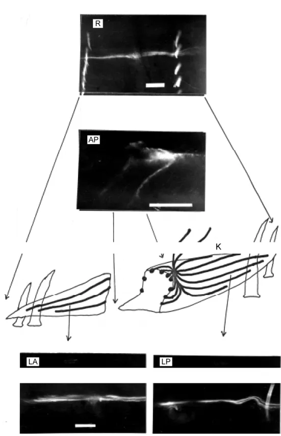
Only 9 of the 23 modulatory neurons tested showed consistent morphology. Fig-ures 6 and 7 are two examples of these 9 modulatory neurons after filling with Luci-fer Yellow-CH. Only one longitudinal band of structure could be focused in the radial dimension (R and PK; Figures 6 and 7, spectively). Its lateral dimensional view re-tained all the morphological features of the known INT neuron (Figure 6C in Ref. 3), i.e., two afferent processes in the third seg-mental nerve trunk and two bundles of longi-tudinal processes at the anterior and poste-rior sides of a central junction. Both bundles are divided into several branches. Each of these branches contains numerous fine pro-cesses (LA and LP; Figure 6). The character-istic feature of the morphology of this neu-ron is revealed when the neuneu-ron is viewed in the anteroposterior dimension (AP; Figure 6) at the cross section of a Kästchen (8), where about 20 of these branches are seen distributed on the surface of this Kästchen; hence, the name, “Peri-Kästchen” neuron given in this article. The three-dimensional reconstruction of this neuron was drawn in Figure 6-K. Its relative position with respect
to the M-neurons and effector muscle cell is shown in the radial dimension (Figure 7). Comparing the PK neuron to the known intramural, or intermuscular, neurons (5-7) by replicating the histological methods used in these references, it can be seen that these so-called “intramural”, or “intermuscular”, neurons (dark cells which are actually stain-ed blue by hematoxylin against the light-colored muscle cells which are actually stain-ed pink by eosin) are distributstain-ed both on the surface and inside these three Kästchenen in Figure 8. The neurons on the surface of these Kästchenen may correspond to the longitu-dinal branches of a single PK neuron identi-fied in AP (Figure 6) instead of many indi-vidual neurons assumed in the early works. The neurons inside these Kästchenen are apparently of a separate category because no Lucifer Yellow-CH was diffused into them (AP; Figure 6). We tentatively called them “Intra-Kästchen” (IK) neurons in contrast to the PK neuron.
D iscussio n
Figure 6 - M orphology of the modulatory neuron. R, Radial di-mensional view . The tw o row s of bright objects are setae w ith natural fluorescence. AP, An-teroposterior dimensional view at t he approxim at e posit ion show n in Figure 6-K. K, Recon-struction of a Kästchen in three dimensions (not draw n to scale) w ith PK neuron branches dis-tributed on its surface and the anterior end to the left. The tw o centripetal processes at the top are inside the third segmental nerve (see Ref . 3). Circular muscle layer and epidermis are visible at the bottom of the draw ing. The coelom cavity is show n at the top of the draw -ing. Four setae can be seen, tw o at each end of this Kästchen. LA, Lateral dimensional view of part of an anterior branch of the PK neuron; LP, lateral dimen-sional view of part of a posterior branch of the PK neuron. The calibration bar in all four photo-graphs represents 100 µm.
K R
AP
used as the control, or unconditioned, UR, response in this and in the third (4) article because it is free from the facilitatory influ-ence (Figure 2-2). Since the PK response does facilitate this UR, it must be decided if it can be used as a CR in further experiments. A conditioning stimulus, as defined in a conditioning paradigm of the other animals (10-12), modifies the synaptic excitability of the unconditioning neurons persistently. The necessity of coincidence of the US and SCS for their facilitating action on the UR means that this effect is not persistent. Experiments in the third article (4) in this series were designed to find an SCS with a persistent facilitation on the UR in order to be com-pared to that in the conditioning paradigm of the other animals (10-12).
The morphology of this PK neuron iden-tified by Lucifer Yellow-CH filling solved a riddle left by histological studies (5-7) where each of these dark-stained structures (Figure 8) was counted as an individual intramural neuron. Results in Figures 6, 7 and 8 show that those on the surface of a Kästchen are branches of a single PK neuron while those inside the Kästchen are IK neurons of still unidentified morphology and physiology. Figure 7 - Anatomical positions of
t he ident if ied neurons and muscle cells. All cells w ere filled w ith Lucifer Yellow -CH and pho-tographed in the radial dimen-sion. The horizontal row of bright objects in the middle of this fig-ure are setae w ith natural fluo-rescence. The first pair of seg-mental nerves is faintly visible above the setae. The second pair of segmental nerves is near the low er edge of this figure. PK, Peri-Kästchen neuron; M , me-chanical stimulus-sensitive, or generator, neuron slightly unfo-cused on this plane; E, effector muscle cell. Calibration: 100 µm.
Figure 8 - Comparison of PK neu-rons to intramural or intermus-cular neurons. Anteroposterior dimensional view of a paraffin-embedded 10-µm thick cross-section of the body of the earth-w orm, Amynthas haw ayanus, show ing three Kästchenen. The circular muscle layer and epider-mis are visible at the bottom of the figure, and the coelomic cav-ity at the top. Dark points are neuron-like structures stained blue by hem at oxylin. Light points are muscle cells stained pink-red by eosin. Calibration: 100 µm.
Re fe re nce s
1. Chang YC, Assm é Z, Caffaro ECL & Bartoszeck AB (1998). Identification of the main generator source of longitudinal muscle contraction in the earthw orm ven-tral nerve cord. Brazilian Journal of M edi-cal and Biologiedi-cal Research, 31: 1285-1294.
2. Chang YC & Assmé Z (1989). Neurônios no circuito reflexo periférico da minhoca, Amynthas haw ayanus. Presented at the IV Reunião Anual da Federação de Socie-dades de Biologia Experim ent al, Caxambu, M G, Brasil, 21 (Abstract). 3. Chang YC, M archioro M & Assm é Z
(1991). Tw o groups of peripheral afferent neurons in the earthw orm reflex arc. Comparative Biochemistry and Physiolo-gy, Section A, 100: 563-569.
4. Chang YC, Caffaro ECL, Assm é Z &
Bartoszeck AB (1998). Persistent attenua-tion and enhancement of the earthw orm main muscle contraction generator re-sponse induced by repeated stimulation of a peripheral neuron. Brazilian Journal of M edical and Biological Research, 31: 1303-1311.
5. Dechant E (1906). Beiträge zur Kenntnis des peripheren Nervensystem des Re-genw urmes. Arbeiten von Zoologisches Institut, 16: 361-383.
6. Daw son AB (1920). The intermuscular nerve cells in the earthw orm. Journal of Comparative Neurology, 32: 155-171. 7. Zyeng DH (1930). Distribution of
inter-muscular nerve cells in the earthw orm. Tohoku Imperial University Science Re-ports, Series 4, 5: 449-466.
8. Hesse R (1894). Zur vergleichenden
Anatomie des Oligochaeten. Zeitschrift für Wissenschaftliche Zoologie, 58: 394-439. 9. Assmé Z & Chang YC (1990).
Decremen-tal propagation of reflex spikes along the giant axons of the earthw orm, Amynthas haw ayanus. Brazilian Journal of M edical and Biological Research, 23: 329-332. 10. Carew TJ, Walters ET & Kandel ER (1981).
Classical conditioning in a simple w ith-draw al reflex in Aplysia californica. Jour-nal of Neuroscience, 1: 1426-1437. 11. Lukow iak K & Sahley C (1981). The in
vitro classical conditioning of the gill w ith-draw al reflex of Aplysia californica. Sci-ence, 212: 1516-1518.
12. Sastry BR, Goh JW & Auyeung A (1986). Associative induction of posttetanic and long-term potentiations in CAl neurons of rat hippocampus. Science, 232: 988-990.
PK
M