Relationship between cardiovascular
dysfunction and hyperglycemia in
streptozotocin-induced diabetes in rats
1Instituto de Cardiologia do Rio Grande do Sul,
Fundação Universitária de Cardiologia, Porto Alegre, RS, Brasil
2Fundação Faculdade Federal de Ciências Médicas de Porto Alegre, Porto Alegre,
RS, Brasil
3Instituto de Biociências, Universidade Federal do Rio Grande do Sul, Porto Alegre,
RS, Brasil
4Hospital de Clínicas de Porto Alegre, Porto Alegre, RS, Brasil
5Laboratório de Hipertensão Experimental, Unidade de Hipertensão, Instituto do
Coração, Faculdade de Medicina, Universidade de São Paulo, São Paulo, SP, Brasil B.D. Schaan1,
P. Dall’Ago2,
C.Y. Maeda3,
E. Ferlin4,
T.G. Fernandes3,
H. Schmid2 and
M.C. Irigoyen5
Abstract
Streptozotocin (STZ)-induced diabetes in rats is characterized by cardiovascular dysfunction beginning 5 days after STZ injection, which may reflect functional or structural autonomic nervous system damage. We investigated cardiovascular and autonomic function, in rats weighing 166 ± 4 g, 5-7, 14, 30, 45, and 90 days after STZ injection (N = 24, 33, 27, 14, and 13, respectively). Arterial pressure (AP), mean AP (MAP) variability (standard deviation of the mean of MAP, SDMMAP), heart rate (HR), HR variability (standard deviation of the normal pulse intervals, SDNN), and root mean square of successive difference of pulse intervals (RMSSD) were measured. STZ induced increased glycemia in diabetic rats vs control rats. Diabetes reduced resting HR from 363 ± 12 to 332 ± 5 bpm (P < 0.05) 5 to 7 days after STZ and reduced MAP from 121 ± 2 to 104 ± 5 mmHg (P = 0.007) 14 days after STZ. HR and MAP variability were lower in diabetic vs control rats 30-45 days after STZ injection (RMSSD decreased from 5.6 ± 0.9 to 3.4 ± 0.4 ms, P = 0.04 and SDMMAP from 6.6 ± 0.6 to 4.2 ± 0.6 mmHg, P = 0.005). Glycemia was negatively correlated with resting AP and HR (r = -0.41 and -0.40, P < 0.001) and with SDNN and SDMMAP indices (r = -0.34 and -0.49, P < 0.01). Even though STZ-diabetic rats presented bradycardia and hypoten-sion early in the course of diabetes, their autonomic function was reduced only 30-45 days after STZ injection and these changes were negatively correlated with plasma glucose, suggesting a metabolic origin.
Correspondence
B.D. Schaan Unidade de Pesquisa Instituto de Cardiologia do Rio Grande do Sul Av. Princesa Isabel, 370 90620-001 Porto Alegre, RS Brasil
Fax: +55-51-230-3600
E-mail: [email protected] Publication supported by FAPESP.
Received August 13, 2003 Accepted August 3, 2004
Key words
•Diabetes •Streptozotocin •Heart rate variability
•Rats
•Autonomic nervous system •Arterial pressure variability
Introduction
Many clinical studies have demonstrated that chronic diabetic complications occur late after the onset of the disease, reflecting structural abnormalities in nerves, kidneys,
these complications in autonomic and pe-ripheral nerves (4,5). These findings are con-sistent with the hypothesis that poor meta-bolic control is a major determinant of ner-vous damage. Indeed, other studies of type 1 diabetic patients have shown that autonomic nervous system abnormalities could be re-versed when better metabolic control was obtained (6,7).
The injection of streptozotocin (STZ) in rats leads to the development of a clinical syndrome characterized by hyperglycemia, excessive osmotic diuresis and loss of weight, which is similar to human diabetes. More-over, the STZ-diabetic rat develops the usual chronic microvascular complications (ne-phropathy, peripheral and autonomic neuro-pathy) as observed in diabetic patients (8-10). Studies on 5-day STZ-diabetic rats from our laboratory have shown depressed vagal tone, reduction of vagal effect (11) and im-paired tachycardic response to arterial pres-sure (AP) decreases (12). Fifteen days after STZ injection we observed impairment of the reflex bradycardia and tachycardia pro-duced by vasopressor and vasodepressor agents, respectively (13,14). Similar cardio-vascular changes were described in this mo-del (15) and there is evidence that some of these alterations are reversed by insulin thera-py (16). These findings, associated with the impairment of baroreflex sensitivity, an ex-cellent gauge of autonomic function, sug-gested the early development of autonomic dysfunction in these animals.
Since the autonomic nervous system modulates beat-to-beat fluctuations in heart rate (HR), methods to quantify HR and blood pressure variability have been evaluated as indicators of sympathetic and parasympa-thetic modulation of the cardiovascular sys-tem in humans (17) and in experimental models (18,19). These methods seemed to detect early autonomic dysfunction at a time when other metabolic dysfunctional changes were not clearly observed. Indeed, the rela-tionship between the abnormalities in
auto-nomic modulation and glycemic control ob-served in humans has not been explored in diabetic animals.
The objective of the present study was to investigate cardiovascular and autonomic functions, evaluated by HR and blood pres-sure variability, and their relationship to gly-cemic control in STZ-induced diabetic rats at different times after STZ injection.
Material and Methods
In order to obtain a significant number of animals with diabetes of different duration, we grouped data from all cardiovascular ex-periments performed in STZ-diabetic rats in our laboratory from 2000 to 2001, assem-bling data from 111 male Wistar rats. The experiments were performed by researchers similarly trained in data collection, and con-trol and diabetic rats were always evaluated simultaneously in order to minimize pos-sible circadian and seasonal changes. The animals, weighing 150-280 g, were obtained from the Animal House of Universidade Fe-deral do Rio Grande do Sul, Porto Alegre, RS, Brazil, and kept in small groups with free access to tap water and standard rat chow.
Animals were made diabetic (D) by iv
days, respectively.
Catheters filled with saline were im-planted under anesthesia into the femoral artery and vein (PE-10) for direct measure-ment of AP and drug administration, respec-tively. One day after catheter placement, the arterial cannula was connected to a strain-gauge transducer (P23Db; Gould-Statham, Oxnard, CA, USA) and blood pressure sig-nals were recorded during a 40-min period with a microcomputer equipped with an ana-log-to-digital converter board (CODAS, 2-kHz sampling frequency; Dataq Instruments, Inc., Akron, OH, USA). Rats were conscious and moved freely during the experiments. Recorded data were analyzed on a beat-to-beat basis. To evaluate mean AP (MAP) variability, we used the standard deviation of the mean of MAP (SDMMAP) (20), while HR variability was evaluated by calculating the following indices in the time domain: 1) root mean square of successive differences of pulse intervals (RMSSD), and 2) standard deviation of the normal pulse intervals (SDNN) (18). Each method employed to analyze HR variability reflects different pat-terns of variability: RMSSD reflects high-frequency short-term variations in HR, or vagal activity; SDNN is the square root of the variance, which is mathematically equal to the total power of the signal. The total power corresponds to the contribution of all harmonic components (short-term and
long-term variation) responsible for the variability. Data are reported as means ± SEM. Sta-tistical significance was calculated by the Student t-test to compare unpaired data of D and C during each period after STZ injec-tion. The relationship between metabolic control, measured by plasma glucose and the degree of autonomic modulation, measured by the indices cited above, was evaluated by calculating the Pearson correlation coeffi-cient. Differences were considered to be sig-nificant at P <0.05 for all tests.
Results
Body weight and plasma glucose
Body weights were similar in all experi-mental groups at baseline. Five to 7 days after STZ injection, the weight of diabetic rats was similar to that of controls, although tending to be lower. The weight of the dia-betic animals was lower for all other diadia-betic groups compared to their controls after 14, 30, 45, and 90 days of diabetes. Plasma glucose levels measured after STZ injection were higher than after citrate buffer injection at all times studied (Table 1).
Arterial pressure and heart rate
Groups studied 5-7 and 14, 30, 45, and 90 days after the induction of diabetes
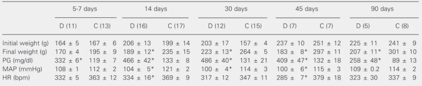
pre-Table 1. Characterization of diabetic (D) and control (C) rats 5-7, 14, 30, 45, and 90 days after STZ injection.
5-7 days 14 days 30 days 45 days 90 days
D (11) C (13) D (16) C (17) D (12) C (15) D (7) C (7) D (5) C (8)
Initial weight (g) 164 ± 5 167 ± 6 206 ± 13 199 ± 14 203 ± 17 157 ± 4 237 ± 10 251 ± 12 225 ± 11 241 ± 9 Final weight (g) 170 ± 4 195 ± 9 189 ± 12* 235 ± 15 223 ± 13* 264 ± 5 183 ± 8* 297 ± 11 207 ± 11* 301 ± 10 PG (mg/dl) 332 ± 6* 119 ± 7 466 ± 42* 133 ± 8 486 ± 40* 131 ± 21 409 ± 47* 132 ± 18 258 ± 48* 89 ± 13 MAP (mmHg) 108 ± 1 112 ± 2 104 ± 5* 121 ± 2 100 ± 4* 114 ± 3 100 ± 6* 115 ± 3 109 ± 0.2 114 ± 2 HR (bpm) 332 ± 5 363 ± 12 334 ± 16* 369 ± 9 317 ± 12 347 ± 11 285 ± 7* 379 ± 18 323 ± 30 337 ± 9
Data are reported as means ± SEM for the number of animals indicated in parentheses. PG = plasma glucose; MAP = mean arterial pressure; HR = heart rate.
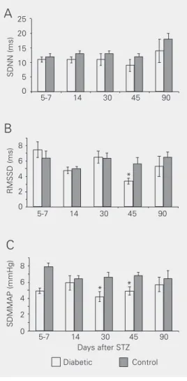
Figure 1. Standard deviation of the normal pulse intervals (SDNN, panel A), root mean square of successive difference of pulse intervals (RMSSD, panel B) and standard deviation of the mean of MAP (SDMMAP, panel C) in diabetic (D) and con-trol (C) animals 5-7 (13C, 11D), 14 (17C, 16D), 30 (15C, 12D), 45 (7C, 7D), and 90 (8D, 5C) days after STZ injection. *P < 0.05 compared to the respective con-trol (Student t-test).
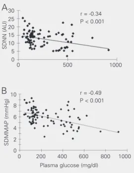
Figure 2. Scattergrams of mean arterial pressure (MAP, N = 85, panel A), and heart rate (HR, N = 87, panel B) versus plasma glu-cose in diabetic and control rats. Correlation was evaluated by the Pearson test.
SDNN (ms)
25 20 15 10 5 0
8 6
4 2 0
8
6
4
2
0
5-7 14 30 45 90
*
5-7 14 30 45 90
5-7 14 30 45 90 Days after STZ
Diabetic Control
* *
A
B
C
RMSSD (ms)
SDMMAP (mmHg)
MAP (mmHg)
160 140 120 100 80 60 40 20 0
500 400 300 200 100 0
r = -0.41 P < 0.001
0 500 1000
HR (bpm)
Plasma glucose (mg/dl)
0 500 1000
A
B
r = -0.40 P < 0.001
sented a significantly lower resting MAP compared to control at each time point of the study. HR was also lower in the D group compared to the C group at all times studied, although the difference was not statistically significant for all periods of diabetes evalu-ated (Table 1).
Autonomic function
The evaluation of autonomic function using indices of HR variability (SDNN and RMSSD) did not identify differences be-tween the groups at 5-7, 14, 30, 45, and 90 days after STZ or citrate injection. The only exception was the RMSSD of the 45-day D animals, which was lower than their C group (P = 0.04) (Figure 1). Also, MAP variability evaluated by the SDMMAP was significant-ly lower in the D vs C groups at 30 and 45
days after induction (4.2 ± 0.6 and 4.9 ± 0.5
vs 6.6 ± 0.6 and 6.8 ± 0.4 mmHg for D and C,
respectively; Table 1).
Relationship between cardiovascular changes and plasma glucose
By plotting the results obtained for all animals, D and C, it was possible to identify a negative relationship between plasma glu-cose levels and MAP (r = -0.41, P < 0.001), HR (r = -0.40, P < 0.001; Figure 2A and B, respectively), SDNN (r = -0.34, P < 0.001) and SDMMAP (r = -0.49, P < 0.001; Figure 3A and B, respectively). There was no rela-tionship between RMSSD and plasma glu-cose levels (r = -0.14, P = 0.15).
Discussion
all of these changes showed a negative cor-relation with plasma glucose.
Bradycardia in this animal model of dia-betes has already been described by us and others (11-13,18,21). Autonomic nervous system dysfunction, indicated by an increase in vagal tone or a decline in sympathetic tone to the heart may reduce HR, as suggested by Jackson and Carrier in 1983 (15), but previ-ous data from our laboratory showed that 5-day diabetic rats had in fact reduced vagal function, whereas their sympathetic tone was not significantly affected (12). Perhaps the enhanced bradycardia induced by electrical vagal stimulation or methacholine adminis-tration could account in part for the final result of lower HR (22). This bradycardia may also be caused by a change in the elec-trophysiological properties of the sinoatrial node, since intrinsic HR is reduced (12). Previous results reported by our group (23) demonstrated that diabetes-induced brady-cardia was attenuated by training, a change that was positively correlated with intrinsic HR. The metabolic improvement character-istic of exercise training may contribute to these changes.
Although some investigators have de-scribed a so-called STZ hypertension, this probably reflects discrepancies between the direct and indirect blood pressure measure-ments (24), since we and others, using the first method, consistently found hypotension in this animal model independently of the duration of diabetes (11-13,18). Osmotic diuresis causing hypovolemia or myocardial dysfunction re-ducing contractile force are putative mechan-isms of hypotension in STZ-diabetic rats (23,25). Interestingly, both mechanisms re-flect metabolic derangement. Moreover, an exaggerated pressor response to chronic hy-perinsulinemia has been reported to occur in spontaneously hypertensive diabetic rats (26), an observation that allows us to expect the opposite change in blood pressure in response to hypoinsulinemia, which is characteristic of the STZ-diabetic rat.
In humans, the earliest detectable feature of diabetic cardiac autonomic neuropathy is a defective parasympathetic control, repre-sented by persistent resting tachycardia and loss of beat-to-beat variation during deep breathing (27), functional changes related to structural changes, as confirmed by post-mortem studies (28). Fazan et al. (19) sug-gested that impairment of cardiac parasym-pathetic nerve function is present early in the course of diabetes in this animal model, as indicated by decreased HR variability. How-ever, other investigators doubt its occurrence before diabetes has been present for some time (29). Indeed, previous results from our group, evaluating MAP variability by the three-dimensional return map, showed that rats with short-term STZ diabetes even pres-ent a normal autonomic control of HR and MAP (18). The present results did not dem-onstrate any change in SDNN, the same index used by Fazan et al. (19) to assess parasympathetic autonomic neuropathy from 5 to 90 days of diabetes duration. Another study, although evaluating spontaneously hypertensive diabetic rats, analyzed the al-terations in AP and HR variability using spectral analysis approaches and found a
Figure 3. Scattergrams of the standard deviations of the nor-mal pulse intervals (SDNN, N = 85, panel A) and of the mean of MAP (SDMMAP, N = 86, panel B) versus plasma glucose in dia-betic and control rats. AU = arbi-trary units. Correlation was evaluated by the Pearson test.
SDNN (AU)
30 25 20 15 10 5 0
10 8
6
r = -0.34 P < 0.001
0 500 1000
SDMMAP (mmHg)
Plasma glucose (mg/dl)
0 200 1000
A
B
r = -0.49 P < 0.0014
2 0
reduced variability of AP without changes in HR variability in diabetic animals (30). This disagreement may be accounted for by the differences in the sensitivity of the methods used to evaluate autonomic dysfunction, i.e., time domain or frequency domain indices. The differences between the current results and previous ones obtained by our group could also be accounted for by different methodologies. Disturbances in autonomic function were observed in 5-day diabetic rats when evaluated by baroreflex sensitivity and after autonomic blockade with propran-olol and atropine (11,12), which could be more sensitive than the indices used in the present study.
Spontaneous fluctuations in MAP and HR have been used to detect autonomic dysfunction in humans (17). The reduced MAP variability, evaluated by SDMMAP, observed in the diabetic groups in the pres-ent study may indicate early functional auto-nomic cardiovascular dysfunction (31). The indices used to evaluate MAP and HR varia-bility were negatively correlated with plasma glucose, suggesting that MAP and HR could be altered by diabetic metabolic decompen-sation. Accordingly, in spontaneously hy-pertensive diabetic rats, reflex bradycardic index and the power of low frequency oscil-lations of systolic arterial pressure are in-versely related to blood glucose, reinforcing the crucial role of metabolic changes in the cardiovascular disorder in this model (30). Hicks et al. (21), in 1998, observed the same tendencies as observed here: a decline in the magnitude of the circadian variation and decreased sympathetic and parasympathetic nervous tone to the heart after STZ injection, changes which were reversed by insulin treat-ment.
It is unlikely that bradycardia and hypo-tension could be the result of autonomic neural structural injury; instead, they could represent autonomic neural dysfunction caused by metabolic changes induced by STZ diabetes. This is supported by the
nega-tive correlation observed between MAP and HR with plasma glucose. Indeed, other in-vestigators have demonstrated that brady-cardia and hypotension induced by experi-mental diabetes can be prevented by insulin treatment (16,30). Our group observed re-duced intrinsic HR in short-term STZ diabe-tes, which could be the result of metabolic injury to pacemaker cells (11). Reduced motor nerve conduction velocity is also nor-malized by insulin treatment in this animal model (32). These data suggest that these early phenomena constitute a stage of neuro-logical dysfunction distinct from “true” neu-ropathy (28). In fact, other investigators did not find structural abnormalities in the vagus nerve, which consists of parasympathetic preganglionic fibers, or enteric ganglia, which consist of parasympathetic neurons in STZ-diabetic rats with a 6- to 12-month duration of diabetes (33,34). Conversely, relevant structural changes characterized by axonal dystrophy did occur in the sympathetic pre-ganglionic nerve fibers, but these changes were observed 12 months after STZ injec-tion (34).
Several mechanisms have been consid-ered to be involved in the pathogenesis of diabetic neuropathy in the STZ-diabetic rat, but hyperglycemia is always implicated. Structural changes occurring in peripheral nerves resemble those of human diabetic neuropathy and are preceded by hyperglyce-mia-induced biochemical abnormalities. Recent evidence has emphasized the impor-tance of vascular dysfunction, driven by metabolic insults to the nerves, as a cause of diabetic neuropathy. Non-enzymatic glyco-sylation of myelin components, reduction of endoneural blood flow, increased oxygen free radical activity, or production and dep-rivation of the nerve growth factor are also involved (32,35).
glycemic control in type 1 diabetic patients, and comparable results were also published by Muhr-Becker et al. (37) in a study evalu-ating cardiac sympathetic dysinnervation scintigraphically in a similar group of pa-tients.
A direct effect of hyperglycemia on vas-cular and myocardial cells should also be considered as a cause for cardiovascular dys-function in this model. The protein kinase C (PKC) pathway, which is activated by hy-perglycemia, has been recently recognized as an important mechanism in the develop-ment of diabetic complications including car-diomyopathy and angiopathy (38). Up-regu-lation of PKCß2 was demonstrated in the heart and aorta at both the transcriptional and translational levels during the early stages of experimental diabetes, suggesting its role
in the diabetic injury to the cardiovascular system (39).
Our results demonstrate some well-known cardiovascular alterations presented by a commonly used diabetic model, the STZ-diabetic rat, such as hypotension and brady-cardia, and transitory autonomic index de-rangements. Considering data reported by other investigators who demonstrated that autonomic nerve structural lesions do not appear as early as these functional changes, we question whether these changes could be assigned to early development of autonomic neuropathy in this model, but we accept the presence of reversible neurological dysfunc-tion. The negative correlation observed be-tween cardiovascular dysfunction and plasma glucose is in accordance with these observa-tions.
References
1. Pirart J (1984). Glycaemic control and prevention of complications.
Minerva Endocrinology, 9: 55-58.
2. The Diabetes Control and Complications Trial Research Group (1993). The effect of intensive treatment of diabetes on the devel-opment and progression of long term complications in insulin-de-pendent diabetes mellitus. New England Journal of Medicine, 329: 977-986.
3. UK Prospective Diabetes Study (UKPDS) Group (1998). Intensive blood-glucose control with sulphonylureas or insulin compared with conventional treatment and risk of complications in patients with type 2 diabetes (UKPDS 33). Lancet, 352: 837-853.
4. Pfeifer MA, Weinberg CR, Cook DL, Reenan A, Halter JB, Ensinck JW & Porte Jr D (1984). Autonomic neural dysfunction in recently diagnosed diabetic subjects. Diabetes Care, 7: 447-453.
5. Young RJ, Ewing DJ & Clarke BF (1983). Nerve function and meta-bolic control in teenage diabetics. Diabetes, 32: 142-147.
6. Hreidarsson AB (1981). Acute, reversible autonomic nervous sys-tem abnormalities in juvenile insulin-dependent diabetes: A pupillographic study. Diabetologia, 20: 475-481.
7. Ferreira SR, Cesarini PR, Vivolo MA & Zanella MT (1998). Abnormal nocturnal blood pressure fall in normotensive adolescents with insulin-dependent diabetes is ameliorated following glycemic im-provement. Brazilian Journal of Medical and Biological Research, 31: 523-528.
8. Jensen PK, Sandahl Christiansen J, Steven K & Parving H-H (1981). Renal function in streptozotocin-diabetic rats. Diabetologia, 21: 409-414.
9. Cameron NE, Cotter MA & Maxfield EK (1993). Anti-oxidant treat-ment prevents the developtreat-ment of peripheral nerve dysfunction in streptozotocin-diabetic rats. Diabetologia, 36: 299-304.
10. Schmidt RE & Plurad SB (1986). Ultrastructural and biochemical characterization of autonomic neuropathy in rats with chronic strep-tozotocin diabetes. Journal of Neuropathology and Experimental Neurology, 45: 525-544.
11. Maeda CY, Fernandes TG, Timm HB & Irigoyen MC (1995). Auto-nomic dysfunction in short-term experimental diabetes. Hyperten-sion, 26: 1000-1004.
12. Maeda CY, Fernandes TG, Lulhier F & Irigoyen MC (1995). Strepto-zotocin diabetes modifies arterial pressure and baroreflex sensitivi-ty in rats. Brazilian Journal of Medical and Biological Research, 28: 497-501.
13. Dall’Ago P, Fernandes TG, Machado UF, Belló AA & Irigoyen MC (1997). Baroreflex and chemoreflex dysfunction in streptozotocin-diabetic rats. Brazilian Journal of Medical and Biological Research, 30: 119-124.
14. De Angelis K, Schaan BD, Maeda CY, Dall’Ago P, Wichi RB & Irigoyen MC (2002). Cardiovascular control in experimental diabe-tes. Brazilian Journal of Medical and Biological Research, 35: 1091-1100.
15. Jackson CV & Carrier GO (1983). Influence of short-term experi-mental diabetes on blood pressure and heart rate in response to norepinephrine and angiotensin II in the conscious rat. Journal of Cardiovascular Pharmacology, 5: 260-265.
16. Chang KSK & Lund DD (1986). Alterations in the baroreceptor reflex control of heart rate in streptozotocin diabetic rats. Journal of Mo-lecular and Cellular Cardiology, 18: 617-624.
18. Schaan BD, Maeda CY, Timm H, Medeiros S, Moraes R, Ferlin E, Fernandes TG, Ribeiro JP, Schmid H & Irigoyen MC (1997). Time course of changes in heart rate and blood pressure variability in streptozotocin-induced diabetic rats treated with insulin. Brazilian Journal of Medical and Biological Research, 30: 1081-1086. 19. Fazan R, Ballejo G, Salgado MC, Moraes MFD & Salgado HC (1997).
Heart rate variability and baroreceptor function in chronic diabetic rats. Hypertension, 30: 632-635.
20. Mancia G & Zanchetti A (1986). Blood pressure variability. In: Zanchetti A & Tarazi RC (Editors), Handbook of Hypertension. Vol. 7.
Pathophysiology of Hypertension. Cardiovascular Aspects. Elsevier Science Publishers B.V., Amsterdam, The Netherlands, 125-152. 21. Hicks KK, Seifen E, Stimers JR & Kennedy RH (1998). Effects of
streptozotocin-induced diabetes on heart rate, blood pressure and cardiac autonomic nervous control. Journal of the Autonomic Ner-vous System, 69: 21-30.
22. Dall’Ago P, Silva VO, De Angelis KL, Irigoyen MC, Fazan Jr R & Salgado HC (2002). Reflex control of arterial pressure and heart rate in short-term streptozotocin diabetic rats. Brazilian Journal of Medi-cal and BiologiMedi-cal Research, 35: 843-849.
23. De Angelis KLD, Oliveira AR, Dall’Ago P, Peixoto LRA, Gadonsky G, Lacchini S, Fernandes TG & Irigoyen MC (2000). Effects of exercise training on autonomic and myocardial dysfunction in streptozotocin-diabetic rats. Brazilian Journal of Medical and Biological Research, 33: 635-641.
24. Kusaka M, Kishi K & Sokabe H (1987). Does so-called streptozotocin (STZ) hypertension exist in rats? Hypertension, 10: 517-521. 25. Hebden RA, Gardiner SM, Bennett T & MacDonald IA (1986). The
influence of streptozotocin-induced diabetes mellitus on fluid and electrolyte handling in rats. Clinical Science, 70: 111-117. 26. Brands MW, Garrity CA, Holman MG & Hall JE (1994). Exaggerated
pressor and chronotropic response to chronic hyperinsulinemia in SH versus WKY rats. American Journal of Hypertension, 7: 75-81. 27. Ewing DJ, Campbell W & Clarke BF (1980). The natural history of
diabetic autonomic neuropathy. Quarterly Journal of Medicine, 193: 95-108.
28. Duchen LW, Anjorin A, Watkins P & MacKay JD (1980). Pathology of autonomic neuropathy in diabetes mellitus. Annals of Internal Medicine, 92: 301-303.
29. Hounsom L & Tomlinson DR (1997). Does neuropathy develop in
animal models? Clinical Neuroscience, 4: 380-389.
30. Farah VMA, De Angelis K, Fiorino P, Joaquim LF, Ayala E, Fazan Jr R, Morris M & Irigoyen MC (2003). Autonomic modulation of arterial pressure and heart rate variability in hypertensive diabetic rats.
Annals of the XV Scientific Meeting of the Inter-American Society of Hypertension, San Antonio, TX, USA, April 27-30,92 (P127). 31. Mésangeau D, Laude D & Elghozi J-L (2000). Early detection of
cardiovascular autonomic neuropathy in diabetic pigs using blood pressure and heart rate variability. Cardiovascular Research, 45: 889-899.
32. Greene DA, De Jesus PVJ & Winegrad AI (1975). Effects of insulin and dietary myoinositol on impaired peripheral motor nerve conduc-tion velocity in acute streptozotocin diabetes. Journal of Clinical Investigation, 55: 1326-1336.
33. Schmidt RE, Plurad SB & Modert CW (1983). Experimental diabetic autonomic neuropathy characterization in streptozotocin-diabetic Sprague-Dawley rats. Laboratory Investigation,49: 538-552. 34. Kniel PC, Junker U, Perrin IV, Bestetti GE & Rossi GL (1986). Varied
effects of experimental diabetes on the autonomic system of the rat. Laboratory Investigation, 54: 523-530.
35. Lee PG, Hohman TC, Cai F, Regalia J & Helke CJ (2001). Streptozo-tocin-induced diabetes causes metabolic changes and alterations in neurotrophin content and retrograde transport in the cervical vagus nerve. Experimental Neurology, 170: 149-161.
36. Burger AJ, Weinrauch LA, D’Elia JA & Aronson D (1999). Effect of glycemic control on heart rate variability in type I diabetic patients with cardiac autonomic neuropathy. American Journal of Cardiol-ogy, 84: 687-691.
37. Muhr-Becker D, Weiss M, Tatsch K, Wolfram G, Standl E & Schnell O (1999). Scintigraphically assessed cardiac sympathetic dysinner-vation in poorly controlled type 1 diabetes mellitus: one-year follow-up with improved metabolic control. Experimental and Clinical En-docrinology and Diabetes, 107: 306-312.
38. Idris I, Gray S & Donnelly R (2001). Protein kinase C activation: isozyme-specific effects on metabolism and cardiovascular compli-cations in diabetes. Diabetologia, 44: 659-673.