Re fle x co ntro l o f arte rial pre ssure and
he art rate in sho rt-te rm stre pto zo to cin
diabe tic rats
1Departamento de Fisiologia, Instituto de Ciências Básicas da Saúde,
Universidade Federal do Rio Grande do Sul, Porto Alegre, RS, Brasil
2Unidade de Hipertensão e Divisão de Experimentação, Instituto do Coração,
Faculdade de Medicina, Universidade de São Paulo, São Paulo, SP, Brasil
3Departamento de Fisiologia, Faculdade de Medicina de Ribeirão Preto,
Universidade de São Paulo, Ribeirão Preto, SP, Brasil P. Dall’Ago1,
V.O .K. Silva1,
K.L.D. De Angelis1,
M.C. Irigoyen1,2,
R. Fazan Jr.3 and
H.C. Salgado3
Abstract
Impaired baroreflex sensitivity in diabetes is well described and has been attributed to autonomic diabetic neuropathy. In the present study conducted on acute (10-20 days) streptozotocin (STZ)-induced dia-betic rats we examined: 1) cardiac baroreflex sensitivity, assessed by the slope of the linear regression between phenylephrine- or sodium nitroprusside-induced changes in arterial pressure and reflex changes in heart rate (HR) in conscious rats; 2) aortic baroreceptor function by means of the relationship between systolic arterial pressure and aortic depressor nerve (ADN) activity, in anesthetized rats, and 3) bradycar-dia produced by electrical stimulation of the vagus nerve or by the iv
injection of methacholine in anesthetized animals. Reflex bradycardia (-1.4 ± 0.1 vs -1.7 ± 0.1 bpm/mmHg) and tachycardia (-2.1 ± 0.3 vs
-3.0 ± 0.2 bpm/mmHg) were reduced in the diabetic group. The gain of the ADN activity relationship was similar in control (1.7 ± 0.1% max/ mmHg) and diabetic (1.5 ± 0.1% max/mmHg) animals. The HR response to vagal nerve stimulation with 16, 32 and 64 Hz was 13, 16 and 14% higher, respectively, than the response of STZ-treated rats. The HR response to increasing doses of methacholine was also higher in the diabetic group compared to control animals. Our results confirm the baroreflex dysfunction detected in previous studies on short-term diabetic rats. Moreover, the normal baroreceptor function and the altered HR responses to vagal stimulation or methacholine injection suggest that the efferent limb of the baroreflex is mainly responsible for baroreflex dysfunction in this model of diabetes.
Co rre spo nde nce H.C. Salgado
Departamento de Fisiologia FMRP, USP
Av. Bandeirantes, 3900 14049-900 Ribeirão Preto, SP Brasil
Fax: + 55-16-633-0017 E-mail: hcsalgad@ fmrp.usp.br
Research supported by CNPq, CAPES, FAPERGS, FAPESP and PRO NEX.
Received March 5, 2001 Accepted March 18, 2002
Ke y words
•Baroreflex
•Baroreceptors
•Streptozotocin
•Vagus nerve
Intro ductio n
Autonomic neuropathy is the most seri-ous complication of diabetes in terms of morbidity and mortality (1). It has been fre-quently reported that diabetes can affect both somatic (mainly sensitive) and autonomic nerves. The arterial baroreceptors are an
still controversial whether both components (tachycardia and bradycardia) of the barore-flex are affected and whether or not the time course of diabetes affects the degree of baro-reflex dysfunction. Reflex tachycardia due to a decrease in AP was found to be attenu-ated as early as five days after the onset of experimental diabetes induced by streptozo-tocin (STZ) in rats (6). However, the reflex bradycardia due to an increase in AP was found to be preserved in this early stage of experimental diabetes. Conversely, reflex bradycardia is impaired in chronic alloxan diabetes in rabbits while reflex tachycardia is preserved in this model of diabetes (5).
Most of the reports showing an impair-ment of baroreflex sensitivity have attrib-uted it to autonomic neuropathy, while the afferent arm of the baroreflex, i.e., the arteri-al (carotid and aortic) baroreceptors, has received much less attention. McDowell et al. (7) demonstrated that the slope of the pressure-aortic nerve activity curve and the slope of the pressure-renal nerve activity curve were not affected by chronic alloxan diabetes in rabbits. These findings indicate that the baroreceptor function as well as the reflex control of renal sympathetic activity are preserved in this model of experimental diabetes. Fazan Jr. et al. (8) also showed normal aortic baroreceptor function in rats with chronic STZ-induced diabetes.
In the present investigation we examined the baroreflex control of heart rate (HR) in rats with short-term diabetes induced by STZ, as well as the aortic baroreceptor function and the HR response to vagal stimulation or muscarinic agonist injections.
Mate rial and Me thods
Animal preparation
The experiments were performed on male Wistar rats weighing 200-250 g, housed in individual cages with free access to water and food. Diabetes was induced by a single
intravenous injection of STZ (50 mg/kg; Sigma, St. Louis, MO, USA) after an over-night fast (8-10 h). Control rats received only vehicle (10 mM citrate buffer, pH 4.5) after a similar fasting period. Data were ob-tained 10 to 20 days after STZ or citrate buffer administration.
Basal he modynamics and barore fle x
Twenty-four hours before the experiments, control (N = 11) and diabetic (N = 11) rats, lightly anesthetized with ether, were implanted with catheters into the femoral artery and vein for AP measurement and drug administration, respectively. On the day of the experiment, the arterial catheter of the conscious freely mov-ing rat was connected to a pressure transducer (P23Gb, Gould-Statham, Oxnard, CA, USA) and pulsatile AP was sampled (1 kHz) for approximately 30 min with a personal com-puter equipped with an analog-to-digital inter-face (Dataq Instruments, Akron, OH, USA). After the basal recording of AP, the animals received increasing doses of phenylephrine (0.25 to 32 µg/kg) or sodium nitroprusside (0.05 to 1.6 µg/kg) given in intravenous bolus injections (0.1 ml). An appropriate interval between doses was allowed for AP and HR to return to basal levels. The values of diastolic and systolic arterial pressure (SAP) were de-tected with Advanced CODAS, Dataq soft-ware and beat by beat values of mean arterial pressure (MAP) and HR were calculated. The maximal response of MAP to phenylephrine or sodium nitroprusside, and the correspond-ing maximal reflex change in HR were col-lected for each dose of the drug. The slope of the linear regression calculated on the basis of reflex changes in HR related to the induced changes in MAP was used as the index of baroreflex sensitivity.
Vagal ne rve stimulation and bradycardic
re sponse s to me thacholine
stimulation (5 V, 2 ms, 2 to 64 Hz, for 10 s) of the right vagus nerve was studied in rats anesthetized with thiopental (35 mg/kg, iv). The interval between stimulations was de-termined on the basis of the time required for HR to return to basal levels. Body tempera-ture was maintained at 37ºC by external heating.
After vagal stimulation, the sensitivity of the muscarinic receptors of the heart was tested by evaluating HR responses to intra-venous injections of increasing doses (5, 7.5 and 10 µg/kg) of methacholine.
Barore ce ptor re cording
In additional control (N = 14) and diabetic (N = 17) groups of rats, whole aortic nerve activity was recorded as described elsewhere (8,9). Briefly, under sodium pentobarbital (40 mg/kg, ip) anesthesia the aortic nerve was isolated and placed on a bipolar stainless steel pair of electrodes. Nerve activity was ampli-fied (Princeton Applied Research Amplifier, model 113, Princeton, NJ, USA) and moni-tored on an oscilloscope (Tektronix, model 5113, Beaverton, OR, USA) associated with a loudspeaker. Action potentials that exceeded a threshold level set right above the noise level of each nerve activity were counted with a nerve traffic analyzer (University of Iowa Bio-engineering, Iowa City, IA, USA) every 200 ms. Integrated nerve activity was sampled (100 Hz) with a personal computer simultaneously with pulsatile carotid pressure. The barorecep-tor firing range was assessed by rapid changes (20 to 30 s) in AP due to withdrawal and reinfusion of blood into the femoral artery of the rats. To avoid the influence of hysteresis, only the values obtained during the increase in pressure (rate of 6-7 mmHg/s) were used. Aortic nerve activity (spikes/s) was normal-ized (% of maximal activity) and plotted against SAP to obtain pressure-nerve activity curves. Using a nonlinear regression based on the Levemberg-Marquadt algorithm (Marquadt, 1964) we calculated the maximum gain of the
pressure-nerve activity curve and the SAP at 50% of maximal nerve activity (SAP50) as the parameters of baroreceptor function.
Blood analysis
Immediately after the experiments, blood was collected from the arterial catheter to measure blood glucose and insulin by a col-orimetric enzymatic test and by radioimmu-noassay, respectively.
Statistical analysis
Data from pressure-nerve activity curves, vagal stimulation and methacholine injec-tions were processed by multivariate analy-sis of variance (MANOVA) for repeated measures. If differences were observed a
post hoc test (Newman-Keuls or Tukey) was employed. The unpaired Student t-test was used to compare the other parameters be-tween control and diabetic rats. Changes were considered significant at P<0.05.
Re sults
All rats given STZ exhibited severe hy-perglycemia (447 ± 49 vs 126 ± 3 mg/dl, P = 0.001) and insulinopenia (16 ± 1 vs 57 ± 11 µU/ml, P = 0.002) associated with a de-crease in body weight (200 ± 8 vs 268 ± 5 g, P = 0.001) as compared to control rats.
Resting HR (322 ± 5 vs 341 ± 6 bpm; P = 0.03) and resting MAP (93 ± 2 vs 104 ± 3 mmHg; P<0.01) were significantly reduced in diabetic rats.
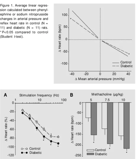
The reflex bradycardia elicited by phen-ylephrine (-1.4 ± 0.1 vs -1.7 ± 0.1 bpm/ mmHg; P = 0.03) as well as the reflex tachy-cardia elicited by sodium nitroprusside (-2.1 ± 0.3 vs -3.0 ± 0.2 bpm/mmHg; P = 0.02) were significantly reduced in the diabetic group as indicated by the slope of the linear regression relating changes in HR to changes in MAP (Figure 1).
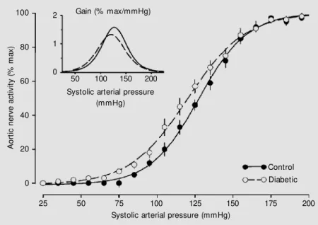
stimula-max/mmHg) and diabetic (1.5 ± 0.1% max/ mmHg) animals. The pressure (aortic de-pressor nerve) activity curve for each group is shown in Figure 3. As indicated by the SAP50 and the gain, the curve for STZ-treated animals was similar to that for control rats.
D iscussio n
The high blood glucose and low insulin levels presented by STZ-treated rats confirm the efficacy of STZ in producing diabetes in rats.
Basal levels of AP and HR were reduced in STZ-treated rats. The literature has re-ported the occurrence of hypertension (10), normotension (11) or hypotension (12,13) in experimental diabetes. A reduction in rest-ing HR is usually accompanied by a reduc-tion in AP in STZ-induced diabetes (13). The mechanisms underlying the association of diabetes and bradycardia are not known, but may reflect changes in the electrophysi-ological properties of the sino-atrial node as indicated by a depressed spontaneous activ-ity of the pacemaker observed in isolated hearts (14). A possible mechanism to ex-plain the hemodynamic changes observed in diabetic rats may be similarly related to al-terations in autonomic control. Indeed, we have previously demonstrated that sympa-thetic and parasympasympa-thetic tone to the heart was reduced in short-term experimental dia-betes (12). These changes were accompa-nied by changes in AP and HR variability and were prevented by treatment with insu-lin (15).
Impairment of cardiac baroreflex in dia-betic patients (4) and experimental models of diabetes has been demonstrated in a num-ber of studies (5,7).
We may hypothesize that this impair-ment is a consequence of the reduced sensi-tivity of sensory baroreceptor endings to AP changes or a consequence of a dysfunction in central baroreflex integration. In addition, an alteration of efferent pathways, including
Figure 1. Average linear regres-sion calculated betw een phenyl-ephrine or sodium nitroprusside changes in arterial pressure and reflex heart rate in control (N = 11) and diabetic (N = 11) rats. * P< 0.05 compared to control (Student t-test).
∆ H e a rt r a te ( b p m ) 100 50 0 -50 -100
-40 -20 0 20 40
∆ M ean arterial pressure (mmHg)
* * ∆ H e a rt r a te ( % ) 0 -20 -40 -60 -80 -100 -120 Control Diabetic
1 10 100
Stimulation frequency (Hz)
∆ H e a rt r a te ( b p m ) -50 -100 -150 -200 -250
5 7.5 10
Control Diabetic A A A A
A BBBBB
*
*
*
Figure 2. Effect of electrical stimulation of the right vagal nerve (A) on heart rate in control (N = 11) and diabetic rats (N = 11). B, Heart rate responses produced by increasing doses of methacholine (5, 7.5 and 10 µg/kg) in control and diabetic rats. Data are reported as means ± SEM . * P<0.05 compared to control (multivariate analysis of variance follow ed by Tukey post-test).
tion with 16, 32 and 64 Hz was 13, 16 and 14% higher than in STZ-treated rats when compared to control. The HR response to 2, 4 and 8 Hz was similar in normal and dia-betic rats (Figure 2A).
The HR response to increasing doses of methacholine (5, 7.5 and 10 µg/kg) was sig-nificantly higher in the diabetic group (-152 ± 29, -192 ± 13, -213 ± 11 vs -73 ± 20, -124 ± 26, -157 ± 18 bpm in controls, respec-tively; P<0.05), see Figure 2B.
SAP50 did not differ between diabetic (123 ± 3 mmHg) and control (128 ± 2 mmHg) rats. The gain of the pressure-nerve activity curve was also similar in control (1.7 ± 0.1%
0
vagal efferent neurons, or changes in the responsiveness of muscarinic receptors to acetylcholine may also be taken into consid-eration.
An interesting finding of the present study was that short-term (10 to 20 days) STZ-induced diabetes showed an impairment of the reflex control of HR associated with normal baroreceptor function. In addition, the bradycardia induced by electrical vagal stimulation or methacholine administration was enhanced in diabetic rats. Moreover, the resting bradycardia and hypotension ob-served in the present study were similar to those obtained in previous ones.
The preserved baroreceptor function dur-ing short-term (10 to 20 days) diabetes ob-served in the present study agrees with previ-ous observations of normal baroreceptor function in rats with chronic STZ-induced diabetes (8) or in rabbits with alloxan-in-duced diabetes (7). Therefore, the attenu-ated HR response due to baroreceptor acti-vation (increase in AP due to phenylephrine) or deactivation (decrease in AP due to so-dium nitroprusside) in short-term diabetes cannot be related to changes in the afference of the reflex.
Studies in humans have clearly demon-strated that the baroreflex-mediated brady-cardia in response to an increase in AP is impaired in diabetic subjects (16-18). In con-trast, the reflex tachycardia induced by a decrease in AP is preserved (18) or impaired (13). The relative role played by the sympa-thetic and parasympasympa-thetic nervous system in these changes is not completely under-stood.
Animal models used to investigate the mechanism(s) of baroreflex dysfunction in diabetes have provided different results. Re-flex bradycardia due to the increase in AP has been found to be normal (6), reduced (19) or enhanced (20). The differences in baroreflex responses reported by different authors may be due to different animal mod-els, drugs used and time course of
develop-ment of diabetes. In fact, we observed that 5 days after STZ administration the reflex tach-ycardia was reduced while the reflex brady-cardia was not (6). However, 15 days after STZ administration we observed an attenua-tion of both responses (13). The present study confirms these previous findings, sug-gesting that in conscious rats the time course of diabetes may play a role in baroreflex dysfunction. In addition, Fazan Jr. et al. (21) showed a progressive reduction in AP and HR variability with the progress of diabetes in rats.
Although the parasympathetic tone evalu-ated by pharmacological blockade was de-pressed as early as at 5 days of STZ-induced diabetes (12), only the sympathetic control of baroreflex-mediated changes in HR (tach-ycardia) was reduced at that time. However, the impairment in baroreflex-mediated brady-cardia in response to an increase in AP ob-served in the present study may indicate that a parasympathetic derangement was attenu-ating the reflex control of HR in diabetes.
The bradycardia elicited by electrical stimulation of vagal efferents in the range of 16 to 64 Hz induced a greater response in
A
o
rt
ic
n
e
rv
e
a
c
ti
v
it
y
(
%
m
a
x
)
100
80
60
40
20
0
25 50 75 100 125 150 175 200
Systolic arterial pressure (mmHg)
Control Diabetic 2
1
0
Gain (% max/mmHg)
Systolic arterial pressure (mmHg)
50 100 150 200
diabetic rats compared to control. However, at lower frequencies of stimulation (i.e., 2 to 8 Hz) we did not find any difference. These results suggest that the vagal fibers heading to the heart are not responsible for the re-duced reflex bradycardia in diabetic rats. An efficient vagal discharge may induce a greater bradycardia in STZ-induced diabetic rats. Moreover, the greater bradycardia observed in diabetic rats in response to methacholine could be explained by several possible de-rangements such as a possible change in the number and/or affinity of the muscarinic receptors in the heart of the diabetic rats.
The study of McDowell et al. (7) showed no differences in the response to vagal elec-trical stimulation at frequencies up to 16 Hz. In contrast, we did not observe differences in the lower frequency range. It is possible that these differences may be attributed to the experimental models used, i.e., rats treated with STZ versus rabbits treated with alloxan. Moreover, the bradycardia obtained in the present study was greater in response not
only to the higher frequency range of stimu-lation but also to the agonist of the musca-rinic receptors.
The reduced baroreflex tachycardia ob-served in diabetic rats may be related to a decreased sympathetic tone to the heart. Moreover, the changes observed in vagal tone may be contributing to the sympathetic-mediated reflex tachycardia since changes in sympathetic function at different levels of parasympathetic tone have been demon-strated (12).
The altered HR responses to vagal stimu-lation or methacholine injection with normal baroreceptor function suggest that the effer-ent limb of the baroreflex is the main factor responsible for baroreflex dysfunction in this model of diabetes.
Ackno wle dgm e nts
The authors acknowledge the technical assistance of Jaci A. Castania.
Re fe re nce s
1. Ew ing DJ, Campbell IW & Clarke BF (1980). The natural history of diabetic au-tonomic neuropathy. Quarterly Journal of M edicine, 49: 95-108.
2. Ganguly PK, Beamish RE, Dhalla KS, Innes IR & Dhalla NS (1987). Norepinephrine storage, distribution, release in diabetic cardiom yopathy. Am erican Journal of Physiology, 252: E734-E739.
3. Lund DD, Subieta AR, Pardini BJ & Chang KSK (1992). Alterations in cardiac para-sympathetic indices in STZ-induced dia-betic rats. Diabetes, 41: 160-166. 4. Eckeberg DL, Harkins SW, Fritsch JM ,
M usgrave GE & Gardner DF (1986). Baro-reflex control of plasma norepinephrine and heart period in healthy subjects and diabetic patients. Journal of Clinical Inves-tigation, 78: 366-374.
5. M cDow ell TS, Chapleau M W, Hajduczok G & Abboud FM (1994). Baroreflex dys-function in diabetes mellitus, I: selective impairment of parasympathetic control of heart rate. American Journal of Physiolo-gy, 266: H235-H243.
6. M aeda CY, Fernandes TG, Lulhier F & Irigoyen M C (1995). Streptozotocin diabe-tes modifies arterial pressure and barore-flex sensitivity in rats. Brazilian Journal of M edical and Biological Research, 28: 497-501.
7. M cDow ell TS, Hajduczok G, Abboud FM & Chapleau M W (1994). Baroreflex dys-function in diabetes mellitus. II: Site of baroreflex impairment in diabetic rabbits.
American Journal of Physiology, 266: H244-H249.
8. Fazan Jr R, Ballejo G, Salgado M CO, M oraes M FD & Salgado HC (1997). Heart rate variability and baroreceptor function in chronic diabetic rats. Hypertension, 30: 632-635.
9. Dias da Silva VJ, Vargas da Silva S, Salgado M CO & Salgado HC (1994). Chronic con-verting enzyme inhibition facilitates baro-receptor ressetting to hypertensive lev-els. Hypertension, 23: I-68-I-72.
10. Katayama S & Lee JB (1985). Hyperten-sion in experimental diabetes mellitus, re-nin-prostaglandin interaction.
Hyperten-sion, 7: 554-561.
11. Hicks KK, Seifen E, Stimers JR & Kennedy RH (1997). Diabetes w ith and w ithout ke-toacidosis on right atrial pacemaker rate and autonomic responsiveness. American Journal of Physiology, 273: H1888-H1893. 12. M aeda CY, Fernandes TG, Timm H & Irigoyen M C (1995). Autonomic dysfunc-tion in short-term experimental diabetes.
Hypertension, 26: 1000-1004.
13. Dall’Ago P, Fernandes TG, M achado UF, Bello AA & Irigoyen M C (1997). Barore-flex and chemoreBarore-flex dysfunction in strep-tozotocin-diabetic rats. Brazilian Journal of M edical and Biological Research, 30: 119-124.
14. Akiyama N, Okomura K, Watanabe Y, Ha-shimoto H, Ito T, Ogaw a K & Satake T (1989). Altered acetylcholine and norepi-nephrine concentrations in diabetic rat heart. Diabetes, 38: 231-236.
rate and blood pressure variability in strep-tozotocin-induced diabetic rats treated w ith insulin. Brazilian Journal of M edical andBiological Research, 30: 1081-1086. 16. Hilsted J (1982). Pathophysiology in
dia-betic autonomic neurophathy: cardiovas-cular, hormonal, and metabolic studies.
Diabetes, 31: 730-737.
17. Page M M & Watkins PJ (1978). Cardio-respiratory arrest and diabetic autonomic neuropathy. Lancet, 1: 15-16.
18. Hom m a S, Yam azaki Y & Karakida T
(1993). Blood pressure and heart rate re-lationships during cervical sympathetic and vagus nerve stimulation in streptozo-tocin diabetic rats. Brain Research, 629: 342-344.
19. Van Buren T, Vleeming W, Krutzen M M , Van de Kuil T, Gipsen WH & De Wildt DJ (1998). Vascular responses of isolated mesenteric resistance and basilar arteries from short- and long-term diabetic rats.
Naunyn-Schmiedebergs Archives of Phar-macology, 358: 663-670.
20. Chang KSK & Lund DD (1986). Alterations in the baroreceptor reflex control of heart rate in streptozotocin diabetic rats. Jour-nal of M olecular and Cellular Cardiology, 18: 617-624.