Ure ase s display bio lo gical e ffe cts
inde pe nde nt o f e nzymatic activity.
Is the re a co nne ctio n to dise ase s
cause d by ure ase -pro ducing bacte ria?
1Programa de Pós-Graduação em Biologia Celular e Molecular,
Centro de Biotecnologia, 2Departamento de Biofísica, Instituto de Biociências,
Universidade Federal do Rio Grande do Sul, Porto Alegre, RS, Brasil D. O livera-Severo1*,
G.E. Wassermann1*
and C.R. Carlini1,2
Abstract
Ureases are enzymes from plants, fungi and bacteria that catalyze the hydrolysis of urea to form ammonia and carbon dioxide. While fungal and plant ureases are homo-oligomers of 90-kDa subunits, bacterial ureases are multimers of two or three subunit complexes. We showed that some isoforms of jack beanurease, canatoxin and the classical urease, bind to glycoconjugates and induce platelet aggregation. Canatoxin also promotes release of histamine from mast cells, insulin from pancreatic cells and neurotransmitters from brain synaptosomes.
In vivo it induces rat paw edema and neutrophil chemotaxis. These effects are independent of ureolytic activity and require activation of eicosanoid metabolism and calcium channels. Helicobacter pylori, a Gram-negative bacterium that colonizes the human stomach mucosa, causes gastric ulcers and cancer by a mechanism that is not under-stood. H. pylori produces factors that damage gastric epithelial cells, such as the vacuolating cytotoxin VacA, the cytotoxin-associated protein CagA, and a urease (up to 10% of bacterial protein) that neutralizes the acidic medium permitting its survival in the stomach.
H. pylori whole cells or extracts of its water-soluble proteins promote inflammation, activate neutrophils and induce the release of cytokines. In this paper we review data from the literature suggesting that H. pylori urease displays many of the biological activities observed for jack bean ureases and show that bacterial ureases have a secretagogue effect modulated by eicosanoid metabolites through lipoxygenase pathways. These findings could be relevant to the elucidation of the role of urease in the pathogenesis of the gastrointestinal disease caused by H. pylori.
Co rre spo nde nce
C.R. Carlini
Departamento de Biofísica Instituto de Biociências, UFRGS 91501-970 Porto Alegre, RS Brasil
Fax: + 55-51-3316-7003 E-mail: ccarlini@ ufrgs.br
Research supported by CNPq, CAPES and FAPERGS. D. O livera-Severo and G.E. Wassermann were recipients of fellowships from CAPES.
*These authors contributed equally to this study.
Received O ctober 27, 2005 Accepted March 30, 2006
Ke y words
•Urease
•Canatoxin
•Helicobacter pylori
•Inflammation
•Neutrophils
•Eicosanoids
Intro ductio n
Ureases (urea amidohydrolase; EC 3.5. 1.5) are nickel-dependent enzymes (1) that catalyze the hydrolysis of urea to form am-monia and carbon dioxide. They have been isolated from a wide variety of organisms
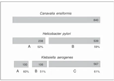
are similar in sequence to the small subunits of bacterial enzymes (e.g., UreA and UreB of Klebsiella aerogenes). The large subunits of bacterial ureases (e.g., UreC of K. aeroge-nes) resemble the carboxy-terminal portions of plant and fungal subunits. So far only bacterial ureases have had their 3-D crystal-lographic structure successfully resolved, e.g., K. aerogenes (1FWJ), Bacillus pasteurii
(4UBP), and Helicobacter pylori (1E9Z) (2, 3). However, the high sequence similarity of all ureases indicates they are variants of the same ancestral protein and are likely to pos-sess similar tertiary structures and catalytic mechanisms (2,3). Despite their highly con-served structures and enzymatic action, little is known about the physiological role of ureases in the source organisms. Urease ac-tivity enables bacteria to use urea as a sole nitrogen source. Some bacterial ureases play an important role in the pathogenesis of human and animal diseases such as those from Proteusmirabilisand H. pylori (2).
The wide distribution of ureases in
legumi-nous seeds as well as the accumulation pattern of the protein during seed maturation suggest an important physiological role. Soybean mu-tants lacking the embryo-specific highly ac-tive isoform of urease do not exhibit any of the abnormalities associated with loss of the less active ubiquitous isoform, suggesting that this enzyme probably does not have an essential physiological role (4). In vitro cultures of developing soybean cotyledons have indicated that ureases do not play an important role in embryo nutrition since urea is an extremely poor nitrogen source (4). The obvious ques-tion from this observaques-tion is why would the developing soybean embryo invest in produc-ing a very active ureolytic protein when it usually does not “encounter” urea. Polacco and Holland (4) have proposed that plant ure-ases may have a role in plant defense against predators due to the high toxicity of the ammo-nia released.
Plant-de rive d ure ase s: canatoxin and jack be an ure ase
The jack bean Canavalia ensiformis is the source of interesting proteins which have contributed significantly to modern Bio-chemistry. One of these is no doubt urease, the first protein ever crystallized (5), and the first nickel-containing enzyme described (1). In 1981, we isolated a toxic protein, named canatoxin, which accounts for 0.5% of seed dry weight of jack beans (6). Canatoxin is lethal to rats and mice by intraperitoneal injection, but it is inactive when given orally (7). Canatoxin, which consists of a noncova-lently linked dimer of 95-kDa acidic poly-peptide chains, was characterized recently as a variant form of the classical more abun-dant jack bean urease (8). RT-PCR applied to mRNA isolated from C. ensiformis tissues and Southern blots confirmed the presence of a family of urease-related genes with at least two members sharing 86% similarity (9). Jack bean ureases presented differential behavior in immobilized metal affinity chro-Canavalia ensiformis
Helicobacter pylori
Klebsiella aerogenes
840
539
59%
567
61% 238
106 100
52%
A B
A 60% B 51% C
matography enabling separation of the isoen-zymes (8,10). A particle-induced X-ray emis-sion technique applied to determine the metal content of the isoforms showed that cana-toxin displays ca. 1 atom of nickel and 1 of zinc per monomer, contrasting with 2 atoms of nickel and absence of zinc in the mono-mer of the major form of urease in C. ensi-formis (8,11).
Inse cticidal prope rtie s of plant ure ase s
The insecticidal properties of plant ure-ases were first described for canatoxin (12) and later for C. ensiformis major urease and soybean embryo-specific urease (13). The kissing bug Rhodnius prolixus, and three economically important crop pests, the cow-pea weevil Callosobruchus maculatus, the green stinkbug Nezara viridula and the cot-ton stainer bug Dysdercus peruvianus, are susceptible to the lethal effect of these pro-teins when they are added to their diets at 0.02 to 0.1% (w/w) levels (14,15). Suscep-tible insects have cathepsins of type B and D as their main digestive enzymes. Canatoxin and urease are hydrolyzed by these enzymes to release an internal entomotoxic peptide of 10 kDa (16). No effects of intact canatoxin/ urease were seen in insects relying on tryp-sin-like digestive enzymes, which apparently degrade the proteins more extensively (12). A recombinant peptide, equivalent to that produced by hydrolysis of canatoxin with insect cathepsins, was obtained by heterolo-gous expression in Escherichia coli. This peptide presented potent insecticidal effects (17) and did not affect mice or neonate rats upon oral or intraperitoneal administration. In contrast, the urease from the soil bacte-rium B. pasteurii is devoid of insecticidal properties, as expected from its three-chain structure, since part of the sequence of the entomotoxic peptide is absent in microbial ureases. In plant ureases this corresponds to a fragment located between the UreB and
UreC chains of B. pasteurii urease (13). Taken together, our results indicate that plant ureases are probably involved in de-fense mechanisms of plants against insect predation and that their insecticidal proper-ties are independent of their enzymatic ac-tivity, being associated with an internal pep-tide of these proteins.
Biological prope rtie s of canatoxin and similaritie s with othe r ure ase s
Canatoxin administered intraperitoneal-ly to rats or mice (LD50 0.4-0.6 and 2-3 mg/ kg, respectively) induces respiratory distress, convulsion, and death (6,18). At subconvul-sant doses canatoxin promotes increased gonadotropin (19) and plasma insulin levels (20) and pro-inflammatory effects in rats (21). In vitro, canatoxin displays potent secre-tagogue activity at nanomolar doses in sev-eral isolated cellular systems, inducing plate-let secretion and aggregation (22,23), secre-tion of labeled dopamine and serotonin from rat brain synaptosomes (23), histamine re-lease from mast cells (24), and secretion of insulin from isolated pancreatic islets (23,25). Thus, canatoxin induces dose-dependent aggregation of platelets from different spe-cies at concentrations as low as 20 nM (22). Rat isolated pancreatic islets secrete insulin when exposed to canatoxin (1 µM), making this protein about 20,000-fold more potent than glucose in provoking insulin release (25). Most of these effects, either in vivo or
in vitro, apparently involve activation of arachidonic acid metabolism mainly through the lipoxygenase pathway, since they are blocked by lipoxygenase inhibitors such as nordihydroguaiaretic acid and esculetin, but not by cyclooxygenase inhibitors (Table 1) (21-23,25,26). Pretreatment of animals with lipoxygenase inhibitors protected them also against the lethal effect of canatoxin (26).
h by an intense cellular infiltration at the site of administration. Pharmacological studies suggested that canatoxin-induced edema is a phenomenon mediated by several compo-nents. Initially histamine, serotonin, platelet aggregating factor, and prostaglandins play a role as agonists while lipoxygenase me-tabolites, probably leukotrienes, may account for the development of an intense cellular infiltration at the inflammatory site. Cana-toxin also induced neutrophil migration into rat peritoneal and pleural cavities and into air pouches (29). This effect was dependent on the resident macrophage population and was inhibited by glucocorticoids but not by non-steroidal anti-inflammatory drugs. It has also been shown that rat macrophage mono-layers treated with canatoxin release a neu-trophil chemotactic factor (29). Mouse peri-and to alter Ca2+ flux across the plasma
membrane of platelets through a verapamil-inhibitable Ca2+ channel (28). Canatoxin does not activate phospholipase C, and the intra-cellular calcium mobilization mediated by inositol 1,4,5-triphosphate does not play a role in platelet activation by this toxin. Pre-incubation of platelets with 8-bromo-gua-nosine 3',5'-cyclic monophosphate inhibited the canatoxin-evoked calcium influx, ara-chidonate release, ATP secretion, and cell aggregation, showing that the calcium in-flux is an early step in the mechanism of platelet activation by canatoxin, being modu-lated by cGMP (28).
Canatoxin displays pro-inflammatory activity (21). Thus, intraplantar injection of 50-300 µg canatoxin induced a dose-depend-ent rat hind-paw edema characterized after 3
Table 1. Modulation of canatoxin-induced effects by inhibitors of the lipoxygenase pathway.
Model/Effect Canatoxin EC50 Inhibitor Dose % inhibition Ref.
Rabbit platelets
Aggregation 300 nM NDGA 0.52 mM 50 22
ETYA 0.02 mM 50 22
BW755C 0.05 mM 50 22
5-HT secretion 300 nM NDGA 0.5 mM 75 23
Esculetin 0.1 mM 85 23
Rat brain synaptosomes
5-HT secretion 500 nM NDGA 0.2 mM 90 23
Esculetin 0.1 mM 90 23
Dopamine secretion 2 µM NDGA 0.5 mM 42 23
Rat pancreatic islets
Insulin secretion 200 nM NDGA 0.2 mM 76 25
Esculetin 0.1 mM 36 25
Rat mast cells
Histamine secretion 0.5 mM Not tested - - 24
Mouse macrophages
Release of lysosomal enzymes 0.3 mM NDGA 0.15 mM No inhibition #
Rat - in vivo
Hypoglycemia 0.4 mg/kg NDGA 125 mg/kg 100 26
Esculetin 125 mg/kg 100 26
Hyperinsulinemia 0.4 mg/kg NDGA 125 mg/kg 100 20
Hypoxia 0.4 mg/kg NDGA 125 mg/kg 72 26
Esculetin 125 mg/kg 50 26
Paw edema 0.3 mg/paw NDGA 100 mg/kg 66 21
Esculetin 50 mg/kg No inhibition 21
Convulsions 0.4 mg/kg NDGA 125 mg/kg 75 26
toneal macrophages release lysosomal en-zymes when exposed to canatoxin through a pathway involving nitric oxide (NO) signal-ing and guanyl cyclase activation (Ghazaleh FA, unpublished data).
In addition to lethality to insects, jack bean urease unexpectedly also displays other relevant biological properties observed for canatoxin, such as activation of blood plate-lets and monovalent lectin activity, but no toxicity when administered intraperitoneal-ly to mice, probabintraperitoneal-ly due to its larger size (540 kDa as opposed to 185 kDa) (8). Cana-toxin as well as urease interact with polysia-logangliosides (GD1b and GT1b) and sialo-proteins (mucin, tireoglobulin, fetuin) on the surface of erythrocytes and in ELISA micro-plates (7,8). This property of binding carbo-hydrates probably “directs” the proteins to cell surfaces enriched with this type of gly-coconjugates and may provide an explana-tion for their selective tissue specificity. Pre-treatment of the proteins with the thiol oxidant
p-hydroxymercuribenzoate (pHMB) irre-versibly abolished the ureolytic activity of urease (IC50 0.5 mM) and of canatoxin (IC50 5 mM) (8). In contrast, pHMB-treated cana-toxin or urease was still fully active to pro-mote platelet aggregation and binding to glycoconjugates. Moreover, the intraperito-neal toxicity of canatoxin was also not af-fected by pHMB treatment, indicating that these biological effects are not related to the enzymatic activity (8,10,13).
In order to determine if ureases from other sources share with jack bean ureases the property of inducing biological effects independent of their ureolytic activity, soy-bean embryo-specific urease and B. pasteurii
urease (30) were tested in rabbit platelets. Both ureases induced platelet aggregation even after being treated with pHMB (13). Furthermore, purified recombinant H. py-lori urease also displays platelet aggregating activity (Figure 2) (Wassermann GE, un-published data).
The pattern of platelet response to all the
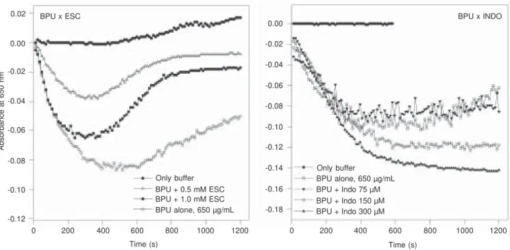
ureases tested so far was very similar, with a collagen-type shape-change reaction. As is the case for canatoxin (22,23), platelet ag-gregation induced by B. pasteurii or H. py-lori ureases also depends on lipoxygenase-derived metabolites. Thus, treatment of plate-lets with indomethacin, a cyclooxygenase inhibitor, potentiates urease-induced aggre-gation while the lipoxygenase inhibitor es-culetin blocks platelet responses to the mi-crobial enzymes (Figure 3).
Taken together, our data indicate that plant and microbial ureases form a group of multifunctional proteins with at least two distinct domains: 1) a thiol-dependent do-main containing the ureolytic active site, and 2) thiol-independent domain(s) involved in toxic effects on insects (and mice, only for canatoxin), binding to glycoconjugates and in the activation of blood platelets.
In addition to the platelet aggregating activity found for B. pasteurii and H. pylori, these and other bacterial ureases may share other biological activities and pro-inflam-matory properties with canatoxin, including the ability to activate lipoxygenases and the metabolism of eicosanoids. If true, these considerations could change our present un-derstanding of the pathogenesis of some dis-eases caused by urease-producing bacteria, such as urolithiasis due to Proteus mirabilis
or gastric ulcers consequent to H. pylori
infection.
Helicobacter p ylori, its ure ase and its implications in gastric dise ase s
affect-A
b
so
rb
a
n
ce
a
t
6
5
0
n
m
0.02
0.00
-0.02
-0.04
-0.12
0 200 400 600 800 1000 1200 0 200 400 600 800 1000 1200
Time (s) Time (s)
-0.02
-0.04
-0.08
-0.16 -0.10
-0.12
-0.14
-0.18 BPU x ESC
Only buffer BPU + 0.5 mM ESC -0.06
-0.08
-0.10
BPU + 1.0 mM ESC BPU alone, 650 µg/mL
Only buffer
BPU alone, 650 µg/mL BPU + Indo 75 µM BPU + Indo 150 µM BPU + Indo 300 µM
Figure 3. Effect of inhibitors of arachidonate metabolism on platelet aggregation induced by Bacillus pasteurii urease. Rabbit platelet-rich plasma suspensions were pretreated for 2 min with the indicated final concentrations of esculetin (ESC, left panel) and indomethacin (INDO, right panel) and then exposed to 0.7 mg/mL B. pasteurii urease (BPU; Sigma). The decrease in absorbance readings at 650 nm indicating platelet aggregation was monitored every 11 s for 20 min with a plate reader (13). Typical experiments are shown.
A
b
so
rb
a
n
ce
a
t
6
5
0
n
m
0.02
0.00
-0.02
-0.04
-0.06
0 200 400 600 800 1000 1200 0 200 400 600 800 1000 1200
Time (s) Time (s)
0.04
0.02
0.00
-0.02
-0.10 -0.04
-0.06
-0.08
-0.12
BPU HPU
Only buffer
16 µg/mL 40 µg/mL
162 µg/mL 650 µg/mL
Only buffer
40 µg/mL
162 µg/mL 650 µg/mL
Figure 2. Dose-response curve of platelet aggregation induced by purified Bacillus pasteurii (BPU, left panel) and Helicobacter pylori ureases (HPU, right panel). Rabbit platelet-rich plasma suspensions were challenged with different concentrations of purified B. pasteurii (Sigma, St. Louis, MO, USA) or recombinant H. pylori (Ref. 44) ureases. The decrease in absorbance at 650 nm indicating platelet aggregation was monitored every 11 s for 20 min with a plate reader (13). Typical experiments are shown.
-0.06
ing 40 to 60 persons per 100,000 yearly. Population-based intervention studies have shown the beneficial effect of early treat-ment of H. pylori in the prevention of gastric cancer. Notably, the most common lym-phoma of the stomach, mucosa-associated lymphoid tissue (MALT) lymphoma, is strongly associated with H. pylori infection and H. pylori eradication therapy is today the widely accepted initial treatment of stage I gastric MALT lymphoma, leading to re-mission rates of as much as 70% (34).
H. pylori is estimated to infect half of the world’s population, with peaks of 90% of the population infected in countries with poor sanitary conditions and of low socio-economic level (33,34). The prevalence of the organism in the Brazilian population ranges from 34% among urban children in the richer Southeast region to over 82% among adults in the poorer Northeast region (35,36). For the discovery of H. pylori and demonstration of its association with gastric disease, the Australian gastroenterologists Warren and Marshall (31) received the 2005 Nobel Prize in Medicine.
H. pylori-induced gastroduodenal disease depends on the inflammatory response of the host and on the production of main viru-lence factors, such as the vacuolating cyto-toxin VacA and the cytocyto-toxin-associated pro-tein CagA, and of urease, all of which cause damage to gastric epithelial cells (37-39). H. pylori resides within the mucus and on the apical surface of epithelial cells, where it attaches firmly via adhesin molecules. All
H. pylori isolates, as well as each of the gastric Helicobacter species identified to date, produce large quantities of the enzyme urease, which may account for 10-15% of the bacterial protein. Urease and urea influx through UreI, a pH-gated urea channel, have been shown to be essential for gastric colo-nization and for acid survival in vivo. Intra-bacterial urease generation of ammonia and a membrane-anchored periplasmic carbonic anhydrase regulate inner membrane
poten-tial and periplasmic pH to approximately 6.1 under acidic conditions, allowing adequate bioenergetics for survival and growth (40). Upon bacterial autolysis urease is released and adsorbed onto the extracellular surface of viable bacteria where it represents about 30% of the total cell urease content (38-39). The native urease of H. pylori has a molecular mass of approximately 540 kDa and is a nickel-containing hexameric mole-cule consisting of two subunits, UreA (26.5 kDa) and UreB (60.3 kDa), in a 1:1 molar ratio (41,42). The residues involved in the coordination of the two active site Ni2+ ions are completely conserved between H. pylori
and K. aerogenes ureases (42). The circular genome of H. pylori encodes about 1500 genes, depending on the strain (43). The biosynthesis of urease is coordinated by a gene cluster composed of two structural genes encoding the UreA and UreB subunits and five accessory proteins which are respon-sible for Ni2+ uptake and insertion into the active site of the apoenzyme (44). Active recombinant H. pylori urease was produced in E. coli transformed with pHP8080, a plas-mid encoding the whole operon with the two subunit structural genes and the NixA nickel transporter (44).
Pathoge ne sis of gastroduode nal dise ase s and Helicobacter p ylori
The exact mechanisms by which H. pylori
stomach of gnotobiotic piglets resulted in pref-erential colonization of the urease-positive bacteria (46), suggesting that neutralization of the gastric acidity is not the sole role of urease for colonization.
Several reports have demonstrated H. py-lori-induced apoptosis in gastric epithelial cells
in vivo and in vitro (47). Neutrophil apoptosis and subsequent clearance by phagocytes are critical to the resolution of acute inflammation (48), a process modulated by the NF-kappaB pathway and inflammatory regulators such as interleukin-8 (IL-8), lipopolysaccharide, or leukotriene B4 (48,49).
Although this organism is known to be noninvasive, H. pylori infection elicits gas-tric mucosal infiltration of inflammatory cells, especially neutrophils (38,50). Histo-logical observation in humans has indicated that the degree of H. pylori infection and the severity of mucosal injury are directly corre-lated with the extent of neutrophil infiltra-tion into the mucosa. Superfusion of the exposed mesentery with aqueous extracts of
H. pylori, which are rich in urease and de-void of significant contamination by lipo-polysaccharide from the cell wall, resulted in a three-fold increase in adherent leuko-cytes within the venules and a four-fold increase in those that had emigrated into the interstitium resulting in gastric mucosal in-jury (50). Kim and co-workers (51) reported that supernatants of H. pylori cultures en-hance neutrophil degranulation and adhe-sion capacity and up-regulate IL-8 expres-sion by activated neutrophils. Purified H. pylori urease was shown to directly activate primary human blood monocytes and to stimulate dose-dependent production of in-flammatory cytokines (IL-1b, IL-6, IL-8, and tumor necrosis factor-alpha) (52). Re-cently, Enarsson et al. (53) reported that H. pylori induced significant T-cell migration in a model system using human umbilical vein endothelial cells. CD4+ and CD8+ T cells migrated to the same extent in response to H. pylori. Although the presence of a
functional cag pathogenicity island contrib-uted to the transendothelial migration, puri-fied H. pylori urease alone induced a migra-tion effect similar to that of whole live bacte-ria. On the other hand, mutant H. pylori nega-tive for urease A subunit still promoted sig-nificant cell migration (53), suggesting that the ability of the bacterial urease to induce this effect may rely only on its B chain.
Inducible NO-synthesizing enzyme (iNOS) is expressed after activation by pro-inflam-matory cytokines in macrophages, endothe-lial cells and other cells. NO acts as a mes-senger in various inflammatory pathways and contributes to defense mechanisms against microorganisms but can also have cytotoxic effects. iNOS levels are up-regu-lated in a subgroup of patients with chronic active gastritis (54). Recombinant H. pylori
urease was shown to stimulate directly mac-rophage iNOS expression (55).
Ischemic lesions due to vascular insuffi-ciency may lead to the development of ul-cers within the gastric mucosa. Studies us-ing fluorescent in vivo microscopy have shown that H. pylori infection alters blood flow, the endothelial lining of the vessels, and leukocyte activity and often induces the formation of circulating or adherent platelet aggregates, consistent with epidemiological studies that suggest a possible association between H. pylori infection and the inci-dence of cardiovascular diseases, as reviewed by Kalia and Bardhan in 2003 (56). So far, there are no published studies on the effects of purified H. pylori urease on platelets. On the other hand, it is well known that platelets participate in the inflammatory response by modulating the activity of other inflamma-tory cells and as a storage site of vasoactive substances and inflammatory mediators such as histamine, serotonin, platelet aggregating factor, thromboxane A2 and other eicosan-oids, as well as by generating cytotoxic su-peroxide and hydroxyl radicals which may induce microcirculatory disturbances (56).
secretion but is also a major vasoactive me-diator in microcirculatory physiology. A major source of histamine within the gas-trointestinal tract is the mast cell and it was demonstrated that H. pylori, and in particu-lar its cell wall materials, could potentiate secretogogue-induced histamine release from isolated mast cells (57).
H. pylori adhesion to the gastric mucosa represents the initial contact between the bacterium and its host. Numerous adhesive properties of H. pylori have been described, including hemagglutination, attachment to epithelial cells, and binding to oligosaccha-rides or proteins of the extracellular matrix. Several research groups have reported that
H. pylori cells contain proteins that bind Neu5Ac (39,58).
Adhesins are bacterial proteins, glyco-conjugates, or lipids involved in the initial stages of colonization mediating the interac-tion between the bacterium and the host cell surface. It is predicted by genome sequenc-ing that H. pylori possesses a supergene family of 32 genes encoding putative outer membrane proteins. Among these, the bac-terial adhesin BabA2 has been identified to bind human blood group antigen Lewis b in the gastrointestinal mucosa. Two other mem-bers of this family, Alp A and B, are neces-sary for H. pylori to attach to human gastric tissue. A sialic acid-binding adhesin, SabA, was identified using a sialyl-Lex saccharide as a probe. The adhesion of H. pylori to fibronectin and lactoferrin is not dependent on BabA or SabA activities because the
babA/sabA double mutant still binds to these proteins. Thus, the presence of an additional binding activity of H. pylori has been sug-gested (58,59).
In general, adhesin receptors are carbo-hydrate moieties on glycoproteins or glyco-sphingolipids. Extracellular matrix proteins such as laminin and collagen type IV have been proposed as receptors for H. pylori. For cellular receptors, phosphatidylethanola-mine, laminin, and sialic acid-containing
molecules are regarded as potential recep-tors other than Lewis b. H. pylori-binding gangliosides and sialylated glycoproteins are present in relatively high amounts in human neutrophils (59). Bacterial binding to nor-mal gastric cells may be through nonsialyl-ated receptors, like Lewis b antigenic struc-tures, or lactotetraose. However, the level of sialylated structures increases accompany-ing inflammation. Sialyllactose has been re-ported to inhibit binding of H. pylori to cultured gastrointestinal epithelial cells and chronic atrophic gastritis in mice has been shown to be associated with increased syn-thesis of Neu5Aca3Gal structures (58,59).
It has become increasingly clear that ure-ase has other functions in the physiology of
H. pylori besides alkalization of the medi-um. Icatlo’s group (60) has shown that puri-fied H. pylori urease binds to gastric mucin and sulfated cell membrane glycolipids in an acidic setting. This property is expressed independently of its ureolytic activity which requires pH above 5.0. The interaction of urease with sulfated glycoproteins, heparin and heparinoids at pH 4.0 was shown to be dose- and time-dependent, and affected by the pH and salt concentration of the medium. Reports of antibiotic-resistant H. pylori
clinical isolates are increasing. Therefore, specific drugs targeting factors important for bacterial colonization, such as urease and chemotaxis, may be useful to minimize generation of drug-resistant bacteria. Carbo-hydrates and their chemical analogs are rel-evant candidates for anti-adhesion therapy (58-60).
Pe rspe ctive s
lines of evidence that point in this direction for H. pylori urease. These include a) pro-inflammatory activity accompanied by mono-nuclear phagocyte activation and neutrophil chemotaxis; b) platelet aggregating activity; c) histamine release from mast cells; d) lec-tin-like activity towards sialic-acid-contain-ing glycoconjugates. Most studies reported so far were carried out with whole H. pylori
cells or non-fractionated aqueous extracts and therefore the conclusion of involvement of urease in these phenomena is merely cir-cumstantial.
As reviewed here, similar findings have been reported for the plant urease canatoxin. Thus, it is possible that H. pylori urease may
also have other biological activities presented by canatoxin, particularly the secretagogue effect modulated by eicosanoid metabolites through lipoxygenase pathways, dependent on verapamil inhibitable calcium channels, and involving cGMP and NO signaling. Another important aspect to be investigated is whether or not the biological activities displayed by H. pylori urease depend on its ureolytic activity. If proven to be true, these findings could be extremely relevant to the elucidation of mechanisms leading to gas-trointestinal disease caused by this bacte-rium and should be taken into consideration in the development of more efficient thera-peutic approaches.
Re fe re nce s
1. Dixon NE, Gazzola TC, Blakeley RL, Zermer B. Jack bean urease (EC 3.5.1.5). A metalloenzyme. A simple biological role for nickel? J Am Chem Soc 1975; 97: 4131-4133.
2. Mobley HL, Island MD, Hausinger RP. Molecular biology of micro-bial ureases. Microbiol Rev 1995; 59: 451-480.
3. Sirko A, Brodzik R. Plant ureases: roles and regulation. Acta Biochim Pol 2000; 47: 1189-1195.
4. Polacco JC, Holland MA. Roles of urease in plant cells. Int Rev Cytol
1993; 145: 65-103.
5. Sumner JB. The isolation and crystallization of the enzyme urease.
J Biol Chem 1926; 69: 435-441.
6. Carlini CR, Guimarães JA. Isolation and characterization of a toxic protein from Canavalia ensiformis (jack bean) seeds, distinct from concanavalin A. Toxicon 1981; 19: 667-675.
7. Carlini CR, Guimarães JA. Plant and microbial toxic proteins as hemilectins: emphasis on canatoxin. Toxicon 1991; 29: 791-806. 8. Follmer C, Barcellos GB, Zingali RB, Machado OL, Alves EW,
Barja-Fidalgo C, et al. Canatoxin, a toxic protein from jack beans ( Canava-lia ensiformis), is a variant form of urease (EC 3.5.1.5): biological effects of urease independent of its ureolytic activity. Biochem J
2001; 360: 217-224.
9. Pires-Alves M, Grossi-de-Sa MF, Barcellos GB, Carlini CR, Moraes MG. Characterization and expression of a novel member (JBURE-II) of the urease gene family from jackbean [Canavalia ensiformis (L.) DC]. Plant Cell Physiol 2003; 44: 139-145.
10. Follmer C, Wassermann GE, Carlini CR. Separation of jack bean (Canavalia ensiformis) urease isoforms by immobilized metal af-finity chromatography and characterization of insecticidal properties unrelated to ureolytic activity. Plant Sci 2004; 167: 241-246. 11. Follmer C, Carlini CR, Yoneama ML, Dias JF. PIXE analysis of
urease isoenzymes isolated from Canavalia ensiformis seeds. Nucl Instrum Methods Phys Res B 2002; 189: 482-486.
12. Carlini CR, Oliveira AE, Azambuja P, Xavier-Filho J, Wells MA. Biological effects of canatoxin in different insect models: evidence for a proteolytic activation of the toxin by insect cathepsin-like
en-zymes. J Econ Entomol 1997; 90: 340-348.
13. Follmer C, Real-Guerra R, Wasserman GE, Olivera-Severo D, Carlini CR. Jackbean, soybean and Bacillus pasteurii ureases: bio-logical effects unrelated to ureolytic activity. Eur J Biochem 2004; 271: 1357-1363.
14. Staniscuaski F, Ferreira-Dasilva CT, Mulinari F, Pires-Alves M, Carlini CR. Insecticidal effects of canatoxin on the cotton stainer bug
Dysdercus peruvianus (Hemiptera: Pyrrhocoridae). Toxicon 2005; 45: 753-760.
15. Carlini CR, Grossi-de-Sa MF. Plant toxic proteins with insecticidal properties. A review on their potentialities as bioinsecticides. Toxi-con 2002; 40: 1515-1539.
16. Ferreira-Dasilva CT, Gombarovits ME, Masuda H, Oliveira CM, Carlini CR. Proteolytic activation of canatoxin, a plant toxic protein, by insect cathepsin-like enzymes. Arch Insect Biochem Physiol
2000; 44: 162-171.
17. Mulinari F, Freitas-Silva MA, Grossi-de-Sá MF, Moraes MG, Kurtenbach E, Carlini CR. Toxina Praguicida, Construção Gênica e Método de Controle de Pragas. Patent No. 001120/RS. Patent registered at National Institute for Intellectual Property (INPI), Brazil; 8-4-2004.
18. Carlini CR, Gomes C, Guimarães JA, Markus RP, Sato H, Trolin G. Central nervous effects of the convulsant protein canatoxin. Acta Pharmacol Toxicol 1984; 54: 161-166.
19. Ribeiro-DaSilva G, Pires-Barbosa R, Carlini CR. Effect of canatoxin on the circulating levels of gonadotropins and prolactin in rats. Braz J Med Biol Res 1989; 22: 387-395.
20. Ribeiro-DaSilva G, Prado JF. Increased insulin circulating levels induced by canatoxin in rats. Toxicon 1993; 31: 1131-1136. 21. Benjamin CF, Carlini CR, Barja-Fidalgo C. Pharmacological
charac-terization of rat paw edema induced by canatoxin, the toxic protein from Canavalia ensiformis (jack bean) seeds. Toxicon 1992; 30: 879-885.
for the involvement of the platelet lipoxygenase pathway. Br J Pharmacol 1985; 84: 551-560.
23. Barja-Fidalgo C, Guimarães JA, Carlini CR. Lipoxygenase-medi-ated secretory effect of canatoxin, the toxic protein from Canavalia ensiformis seeds. Toxicon 1991; 29: 453-459.
24. Grassi-Kassisse DM, Ribeiro-DaSilva G. Canatoxin triggers hista-mine secretion from rat peritoneal mast cells. Agents Actions 1992; 37: 204-209.
25. Barja-Fidalgo C, Guimarães JA, Carlini CR. Canatoxin, a plant protein, induces insulin release from isolated pancreatic islets. En-docrinology 1991; 128: 675-679.
26. Ribeiro-DaSilva G, Pires-Barbosa R, Prado JF, Carlini CR. Convul-sions induced by canatoxin in rats are probably a consequence of hypoxia. Braz J Med Biol Res 1989; 22: 877-880.
27. Alves EW, Ferreira AT, Ferreira CT, Carlini CR. Effects of canatoxin on the Ca(2+)-ATPase of sarcoplasmic reticulum membranes. Toxi-con 1992; 30: 1411-1418.
28. Ghazaleh FA, Francischetti IM, Gombarovits ME, Carlini CR. Stimu-lation of calcium influx and platelet activation by canatoxin: methoxyverapamil inhibition and downregulation by cGMP. Arch Biochem Biophys 1997; 339: 362-367.
29. Barja-Fidalgo C, Carlini CR, Guimarães JA, Flores CA, Cunha FQ, Ferreira SH. Role of resident macrophages in canatoxin-induced in vivo neutrophil migration. Inflammation 1992; 16: 1-12.
30. Benini S, Gessa C, Ciurli S. Bacillus pasteurii urease: A heteropoly-meric enzyme with a binuclear nickel active site. Soil Biol Biochem
1996; 28: 819-821.
31. Warren JR, Marshall BJ. Unidentified curved bacilli on gastric epi-thelium in active chronic gastritis. Lancet 1983; 1: 1273-1275. 32. Yoshiyama H, Nakazawa T. Unique mechanism of Helicobacter
pylo-ri for colonizing the gastric mucus. Microbes Infect 2000; 2: 55-60. 33. Hopkins RJ, Girardi LS, Turney EA. Relationship between
Helico-bacter pylori eradication and reduced duodenal and gastric ulcer recurrence: a review. Gastroenterology 1996; 110: 1244-1252. 34. Fischbach W, Chan AO, Wong BC. Helicobacter pylori and gastric
malignancy. Helicobacter 2005; 10 (Suppl 1): 34-39.
35. Oliveira AM, Queiroz DM, Rocha GA, Mendes EN. Seroprevalence of Helicobacter pylori infection in children of low socioeconomic level in Belo Horizonte, Brazil. Am J Gastroenterol 1994; 89: 2201-2204.
36. Mitchell A, Silva TM, Barrett LJ, Lima AA, Guerrant RL. Age-specific
Helicobacter pylori seropositivity rates of children in an impover-ished urban area of northeast Brazil. J Clin Microbiol 2003; 41: 1326-1328.
37. Covacci A, Telford JL, Del Giudice G, Parsonnet J, Rappuoli R.
Helicobacter pylori virulence and genetic geography. Science 1999; 284: 1328-1333.
38. Montecucco C, Papini E, de Bernard M, Zoratti M. Molecular and cellular activities of Helicobacter pylori pathogenic factors. FEBS Lett 1999; 452: 16-21.
39. Rieder G, Fischer W, Haas R. Interaction of Helicobacter pylori with host cells: function of secreted and translocated molecules. Curr Opin Microbiol 2005; 8: 67-73.
40. Sachs G, Weeks DL, Wen Y, Marcus EA, Scott DR, Melchers K. Acid acclimation by Helicobacter pylori. Physiology 2005; 20: 429-438. 41. Hu LT, Mobley HL. Purification and N-terminal analysis of urease
from Helicobacter pylori. Infect Immun 1990; 58: 992-998. 42. Ha NC, Oh ST, Sung JY, Cha KA, Lee MH, Oh BH. Supramolecular
assembly and acid resistance of Helicobacter pylori urease. Nat Struct Biol 2001; 8: 505-509.
43. Tomb JF, White O, Kerlavage AR, Clayton RA, Sutton GG,
Fleisch-mann RD, et al. The complete genome sequence of the gastric pathogen Helicobacter pylori. Nature 1997; 388: 539-547. 44. McGee DJ, May CA, Garner RM, Himpsl JM, Mobley HL. Isolation of
Helicobacter pylori genes that modulate urease activity. J Bacteriol
1999; 181: 2477-2484.
45. Tsuda M, Karita M, Morshed MG, Okita K, Nakazawa T. A urease-negative mutant of Helicobacter pylori constructed by allelic ex-change mutagenesis lacks the ability to colonize the nude mouse stomach. Infect Immun 1994; 62: 3586-3589.
46. Eaton KA, Brooks CL, Morgan DR, Krakowka S. Essential role of urease in pathogenesis of gastritis induced by Helicobacter pylori in gnotobiotic piglets. Infect Immun 1991; 59: 2470-2475.
47. Cover TL, Krishna US, Israel DA, Peek Jr RM. Induction of gastric epithelial cell apoptosis by Helicobacter pylori vacuolating cytotoxin.
Cancer Res 2003; 63: 951-957.
48. Savill J, Dransfield I, Gregory C, Haslett C. A blast from the past: clearance of apoptotic cells regulates immune responses. Nat Rev Immunol 2002; 2: 965-975.
49. Hebert MJ, Takano T, Holthofer H, Brady HR. Sequential morpho-logic events during apoptosis of human neutrophils. Modulation by lipoxygenase-derived eicosanoids. J Immunol 1996; 157: 3105-3115.
50. Shimoyama T, Fukuda S, Liu Q, Nakaji S, Fukuda Y, Sugawara K.
Helicobacter pylori water soluble surface proteins prime human neutrophils for enhanced production of reactive oxygen species and stimulate chemokine production. J Clin Pathol 2003; 56: 348-351. 51. Kim JS, Jung HC, Kim JM, Song IS, Kim CY. Helicobacter pylori
water-soluble surface proteins activate human neutrophils and up-regulate expression of CXC chemokines. Dig Dis Sci 2000; 45: 83-92.
52. Harris PR, Mobley HL, Perez-Perez GI, Blaser MJ, Smith PD. Heli-cobacter pylori urease is a potent stimulus of mononuclear phago-cyte activation and inflammatory cytokine production. Gastroenter-ology 1996; 111: 419-425.
53. Enarsson K, Brisslert M, Backert S, Quiding-Jarbrink M. Helico-bacter pylori induces transendothelial migration of activated memory T cells. Infect Immun 2005; 73: 761-769.
54. Fu S, Ramanujam KS, Wong A, Fantry GT, Drachenberg CB, James SP, et al. Increased expression and cellular localization of inducible nitric oxide synthase and cyclooxygenase 2 in Helicobacter pylori
gastritis. Gastroenterology 1999; 116: 1319-1329.
55. Gobert AP, Mersey BD, Cheng Y, Blumberg DR, Newton JC, Wilson KT. Cutting edge: urease release by Helicobacter pylori stimulates macrophage inducible nitric oxide synthase. J Immunol 2002; 168: 6002-6006.
56. Kalia N, Bardhan KD. Of blood and guts: association between
Helicobacter pylori and the gastric microcirculation. J Gastroenterol Hepatol 2003; 18: 1010-1017.
57. Yamamoto J, Watanabe S, Hirose M, Osada T, Ra C, Sato N. Role of mast cells as a trigger of inflammation in Helicobacter pylori
infection. J Physiol Pharmacol 1999; 50: 17-23.
58. Dubreuil JD, Giudice GD, Rappuoli R. Helicobacter pylori interac-tions with host serum and extracellular matrix proteins: potential role in the infectious process. Microbiol Mol Biol Rev 2002; 66: 617-629. 59. Miller-Podraza H, Johansson P, Angstrom J, Larsson T, Longard M, Karlsson KA. Studies on gangliosides with affinity for Helicobacter pylori: binding to natural and chemically modified structures. Glyco-biology 2004; 14: 205-217.