Regulation of nephron
acidification by corticosteroids
1Departamento de Fisiologia e Biofísica, Instituto de Ciências Biomédicas,
Universidade de São Paulo, 05508-900 São Paulo, SP, Brasil
2Departamento de Química Biológica, Facultad de Ciencias Exactas y Naturales,
Universidad de Buenos Aires, 1428 Buenos Aires, Argentina G. Malnic1,
M. Ansaldo2,
C.P. Lantos2 and
M.C. Damasco2
Abstract
The present paper reviews work from our laboratories evaluating the importance of adrenal cortical hormones in acidification by proximal and cortical distal tubules. Proximal acidification was determined by stationary microperfusion, and measurement of bicarbonate reabsorp-tion using luminal pH determinareabsorp-tion was performed with H+ -ion-sensitive microelectrodes. Rats were adrenalectomized (ADX) 48 h before the experiments, and corticosteroids (aldosterone (A), corticos-terone (B), and 18-OH corticoscorticos-terone (18-OH-B)) were injected intra-muscularly 100 and 40 min before the experiments. In ADX rats stationary pH increased significantly to 7.03 as compared to sham-operated rats (6.78). Bicarbonate reabsorption decreased from 2.65 ± 0.18 in sham-operated rats to 0.50 ± 0.07 nmol cm-2 s-1 after ADX. The administration of the three hormones stimulated proximal tubule acidification, reaching, however, only 47.2% of the sham values in aldosterone-treated rats. Distal nephron acidification was studied by measuring urine minus blood pCO2 differences (U-B pCO2) in bicar-bonate-loaded rats treated as above. This pCO2 difference is used as a measure of the distal nephron ability to secrete H+ ions into an alkaline urine. U-B pCO2 decreased significantly from 39.9 ± 1.26 to 11.9 ± 1.99 mmHg in ADX rats. When corticosteroids were given to ADX rats before the experiment, U-B pCO2 increased significantly, but reached control levels only when aldosterone (two 3-µg doses per rat) plus corticosterone (220 µg) were given together. In order to control for the effect of aldosterone on distal transepithelial potential differ-ence one group of rats was treated with amiloride, which blocks distal sodium channels. Amiloride-treated rats still showed a significant reduction in U-B pCO2 after ADX. Only corticosterone and 18-OH-B but not aldosterone increased U-B pCO2 back to the levels of sham-operated rats. These results show that corticosteroids stimulate renal tubule acidification both in proximal and distal nephrons and provide some clues about the mechanism of action of these steroids.
Correspondence G. Malnic
Departamento de Fisiologia e Biofísica
ICB, USP
Av. Prof. Lineu Prestes, 1524 05508-900 São Paulo, SP Brasil
Fax: 55 (011) 818-7285
Presented at the International Symposium Neuroendocrine Control of Body Fluid Homeostasis, Ribeirão Preto, SP, Brasil, August 17-20, 1996.
Research supported by FAPESP, CNPq, FINEP/PADCT, Conicyt and Universidad de Buenos Aires.
Received November 29, 1996 Accepted January 6, 1997
Key words
•Aldosterone •Corticosterone
•Bicarbonate reabsorption •Amiloride
•Urine
Introduction
It is known that the main action of miner-alocorticoids on urinary acidification occurs in the distal nephron via stimulation of api-cal vacuolar H+-ATPase (1,2). Both whole
animal studies on the adrenalectomized (ADX) rat and microperfusion studies of adrenalectomized rabbit collecting duct have demonstrated the importance of adrenocor-tical steroids for urinary acid excretion and bicarbonate reabsorption (3,4). The mech-anism of action of these hormones involves the regulation of gene transcription and in-corporation of new transporters (Na+-K+
-ATPase) and Na+ channels into cell
mem-branes (5,6). In addition, rapid, non-genom-ic stimulation of electrolyte transport has been observed in a number of extrarenal cells, including lymphocytes and vascular smooth muscle cells. This effect is mediated by specific membrane receptors for aldoster-one, which lead to the incorporation or acti-vation of pre-existing sodium channels, el-evation of cell sodium and secondary activa-tion of the basolateral Na+-K+-ATPase,
re-sponsible for an increase in electrolyte trans-port within a few minutes after hormone addition (7).
Corticosteroids have been shown to af-fect other acid/base transporters besides H+
-ATPase. Aldosterone stimulates Na+/H+
ex-change in renal early distal amphibian tu-bules (1,2), and in cultured MDCK cells (8), specifically on their basolateral surface (9). These cells are derived from canine kidney, and have several properties in common with distal nephron α-intercalated cells. Aldos-terone has also been shown to stimulate Cl-/HCO
3
exchange, both in MDCK cells (8) and in cardiac cells (10), as well as the H+-K+-ATPase of the apical membrane of
MDCK cells (11). In addition, glucocorti-coids have been shown to stimulate Na+/H+
exchange in OKP cells in culture and in rat distal colon cells (12,13). Glucocorticoids act via stimulation of the expression of
dif-ferent forms of these exchangers, in particu-lar NHE1 and NHE3 (14,15).
The observation that corticosteroid hor-mones act on the Na+/H+ exchanger and H+
-K+-ATPase, in addition to the vacuolar H+
-ATPase led us to study the effect of these hormones on H+-ion secretion in renal
proxi-mal tubules by microperfusion techniques. In addition, we studied the role of corticos-teroid hormones in distal nephron acidifica-tion by the determinaacidifica-tion of urine minus blood pCO2 (U-B pCO2) differences. Urine
pCO2 may have a number of origins,
includ-ing delayed dehydration of carbonic acid, trapping of CO2 in the countercurrent
sys-tem, mixture of urines at different pH values from different nephrons, and ampholyte prop-erties of bicarbonate buffer, among others (16). However, U-B pCO2 has also been
considered to represent the magnitude of distal nephron (mostly collecting duct) H+
-ion secret-ion at high urinary bicarbonate concentrations (17,18). In these experiments, corticosterone (B), aldosterone (A) or 18-OH corticosterone (18-18-OH-B) was given to ADX rats in order to investigate the role of each of them in supporting the normal rate of renal tubule H+ secretion. 18-OH-B is a
natu-ral steroid of the biosynthetic pathway of aldosterone, and has been shown to stimu-late titratable acid excretion and to reduce urine pH in ADX rats (19,20). In the present paper we review some of the work per-formed in our laboratories in this area.
Material and Methods
Microperfusion studies
and 3 µg aldosterone or 6 µg 18-OH-B was given 100 and 40 min before the experiment. These doses lead to blood levels of these hormones that are within the upper limits of the physiological range (22,23). Rats were prepared for micropuncture as previously described (24). Stationary microperfusion was performed with double-barrelled pipettes blocking droplets of 25 mM bicarbonate Ringer solution with castor oil in the tubule lumen. These fluid droplets were punctured with pH-sensitive microelectrodes and the pH was followed from the initial alkaline value (approximately pH 8) to the stationary level. Bicarbonate concentrations were cal-culated from these curves and from arterial blood pCO2, and bicarbonate fluxes were
obtained from their rate of disappearance and tubular geometry (24). Urine was col-lected from the bladder during experiments, and GFR was determined by inulin clear-ance. Sodium and potassium in urine were determined by flame photometry.
Urine-blood pCO2 studies
Adrenalectomy and hormone supplemen-tation were performed as described above. The rats received an infusion of 0.6 M NaHCO3 plus 5% mannitol during the
ex-periments which raised urine pH to about 7.8. Urine pH and pCO2 were determined
with a radiometer model BMS3 MK2 blood micro system (Radiometer, Copenhagen, Denmark). Urine pCO2 was plotted against
urine bicarbonate concentration, and the pCO2 at 150 mM (or 120 mM in experiments
in which amiloride was given) urine bicar-bonate was obtained from the respective regression lines. Comparisons between groups were made on the basis of these values. In one group of rats, a priming dose of amiloride of 0.4 mg/100 g body weight was given, followed by an infusion of 1 mg 100 g-1 h-1, and similar procedures as
de-scribed above were performed.
Results and Discussion
Figure 1 shows the general conditions of the rats in this study. The mean control arte-rial blood pressure of 129.4 ± 5.75 mmHg decreased to 100.9 ± 8.67 mmHg (P<0.05) in ADX, but did not recover in supplemented rats. Urine sodium/potassium ratios increased significantly after ADX, confirming corti-coid hormone depletion. Hormone supple-mentation caused complete recovery only when aldosterone was given. GFR decreased
Figure 1 - General conditions of sham-operated and adrenalectomized (ADX) rats, and the effect of corticosterone (B), aldosterone (A) and 18-hydroxycorticosterone (18-OH-B) on ADX rats. UNa/UK, Urine Na+/K+ concentration ratio; GFR, glomerular filtration rate per kg rat; MABP, mean arterial blood pressure. Data are reported as means ± SEM. Data from Ref. 21, with permission.
MABP (mmHg)
140
120
1.4
UNa
/U
K
1.2
1.0
0.8
0.6
0.4
0.2
0.0
7.0
6.8
6.6
6.4
6.2
6.0
Urine pH
100
80
60
40
20
0
10.0
7.5
5.0
2.5
0.0
GFR (ml min
-1 kg -1)
Blood pressure UNa/UK
GFR Urine pH
from a control value of 7.98 ± 0.36 ml min-1 kg
body weight-1 to 5.77 ± 0.66 ml min-1 kg-1 in
ADX, and recovered only in corticosterone-supplemented rats, as previously shown (25). Urine pH increased markedly from 6.76 ± 0.020 in sham-operated rats to 7.03 ± 0.028 in ADX rats, and recovered entirely only in aldosterone-treated animals. These whole-animal data show that the rats in our experi-ments conform to the findings generally en-countered in corticosteroid-depleted animals, indicating the role of these hormones in urinary acidification.
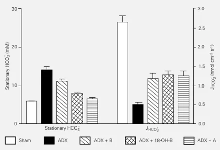
Figure 2 shows results obtained in the experiments of in vivo microperfusion of convolute cortical proximal tubules (S2 seg-ments). Stationary bicarbonate concentra-tions increased markedly from 5.97 ± 0.23 mM in controls to 14.1 ± 0.88 mM in ADX, corresponding to a rise in tubule lumen pH from 6.76 ± 0.020 in controls to 7.03 ± 0.028 in ADX. Stationary bicarbonate returned to normal with aldosterone and almost to nor-mal after supplementation with the other steroids. Bicarbonate reabsorption (JHCO3-)
fell markedly from 2.65 ± 0.18 nmol cm-2 s-1
in sham-operated rats to 0.50 ± 0.07 nmol
cm-2 s-1 in ADX rats. During hormone
sup-plementation recovery was only partial with all steroids, reaching only a value in the range of 1.2 to 1.3 nmol cm-2 s-1. This
reduc-tion in reabsorpreduc-tion rates was mostly due to a delay in the rate of fall in luminal bicarbon-ate concentrations. Half-times of luminal dis-appearance of bicarbonate increased from 3.73 ± 0.17 s in sham-operated rats to 11.43 ± 0.72 s in ADX rats. Proximal acidification was reduced in these experiments by 73% in ADX, which cannot be explained only by inhibition of proximal H+-ATPase, since this
transporter is responsible for at most 30% of proximal bicarbonate reabsorption (26). This finding suggests a role of these hormones in the stimulation of luminal insertion or turn-over of Na+/H+ exchanger molecules.
A recent series of experiments using brush-border membrane vesicles and fluoro-metric pH determination with acridine or-ange have detected a significant reduction in the rate of Na+/H+ exchange in vesicles from
ADX rats, therefore localizing the hormonal effect to the apical (brush-border) membrane of proximal tubule cells. The origin of this reduction was shown to involve a decrease in Vmax of the exchanger, without alteration
of Km for Na+ (Igarreta MP, Calvo JC,
Paladini A and Damasco MC, unpublished data). Also, in these experiments the admin-istration of corticosterone and 18-OH-B to ADX rats reversed the inhibition of the ex-changer.
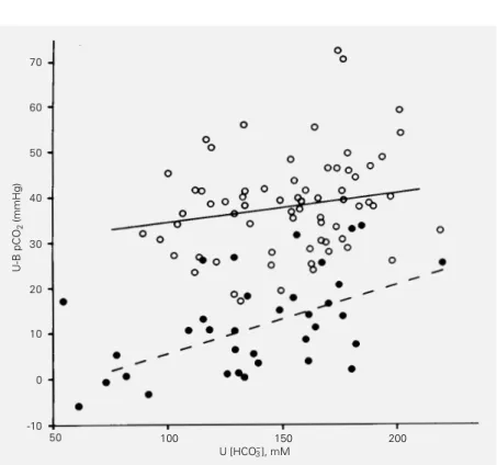
The following series of experiments was performed to analyze the role of several corticosteroid hormones in acid secretion by the distal nephron, using the urine minus blood pCO2 difference (27). Figure 3 shows
a plot relating U-B pCO2 to urine
bicarbon-ate concentrations in control and ADX rats. CO2 is generated when H+ is secreted into a
bicarbonate-containing fluid. This pCO2
dif-ference increases with bicarbonate concen-tration, which is a property of the CO2/HCO3
-buffer system. Therefore, it is important that these differences are measured at
well-de-Figure 2 - Stationary bicarbonate concentrations (HCO3-s) and net bicarbonate reabsorption (JHCO3-) in proximal tubules from sham-operated and ADX rats. Experimental groups as in Figure 1. Data from Ref. 21, with permission.
3.0
2.5
2.0
1.5
1.0
0.5
0.0 30
Stationary HCO
3
- (mM) 20
10
0
Stationary HCO3- JHCO3
-JHCO
3
- (nmol cm -2 s -1)
fined urine bicarbonate levels (28). Differ-ences between H+ secretion in experimental
groups are not related to the slope of these lines, but to the level of the line. It is clear that in ADX rats, U-B pCO2 values were
significantly decreased with respect to con-trols. Figure 4 shows the mean pCO2
differ-ences plotted against mean urine bicarbon-ate concentration in the different experimen-tal groups. Again, the marked reduction in U-B pCO2 in ADX rats is apparent.
Supple-mentation with hormones given within 100 min before the experiments raised these val-ues significantly; however, only when aldos-terone and corticosaldos-terone were given together did the U-B pCO2 values return to the
trol level, indicating that both hormones con-tribute to normal urine pCO2. The role of
corticosterone in maintaining urine pCO2
may be related to its known effect of increas-ing GFR, as discussed above, which leads to an enlarged urinary buffer load, represented mostly by phosphate salts. In the rats studied in these experiments, phosphate excretion fell from 2.30 ± 0.17 µEq/min in sham-operated rats to 1.17 ± 0.20 µEq/min in ADX rats, and recovered to 1.90 ± 0.19 µEq/min when corticosterone was given. This rise in urine phosphate excretion may be due to the increased GFR after corticosterone adminis-tration. It is also known that the phosphate level in urine is an important factor regulat-ing urine pCO2 (29), which may explain the
role of corticosterone in increasing U-B pCO2.
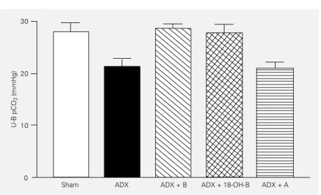
In another group of experiments, we stud-ied the role of amiloride, a blocker of sodium channels which affects the transepithelial potential difference (PD) in hormonal regu-lation of distal H+ secretion as evaluated by
U-B pCO2 (30). Sham-operated and ADX
rats treated as above received amiloride dur-ing the experiments. The administration of amiloride reduced U-B pCO2 from 49.3 ±
2.7 mmHg in controls to 29.8 ± 3.2 mmHg (P<0.01). This finding is commonly attrib-uted to the reduction of collecting duct
trans-Figure 3 - Urine minus blood pCO2 (U-B pCO2) differences plotted against urine bicarbonate concentration (U) incontrol (open circles) and adrenalectomized (filled circles) rats. From Ref. 27, with permission.
Figure 4 - U-B pCO2 plotted against urine bicarbonate concentrations under different experimental conditions. Data are reported as means ± SEM. Symbols as in Figure 1, except H = 18-OH corticosterone. From Ref. 27, with permission.
U-B pCO
2
(mmHg)
70
60
50
40
30
20
10
0
-10
50 100 150 200
U [HCO3- ], mM
U-B pCO
2
(mmHg)
50
40
30
20
10
0
100 150
U [HCO3- ], mM
200
Sham C
ADX
+ B
+ A
+ H
+ B + H
+ A + H
epithelial PD and, possibly, also to the effect of amiloride on Na+/H+ exchange. U-B pCO
2
was further reduced to 21.3 ± 1.6 mmHg (P<0.05) in ADX rats, as shown in Figure 5. Supplementation with corticosterone and 18-OH-B led to recovery toward the pCO2
dif-ference of sham-operated rats. However, in aldosterone-treated rats no change of U-B pCO2 was observed. This finding supports
the view that aldosterone stimulates H+-ion
secretion by a process related to the presence of activated sodium channels, possibly trans-epithelial PD. It is well known that amiloride reduces this PD in late distal tubule and in cortical collecting ducts from 30-50 mV, lumen negative, to near zero, impairing a factor that stimulates the transfer of the posi-tive H+ ion into the lumen. On the other hand,
corticosterone, as discussed above, may act mainly by its effect on GFR and buffer filtra-tion, leading to greater urinary phosphate excretion, an effect which is not affected by amiloride.
What is the mechanism by which corti-costeroid hormones act on acid excretion? A classical explanation proposed by AlAwqati and colleagues (31) based on experiments in turtle bladder assumes that these hormones stimulate H+-ATPase expression or turnover.
In the mammalian nephron, several trans-porters involved in urine acidification have been described. It has been shown that al-dosterone stimulates Na+/H+ exchange in a
number of both amphibian (32) and mam-malian (8,9,33) tissues and cells in culture. Glucocorticoids have also been shown to stimulate Na+/H+ exchange in cells in culture
(13). This stimulation is thought to occur by membrane insertion or activation of trans-porters, which is compatible with our find-ings in the proximal tubule. In these experi-ments there was only a relatively small effect on the transepithelial pH gradient, which depends on the sodium gradient across the apical cell membrane, and a large modifica-tion of the acidificamodifica-tion half-time, which, according to a model of proximal tubule cells, depends on the number or turnover of transport sites within the membrane (34). In proximal tubules, not all H+-ion secretion is
due to Na+/H+ exchange, but some 20-30%
have been shown to depend on vacuolar H+
-ATPase, which of course could also undergo the action of these hormones (26). For the distal nephron, the vacuolar H+-ATPase is
the most prevalent H+-ion transporter,
al-though more recently the importance of the gastric type H+-K+-ATPase has been stressed
(35). The stimulation mechanisms of these ATPases are probably similar to those of Na+-K+-ATPase and sodium channels of the
collecting duct principal cells, although the stimulation of H+-ion transporters by these
hormones has not been investigated in com-parable detail. Additional factors involved in corticosteroid stimulation of distal H+-ion
secretion, as supported by our data, are the transepithelial PD which is known to in-crease along the collecting duct in hormone-stimulated animals due to the higher density of Na+ channels, depolarizing the apical
membrane of collecting duct principal cells (36), and the increase in buffer (phosphate) excretion caused by the higher GFR induced by steroids such as corticosterone.
Figure 5 - U-B pCO2 in amiloride-treated rats. Experimental groups as in Figure 1. Data from Ref. 30, with permission.
U-B pCO
2
(mmHg)
30
20
10
0
References
1. Garg LC & Narang N (1988). Effects of aldosterone on NEM-sensitive ATPase in rabbit nephron segments. Kidney Interna-tional, 34: 13-17.
2. Eiam-Ong S, Kurtzman NA & Sabatini S (1993). Regulation of collecting tubule a-denosine triphosphatases by aldosterone and potassium. Journal of Clinical Investi-gation, 91: 2385-2392.
3. Stone DK, Seldin DW, Kokko JP & Jacobson HR (1983). Mineralocorticoid modulation of rabbit medullary collecting duct acidification. A sodium independent effect. Journal of Clinical Investigation, 72: 77-83.
4. Dubrovsky AHE, Nair RC, Byers MK & Levine DZ (1981). Renal net acid excre-tion in the adrenalectomized rat. Kidney International, 19: 516-528.
5. Verrey F (1995). Transcriptional control of sodium transport in tight epithelia by ad-renal steroids. Journal of Membrane Biol-ogy, 144: 93-110.
6. Verrey F & Beron J (1996). Activation and supply of channels and pumps by aldos-terone. News in Physiological Sciences, 11: 126-133.
7. Wehling M (1995). Aldosterone specific membrane receptors, rapid activation of the sodium-hydrogen exchanger, and car-diovascular implications. Cardiovascular Research, 29: 167-171.
8. Oberleithner H, Vogel U, Kersting U & Steigner W (1990). Madin-Darby canine kidney cells. II. Aldosterone stimulates Na+/H+ and Cl-/HCO
3-exchange. Pflügers Archiv, 416: 533-539.
9. Vilella S, Guerra L, Helmle-Kolb C & Murer H (1992). Aldosterone actions on basolateral Na+/H+ exchange in Madin-Darby canine kidney cells. Pflügers Archiv. European Journal of Physiology, 422: 9-15.
10. Korichneva I, Púceat M, Millanvoye-Van Brussel E, Géraud G & Vassort G (1995). Aldosterone modulates both the Na/H antiport and Cl/HCO3 exchanger in cul-tured neonatal rat cardiac cells. Journal of Molecular and Cellular Cardiology, 27: 2521-2528.
11. Oberleithner H, Steigner W, Silbernagl S, Vogel U, Gstraunthaler G & Pfaller W (1990). Madin-Darby canine kidney cells. III. Aldosterone stimulates an apical H+/ K+ pump. Pflügers Archiv, 416: 540-547.
12. Bastl CP, Bressler L, Schulman G, Mendez M & Cragoe Jr EJ (1991). Low-dose glucocorticoids maintain Na-H ex-change in distal colon of adrenalecto-mized rats. American Journal of Physiolo-gy, 261 (Renal, Fluid and Electrolyte Phys-iology): F545-F553.
13. Baum M, Cano A & Alpern RJ (1993). Glucocorticoids stimulate Na+/H+ anti-porter in OKP cells. American Journal of Physiology, 264 (Renal, Fluid and Electro-lyte Physiology): F1027-F1031.
14. Baum M, Moe OW, Gentry DL & Alpern RJ (1994). Effect of glucocorticoids on renal cortical NHE-3 and NHE-1 mRNA. American Journal of Physiology, 267 ( Re-nal, Fluid and Electrolyte Physiology): F437-F442.
15. Baum M, Amemiya M, Dwarakanath V, Alpern RJ & Moe OW (1996). Glucocorti-coids regulate NHE-3 transcription in OKP cells. American Journal of Physiology, 270 (Renal, Fluid and Electrolyte Physiology): F164-F169.
16. Malnic G (1980). CO2 equilibria in renal tissue. American Journal of Physiology, 239: F307-F318.
17. Stinebaugh BJ, Esquenazi R, Schloeder FX, Suki WN, Goldstein MB & Halperin ML (1980). Control of the urine-blood pCO2 gradient in alkaline urine. Kidney International, 17: 31-39.
18. DuBose Jr TD & Caflisch CR (1985). Vali-dation of the difference in urine and blood carbon dioxide tension during bicarbon-ate loading as an index of distal nephron acidification in experimental models of distal renal tubular acidosis. Journal of Clinical Investigation, 75: 1116-1123. 19. Lantos CP, Damasco MC, Aragones A,
Ceballos NR, Burton G & Cozza EN (1987). Versatile steroid molecules at the end of the aldosterone pathway. Journal of Ste-roid Biochemistry, 27: 791-800. 20. Damasco MC, Diaz F, Cenal JP & Lantos
CP (1979). Acute effects of three natural corticosteroids on the acid-base and elec-trolyte composition of urine in adrenalec-tomized rats. Acta Physiologica et Phar-macologica Latinoamericana, 29: 305-314. 21. Damasco MC & Malnic G (1987). Effect of corticosteroids on proximal tubular acidifi-cation in the rat. Mineral and Electrolyte Metabolism, 13: 26-32.
22. Damasco MC, Vallverdu R, Cenal JP, Debedners MEO & Lantos CP (1983). Ef-fects of 18-hydroxycorticosterone and of aldosterone on acid-base parameters in the arterial blood of adrenalectomized rats. Acta Physiologica et Pharmacologica Latinoamericana, 33: 283-292.
23. Schoeneshofer M, Frenner A & Dulce HJ (1981). Assessment of eleven adrenal ste-roids from a single serum sample by com-bination of automatic high-performance liquid chromatography and radioimmu-noassay (HPLC-RIA). Journal of Steroid Biochemistry, 14: 377-386.
24. Gil FZ & Malnic G (1989). Effect of am-photericin B on renal tubular acidification in the rat. Pflügers Archiv, 413: 280-286. 25. Wilcox CS, Cemerikic DA & Giebisch G
(1982). Differential effects of acute mineralo- and glucocorticoid administra-tion on renal acid eliminaadministra-tion. Kidney In-ternational, 21: 546-556.
26. Ulate G, Fernandez R & Malnic G (1993). Effect of bafilomycin on proximal bicar-bonate absorption in the rat. Brazilian Journal of Medical and Biological Re-search, 26: 773-777.
27. Damasco MC, Ansaldo M & Malnic G (1989). Effects of adrenalectomy and acute replacement by corticosteroids on distal acidification. Canadian Journal of Physiology and Pharmacology, 67: 607-614.
28. Arruda JAL, Nascimento L, Mehta DR, Rademacher DR, Sehy JT, Westenfelder C & Kurtzman NA (1977). The critical im-portance of urinary concentrating ability in the generation of urinary carbon dioxide tension. Journal of Clinical Investigation, 60: 922-935.
29. Stinebaugh BJ, Schloeder FX, Gharafry E, Suki WN, Goldstein MB & Halperin ML (1979). Mechanism by which neutral phosphate infusion elevates urine pCO2. Journal of Laboratory and Clinical Medi-cine, 89: 946-958.
30. Ansaldo M, Damasco MC, De Lavallaz MS, Lantos CP & Malnic G (1992). Role of corticosteroids in distal acidification of a-miloride-treated rats. Canadian Journal of Physiology and Pharmacology, 70: 695-700.
32. Cooper GJ & Hunter M (1994). Na+-H+ exchange in frog early distal tubule: Ef-fect of aldosterone on the set-point. Jour-nal of Physiology, 479: 423-432. 33. Noël J & Pouysségur J (1995). Hormonal
regulation, pharmacology, and membrane sorting of vertebrate Na+/H+ exchanger isoforms. American Journal of Physiolo-gy, 268 (Cell Physiology): C283-C296.
34. Amorena C, Fernandes DT & Malnic G (1984). Factors affecting proximal tubular acidification of non-bicarbonate buffer in the rat. Journal of Physiology, 352: 31-48. 35. Wingo CS & Smolka AJ (1995). Function and structure of H-K-ATPase in the kid-ney. American Journal of Physiology, 269 (Renal, Fluid and Electrolyte Physiology): F1-F16.