Roles of estrogen and progesterone in modulating
renal nerve function in the rat kidney
J.B. Graceli
1, M.A. Cicilini
2, N.S. Bissoli
2, G.R. Abreu
2and M.R. Moyse´s
2 1Departamento de Morfologia, Centro de Cieˆncias da Sau´de, Universidade Federal do Espı´rito Santo, Vito´ria, ES, Brasil 2
Departamento de Cieˆncias Fisiolo´gicas, Centro de Cieˆncias da Sau´de, Universidade Federal do Espı´rito Santo, Vito´ria, ES, Brasil
Abstract
The maintenance of extracellular Na+and Cl–concentrations in mammals depends, at least in part, on renal function. It has
been shown that neural and endocrine mechanisms regulate extracellular fluid volume and transport of electrolytes along nephrons. Studies of sex hormones and renal nerves suggested that sex hormones modulate renal function, although this relationship is not well understood in the kidney. To better understand the role of these hormones on the effects that renal nerves have on Na+and Cl– reabsorption, we studied the effects of renal denervation and oophorectomy in female rats.
Oophorectomized (OVX) rats received 17b-estradiol benzoate (OVE, 2.0 mg?kg-1
?day-1, sc) and progesterone (OVP,
1.7 mg?kg-1
?day-1,sc). We assessed Na+and Cl–fractional excretion (FE
Na+and FECl–, respectively) and renal and plasma catecholamine release concentrations. FENa+, FECl–, water intake, urinary flow, and renal and plasma catecholamine release levels increased in OVXvscontrol rats. These effects were reversed by 17b-estradiol benzoate but not by progesterone. Renal denervation did not alter FENa+, FECl–, water intake, or urinary flow valuesvscontrols. However, the renal catecholamine release level was decreased in the OVP (236.6±36.1 ng/g) and denervated rat groups (D: 102.1±15.7; ODE: 108.7±23.2; ODP: 101.1±22.1 ng/g). Furthermore, combining OVX ++ D (OD: 111.9±25.4) decreased renal catecholamine release levels compared to either treatment alone. OVE normalized and OVP reduced renal catecholamine release levels, and the effects on plasma catecholamine release levels were reversed by ODE and ODP replacement in OD. These data suggest that progesterone may influence catecholamine release levels by renal innervation and that there are complex interactions among renal nerves, estrogen, and progesterone in the modulation of renal function.
Key words: Estrogen; Na+and Cl–reabsorption; Progesterone; Renal nerve
Introduction
Estrogen (E2) and progesterone (P4) are steroid
hormones implicated principally in the control of female reproductive functions by genomic and non-genomic mechanisms (1,2). Among their other actions, E2and P4
can also modulate Na+ and Cl–reabsorption along the mammalian nephron and alter the physiological hydro-electrolyte balance (3). It is known that extracellular volume increases in women during the pre-ovulatory phase of the menstrual cycle when estrogen levels are rising (4). Furthermore, salt and water retention occurs during pregnancy (5) and in postmenopausal women who receive E2 (6). These findings could be associated with
the actions of E2 and P4 on renal function, leading to
hydrosaline retention (5). E2stimulates epithelial sodium
channel mRNA expression in the rat kidney (7), primarily in the proximal and distal renal tubules (1,8). P4is known
to compete with aldosterone for mineralocorticoid recep-tors (expressed mainly in distal tubules), which can lead
to receptor activation and increased Na+reabsorption in this nephron segment (9).
Biochemical studies suggest that there are at least three distinct estrogen receptors (ER) and five progester-one receptors (PR) expressed in the kidney. Two ER (ER-aand ER-b) and two PR (PR-A and PR-B) belong to the ligand-activated transcription factors. The third ER, G-protein coupled ER (GPR30) (10), and the membrane progestin receptorsa andc(11) have been studied and belong to the G-protein coupled receptor superfamily.
There are several mechanisms through which the kidney is able to adjust the hydroelectrolyte balance in the body. Among them is the autonomic nervous system, specifically the renal sympathetic nerves (RSN), and circulating endocrine factors (12,13), such as P4and E2
(1-3,5-9). The RSN innervate renal tubules (the proximal tubule, except for the S1segments, followed by the thick
ascending limb of Henle’s loop, the distal convoluted
Correspondence: M.R. Moyse´s, Departamento de Cieˆncias Fisiolo´gicas, CCS, Universidade Federal do Espı´rito Santo, Av. Marechal Campos, 1468, 29050-755 Vito´ria, ES, Brasil. Fax: ++55-27-3335-7330. E-mail: [email protected]
tubule, and the collecting duct). Additionally, adrenergic innervation of the renal vasculature is distributed throughout the renal cortex and the outer stripe of the renal medulla, with the greatest density in the juxtamedullary region of the inner cortex. This distribution allows for the modulation of multiple renal functions (at least in part by the release of catecholamines), such as renal blood flow (RBF), the glomerular filtration rate (GFR), renin release, and Na+ reabsorption (12,13). Although the involvement of P4and
E2in renal ion reabsorption is clear, the relationship with the
RSN that are involved is not well known.
Catecholamines are phenylalanine-derived amines synthesized and produced within the chromaffin cells of the adrenal medulla and postganglionic fibers of the sympathetic nervous system (PSNS). Catecholamines act as hormones and neurotransmitters (13). The principal source of circulating catecholamines is the adrenal medulla, which responds to sympathetic stimulation. The major source of plasma norepinephrine is spillover from PSNS (14). In the kidney, norepinephrine is released in response to RSN fiber activation and binds to specific post-junctional receptors (15), which induces changes in renal function, such as increased Na+and Cl–reabsorption (13). Investigations suggest that sex hormones can modulate catecholamine metabolism in various tissues (13,14,16). P4
dose-dependently inhibited catecholamine secretion from bovine chromaffin cells (16). In rat oophorectomies, intravenous injection of E2into the nucleus tractus solitarius
(17) and central nucleus of the amygdala (18) decreased RSN activity. Although the participation of P4 and E2 in
modulation of the sympathetic nervous system is clear (17), the role of these hormones in the control of catecholamines involved in the renal tissue is not well understood (16).
Based on the knowledge that female hormones and renal nerves can modulate corporal body Na+ and Cl–
balance by modulating renal function and catecholamine release levels, we proposed a study of the possible involvement of P4and E2in the modulation of catecholamine
levels (renal and plasma) in renal nerves.
Material and Methods
All procedures were conducted in accordance with the Biomedical Research Guidelines for the Care and Use of Laboratory Animals, as stated by the Brazilian College of Animal Experimentation (COBEA). The experimental protocol was approved by the Ethics Committee in Animal Experimentation of Universidade Federal do Espı´rito Santo (CEUA) under No. 012/2008 (for oophorectomy) and No. 014/2008 (for renal denervation).
Experimental animals
Female Wistar rats (,250 g, 3 months old) were maintained in temperature- and humidity-controlled cages with 12:12-h light-dark cycles and free access to water and food. All animals showed regular 4- to 5-day estrous
cycles that were monitored by vaginal smears collected each morning for at least two consecutive weeks before starting the experiments, as previously described (19). All experimental protocols were performed by a single researcher.
The rats were divided into 2 groups: control (C, n=8) and oophorectomized rats (OVX, n=24). Afterwards, we evaluated E2 and P4 plasma concentrations and uterine
weight. The OVX rats were kept for 7 days after surgery and then further subdivided into 3 groups: an OVX group that received daily subcutaneous (sc) injections of corn oil as a vehicle (OVX,sc, n=8); an OVX group that received 17b -estradiol benzoate (OVE, 2.0mg?kg-1?day-1 17b-estradiol benzoate, sc, n=8), and an OVX progesterone-treated group (OVP, 1.7 mg?kg-1?day-1 progesterone, sc, n=8). The 17b-estradiol benzoate and progesterone were pur-chased from Sigma Chemical Co. (USA).
In addition, 5 other groups were included: sham-operated, mimicking bilateral renal denervation (CD, n=8); bilateral sympathetic renal denervated (D, n=8); OVX and bilateral sympathetic renal denervated treated only with the same vehicle (OD, n=8); and OVX and bilateral sympathetic renal denervated, treated with the same doses of 17b-estradiol benzoate (ODE, n=8) and progesterone (ODP, n=8), as described earlier. These groups recovered for 7 days after the surgical procedures and 7 days of daily hormonal treatment. During the 7 days of treatment, the rats were placed individually in metabolic cages for 24-h urine collection and renal function assessment. At the end of the experiment, the animals were killed by decapitation, and blood samples were collected to determine renal parameters and plasma hormone levels of E2 (pg/mL) and P4 (ng/mL). The
kidneys were perfused with saline, then removed and frozen for later catecholamine measurements. This procedure was performed for all groups.
Gonadectomy
Bilateral gonadectomy was performed in female rats under ketamine [30 mg/kg, intramuscular injection, (im)] and xylazine (3 mg/kg,im) anesthesia. The females were subjected to a muscular incision to open the peritoneal cavity, identify the posterior connection of the uterine tubules, and remove the ovaries (19). The peritoneal cavity was closed with 4.0 silk sutures, and the animals were allowed to recover. The female sham group only underwent an incision that was also closed with 4.0 silk sutures. After that, the animals were allowed to recover.
Bilateral renal denervation
adventitia surrounding the renal artery and vein were stripped, and all visible renal nerves were cut under a microscope (Model 902/18140, DFV, Brazil). The vessels were then treated with 95% alcohol containing 10% phenol. After renal denervation, the flank incision was sutured closed, and the procedure was repeated on the opposite side to denervate the right kidney. This renal denervation procedure prevents the renal vasoconstrictor response to suprarenal lumbar sympathetic nerve stimu-lation, prevents the anti-natriuretic response to environ-mental stress, and reduces renal tissue norepinephrine concentrations within 15 days postdenervation (20). After the acute experiments, the kidneys were kept frozen until catecholamine concentrations were measured.
Renal function analysis
Methods for renal function analysis were described previously (18). In brief, the electrolyte balance and GFR were measured at the end of the experiment. For electrolyte measurements, serum and 24-h urine samples were analyzed for Na+, Cl–, and creatinine (Cr)
concen-trations according to standard laboratory procedures using an automatic serum analyzer. Then, fractional excretion (FE) and GFR were calculated in each experi-mental group. Urine was collected to determine urinary flow, and plasma was collected after the animals were killed. The electrolyte FE and GFR were calculated using the following formulas: FE(%)=[(Ux6V)/(GFR6Px)] 6 100 and GFR=(Ucr6V)/Pcr), respectively; where
Ux=electrolyte urinary concentration (mg/mL),
Px=electrolyte plasma concentration (mg/mL), Ucr=Cr
urinary concentration (mg/mL), Pcr=Cr plasma
concen-tration (mg/mL), andV=urinary flow (mL/min).
Renal and plasma catecholamine measurements
Renal and plasma catecholamine release (RECA and PLCA, respectively) levels were measured, as previously
described (19,21). The kidneys were perfused with saline, removed, and frozen for later catecholamine analysis. The same procedure was performed for all groups. The kidneys were weighed and adipose tissue was removed. Each kidney was homogenized in 10 volumes of cold 4 mM ethylenediamine tetraacetic acid solution (buffered with Tris-HCl, pH 7.4). The homogenate was centrifuged at 10,000gfor 10 min at 46C. The resulting supernatant was used for renal catecholamine content measurements. The blood plasma and renal supernatants were subjected to fluorescence spectrophotometry (Hitachi, Model F-2000, Japan) and direct reading (l Ex: at 285 nm andl En: at 325 nm). Noradrenaline (L-norepinephrine hydro-chloride) was used as a standard.
Statistical analysis
All results are reported as means±SE. Comparisons between groups were carried out by analysis of variance and the Tukey test. The significance level was set at P,0.05.
Results
After oophorectomy, renal denervation, or both, we evaluated changes in the hormone plasma levels and the uterine, body, and kidney weights (Table 1). The OVX group showed significantly decreased E2and P4 serum
levels (P,0.05) compared to the control group. Oophorectomy also reduced uterine weight in the OVX group (P,0.01). Uterine atrophy was prevented by E2in
the OVE group, but not in the OVP group (P,0.01). Uterine weights in the OVE rats were not significantly different compared to the control rats. The E2 and P4
concentrations were decreased but were reverted to the original values by 17b-estradiol in the OVE rats and by progesterone in the OVP rats. In addition, the D rats did not show differences in female sex hormone levels or
Table 1. Changes in E2and P4plasma levels, and uterine, kidney, and body weight in female rats.
Groups (n=8) E2(pg/mL) P4(ng/mL) Uterine weight (mg) Kidney weight/body
weight ratio (mg/g)
Body weight (g)
C 98.4 ± 11.2 17.8 ± 2.8 453.8 ± 25.3 3.0 ± 0.1 237.7 ± 7.9
OVX 10.7 ± 1.8** 4.1 ± 0.9** 98.1 ± 3.9** 3.1 ± 0.2 241.4 ± 7.1
OVE 110.1 ± 9.8 6.2 ± 2.8** 407.3 ± 34.8 2.9 ± 0.2 223.5 ± 8.1
OVP 10.2 ± 3.3** 16.9 ± 1.7 159.2 ± 10.9** 2.9 ± 0.2 216.1 ± 9.3
CD 98.4 ± 11.2 17.8 ± 2.8 453.8 ± 25.3 3.0 ± 0.1 237.7 ± 7.9
D 103.1 ± 10.0 17.9 ± 2.7 434.7 ± 42.2 3.3 ± 0.2 238.5 ± 8.3
OD 13.1 ± 1.4++ 4.5 ± 1.1++ 109.2 ± 14.4++ 3.1 ± 0.3 243.3 ± 9.4
ODE 113.0 ± 10.7 8.9 ± 1.6++ 397.6 ± 28.6 3.2 ± 0.2 233.6 ± 8.5
ODP 12.4 ± 2.5++ 18.3 ± 3.1 164.5 ± 13.9+ 3.2 ± 0.2 229.9 ± 9.2
Data are reported as means±SE (n=8 per group). C: control; OVX: ovariectomized; OVE: OVX with 17b-estradiol benzoate (E2, 2.0 mg?kg-1?day-1,sc); OVP: OVX with progesterone replacement (P
4, 1.7 mg?kg-1?day-1,sc), both for 7 days; CD: sham-denervated; D: denervated; OD: D ovariectomized; ODE: OD with 17b-estradiol benzoate; ODP: OD with progesterone, both for 7 days. **P,0.01 compared to C,+P
,0.05 and++P
uterine, body, and kidney weights (Table 1). Additionally, the E2and P4plasma concentrations and uterine weights
were lower in the OD group (P,0.01) compared to the CD group. The ODE rats had normal E2 levels but low P4
levels (P,0.01). Conversely, the ODP rats had normal plasma P4levels but low E2levels and uterine atrophy
(P,0.05). The administration of sex hormones and surgical procedures did not produce significant changes in kidney or body weight (Table 1).
The RECA and PLCA concentrations were different in renal tissue (Figure 1) and blood plasma (Figure 2), respectively. We observed that OVX rats had significantly increased RECA levels (1514.0±183.3 ng/g) compared to C rats (996.2±150.8 ng/g; P,0.01, Figure 1A); OVE rats showed similar RECA values compared to C rats (792.7±84.5 ng/g); and OVP rats showed significantly decreased values (236.6±36.1 ng/g; P,0.01, Figure 1A) compared to C rats. As expected, group D rats showed significantly decreased RECA values (D=102.1±15.7; OD=111.9±25.4; ODE=108.7±23.2; ODP=101.1± 22.1 ng/g, P,0.01), independent of oophorectomy and/ or treatment with estrogen (ODE) or progesterone (ODP), compared to CD rats (1006.7±148.6 ng/g, Figure 1B). We did not observe any differences in RECA values
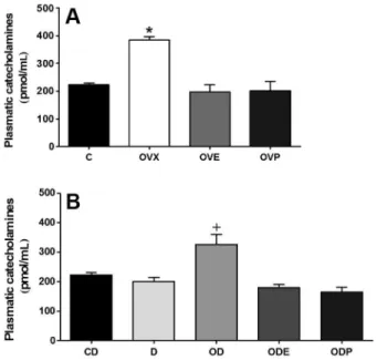
between the C and CD groups. Similar to the RECA results, PLCA levels increased in the OVX group (386.2±10.4 pmol/mL, P,0.01, Figure 2A) compared to the C group (222.2±8.5 pmol/mL). The OVE and OVP group values were similar to the C group (OVE= 197.1±26.2; OVP=202.0±34.6 pmol/mL, respectively, Figure 2A). PLCA levels increased in the OD group (352.2±36.0 pmol/mL, P,0.01) compared to the CD group (223.1±8.7 pmol/mL). The D, ODE and ODP group values (199.8±13.2; 179.3±10.5; 165.5±15.7 pmol/mL, respectively) were similar to that of the CD group (Figure 2B). We did not observe any differences in PLCA levels between the CD and C groups.
We also analyzed renal function parameters (Table 2). FENa+and FECl–were significantly increased in the OVX and OVP groups compared to the C group (in both, P,0.01 and P,0.05, respectively). As expected, both urinary flow and water intake increased in the OVX and OVP groups compared to the C group (P,0.01, P,0.05, P,0.05, and P,0.05, respectively). The renal parameters in the OVE group did not change significantly compared to the C group. Renal denervation did not change the FENa+, FECl–, urinary flow, and water intake compared to the CD group. However, these parameters increased in the OD group (P,0.01, P,0.05, P,0.05, and P,0.05, respec-tively) and the ODP group (P,0.01, P,0.05, P,0.05, and P,0.05, respectively). The ODE group was similar to Figure 1.Renal catecholamine content in female rats.A, Control
(C), ovariectomized (OVX), OVX with 17b-estradiol benzoate (OVE, 2.0 mg?kg-1
?day-1,sc), and OVX with progesterone (OVP, 1.7 mg?kg-1?day-1, sc), both for 7 days. B, Sham-denervated (CD), denervated (D), D ovariectomized (OD), OD with 17b -estradiol benzoate (ODE), and OD with progesterone (ODP), both for 7 days. Data are reported as means±SE (n=8). *P,0.01 and +P
,0.01 compared to C and CD, respectively (ANOVA and Tukey test).
the CD group. No changes in the GFR were observed in any of the groups studied (Table 2).
Discussion
In the present study, we showed a possible relation-ship between renal nerve catecholamine release levels and the roles of E2 and P4 in renal Na+ and Cl–
reabsorption in the rat kidney. Our data support the view that oophorectomy increases RECA levels, PLCA levels, FENa+, and FECl–. In similar animal models, our group showed changes in catecholamine release levels (renal and plasma) and renal function at the different estrous cycle phases (19). As expected, E2treatment reversed
the effect of OVX on RECA levels, PLCA levels, FENa+, and FECl–.
Renal function is modulated (at least in part) by the autonomic nervous system through the PSNS. Catecholamine release levels (a1 predominantly on
adrenergic receptors) from renal tubules and vasculature influence the levels of RBF, GFR, renin release, and Na+ reabsorption (13,22). Our data support studies that have shown that female hormones attenuate sympathetic activation in the central nervous system (17,23). After an acute infusion of intravenous E2in female rats (17) in
the nucleus tractus solitarius, rostral ventrolateral medulla, parabrachial nucleus, central nucleus of the amygdala, and intrathecal space, RSN activity decreased (18). Additionally, there is evidence suggesting that E2
can modulate adrenal and neurogenic catecholamine secretion and inhibit catecholamine release pre-synaptically (24,25). E2 may also upregulate the catalytic activity of
catechol-O-methyltransferase, thereby reducing the avail-ability of catecholamines (26).
On the other hand, we showed that P4 decreased
RECA levels to an even lower concentration than control levels and values similar to those in the renal denervated rats. Predictably, renal denervation decreased RECA levels, which were unchanged by other treatments.
However, it did not change PLCA levels. Our results are consistent with other studies that showed that P4inhibited
catecholamine secretion in bovine chromaffin cells of the adrenal medulla via a dose- and time-dependent mechan-ism and inhibited acetylcholine nicotinic receptors and increased inactivation of Ca2++-voltage-gated channels (16). Additionally, P4has a potentially inhibitory effect on
gamma-aminobutyric acid in the rostral ventrolateral medulla, which decreases central sympathetic activity (27). Previous studies have shown that renal denervation induces diuresis and natriuresis (28,29). However, our results showed similar values of renal function between renal denervated and normal animals. Similarly, DiBona and Sawin (30) showed that renal denervation had no effect on the dynamic autoregulation of RBF in control rats and that renal denervation did not produce changes in Na+excretion (31) or urinary volume in rats fed a normal
Na+diet (20).
Several investigations have shown that E2 and P4
modify body fluids and electrolytes (5,6,10,19). Female sex hormones have been shown to regulate the expression of different transporters in different tissues in rats, such as renal tissue (7,8). E2 replacement in the OVX rats
increases renal epithelial sodium channel mRNA levels compared to males, and P4treatment inhibits the
stimula-tion by E2alone (7). E2increases ClC-2 mRNA expression
in the rat proximal convoluted and proximal straight renal tubules (32). In accordance with previously published data, P4acts as a competitive antagonist of the mineralocorticoid
receptor, which attenuates the effect of aldosterone on Na+reabsorption (5,9). Because oophorectomy reduced Na+ and Cl– transporter expression and hormonal replacement normalized or modulated the expression, it was hypothesized that this downregulation could reduce Na+ and Cl– reabsorption by the nephron and that
hormonal replacement could normalize this possible reduction in Na+ and Cl– reabsorption in this animal model. Increases in FENa+and FECl–were observed, and estrogen replacement normalized these increases. Also, Table 2. Renal function in female rats.
Group (n=8) FENa+(%) FECl–(%) Urinary flow (mL/day) GFR (mL/min) Water intake (mL/day)
C 0.12 ± 0.01 0.39 ± 0.01 10.4 ± 0.8 1.7 ± 0.4 14.6 ± 1.2
OVX 0.21 ± 0.02** 0.62 ± 0.01* 13.7 ± 0.1** 1.5 ± 0.2 17.9 ± 1.1*
OVE 0.12 ± 0.01 0.35 ± 0.08 12.2 ± 0.7 1.7 ± 0.4 14.2 ± 1.3
OVP 0.18 ± 0.05** 0.53 ± 0.02* 14.4 ± 0.8* 2.1 ± 0.8 18.2 ± 1.4*
CD 0.12 ± 0.01 0.39 ± 0.01 10.4 ± 0.8 1.7 ± 0.4 14.6 ± 1.2
D 0.11 ± 0.02 0.41 ± 0.02 11.2 ± 0.9 1.8 ± 0.9 15.3 ± 1.8
OD 0.20 ± 0.01++ 0.67 ± 0.02+ 13.9 ± 0.8+ 1.7 ± 0.4 18.1 ± 1.2+
ODE 0.13 ± 0.02 0.42 ± 0.05 12.2 ± 0.9 1.6 ± 0.5 13.3 ± 1.7
ODP 0.19 ± 0.02++ 0.57 ± 0.03+ 14.2 ± 0.7+ 2.0 ± 0.7 17.7 ± 1.3+
FENa+and FECl–were increased in the OVX rats and not normalized by progesterone replacement.
In addition, E2 and P4 act in renal tissues by
modulating cyclic nucleotide-gated-A1 and Na+-K+ -ATPase (mainly in the renal cortex) by genomic mechan-isms, and these effects could be important for Na+, Cl–, and water balance (33). E2, in vitro, increases the
expression of the NaCl cotransporter and Na+-K+ -ATPase in renal distal tubule cells in culture by non-genomic mechanisms (8). Investigations showed that the kidneys control the expression of nuclear ERs (ER-aand ER-b) and the transmembrane GPR30 (10,11). This effect in the proximal tubules could be mediated directly via its receptors, or it could also act indirectly though the renin-angiotensin-aldosterone system (32).
GFR is considered to be the best and most common parameter for assessing renal function (10,19,33). We did not observe significant changes in the GFR or kidney and body weight following these experimental protocols. Similar to our results, women treated with a GnRH antagonist (to suppress reproductive function for the duration of the study) showed similar urinary volume, osmolality, and GFR after P4 and P4+E2treatment (5)
before, during, and after isotonic saline infusion.
Renal functions are controlled mainly by humoral factors and the sympathetic nervous system (such as
catecholamine release), which differ widely in their time of activation (34). The complex interactions of different hormones, such as sex steroid hormones, atrial natriuretic factor, angiotensin II, aldosterone, and vasopressin, may affect tubular reabsorption and water intake (5,35,36). Furthermore, renal cells after oophorectomy and hormonal replacement adapt and alter the expression of several genes that code for transporter proteins (10,33,34).
The present results show that there is a physiological interaction of E2and P4with renal nerves (catecholamine
release levels) that affects Na+/Cl–reabsorption in rats. Female sex hormones play an important role in maintain-ing Na+/Cl–reabsorption in rats on a normal Na+/Cl-diet. The variations in RECA and PLCA levels related to female sex hormones may influence renal function. On the other hand, changes in the FE of Na+and Cl–may be directly due to the effects of female sex hormones, independent of the main role of the renal nerves while on a normal Na+/Cl– diet. Further investigations are required to better elucidate the mechanisms of E2and P4on the plasma and renal
sympathetic system and on renal function.
Acknowledgments
Research partially supported by CNPq, CAPES, FAPES, and UFES.
References
1. Brunette MG, Leclerc M. Effect of estrogen on calcium and sodium transport by the nephron luminal membranes. J Endocrinol2001; 170: 441-450, doi: 10.1677/joe.0.1700441. 2. Brunette MG, Leclerc M. Renal action of progesterone: effect on calcium reabsorption. Mol Cell Endocrinol2002; 194: 183-190, doi: 10.1016/S0303-7207(02)00113-2. 3. Johnson JA, Davis JO. The effect of estrogens on renal
sodium excretion in the dog. Perspect Nephrol Hypertens 1976; 5: 239-248.
4. Stephenson LA, Kolka MA. Plasma volume during heat stress and exercise in women.Eur J Appl Physiol Occup Physiol1988; 57: 373-381, doi: 10.1007/BF00417979. 5. Stachenfeld NS, Keefe DL, Taylor HS. Responses to a
saline load in gonadotropin-releasing hormone antagonist-pretreated premenopausal women receiving progesterone or estradiol-progesterone therapy.J Clin Endocrinol Metab 2005; 90: 386-394, doi: 10.1210/jc.2004-0941.
6. Dignam WS, Voskian J, Assali NS. Effects of estrogens on renal hemodynamics and excretion of electrolytes in human subjects.J Clin Endocrinol Metab1956; 16: 1032-1042, doi: 10.1210/jcem-16-8-1032.
7. Gambling L, Dunford S, Wilson CA, McArdle HJ, Baines DL. Estrogen and progesterone regulate alpha, beta, and gammaENaC subunit mRNA levels in female rat kidney. Kidney Int 2004; 65: 1774-1781, doi: 10.1111/j.1523-1755.2004.00593.x.
8. Verlander JW, Tran TM, Zhang L, Kaplan MR, Hebert SC. Estradiol enhances thiazide-sensitive NaCl cotransporter density in the apical plasma membrane of the distal
convoluted tubule in ovariectomized rats. J Clin Invest 1998; 101: 1661-1669, doi: 10.1172/JCI601.
9. Myles K, Funder JW. Progesterone binding to mineralocor-ticoid receptors:in vitroandin vivostudies.Am J Physiol 1996; 270: E601-E607.
10. Gava AL, Freitas FP, Meyrelles SS, Silva IV, Graceli JB. Gender-dependent effects of aging on the kidney.Braz J Med Biol Res 2011; 44: 905-913, doi: 10.1590/S0100-879X2011007500101.
11. Lemale J, Bloch-Faure M, Grimont A, El Abida B, Imbert-Teboul M, Crambert G. Membrane progestin receptors alpha and gamma in renal epithelium.Biochim Biophys Acta2008; 1783: 2234-2240, doi: 10.1016/j.bbamcr.2008.07.023. 12. Katholi RE, Woods WT. Afferent renal nerves and
hyperten-sion.Clin Exp Hypertens A1987; 9 (Suppl 1): 211-226, doi: 10.3109/10641968709160175.
13. Johns EJ, Kopp UC, DiBona GF. Neural control of renal function.Compr Physiol2011; 1: 731-767.
14. Currie G, Freel EM, Perry CG, Dominiczak AF. Disorders of blood pressure regulation-role of catecholamine biosynth-esis, release, and metabolism.Curr Hypertens Rep2012; 14: 38-45, doi: 10.1007/s11906-011-0239-2.
15. Sobotka PA, Krum H, Bohm M, Francis DP, Schlaich MP. The role of renal denervation in the treatment of heart failure.Curr Cardiol Rep2012; 14: 285-292, doi: 10.1007/ s11886-012-0258-x.
17. He XR, Wang W, Crofton JT, Share L. Effects of 17beta-estradiol on sympathetic activity and pressor response to phenylephrine in ovariectomized rats.Am J Physiol1998; 275: R1202-R1208.
18. Saleh MC, Connell BJ, Saleh TM. Autonomic and cardio-vascular reflex responses to central estrogen injection in ovariectomized female rats.Brain Res2000; 879: 105-114, doi: 10.1016/S0006-8993(00)02757-8.
19. Gomes HL, Graceli JB, Goncalves WL, dos Santos RL, Abreu GR, Bissoli NS, et al. Influence of gender and estrous cycle on plasma and renal catecholamine levels in rats.Can J Physiol Pharmacol2012; 90: 75-82, doi: 10.1139/y11-102. 20. Franquini JV, Medeiros AR, Andrade TU, Araujo MT, Moyse´s MR, Abreu GR, et al. Influence of renal denervation on blood pressure, sodium and water excretion in acute total obstructive apnea in rats.Braz J Med Biol Res2009; 42: 214-219, doi: 10.1590/S0100-879X2009000200010. 21. Karim MM, Alam SM, Lee SH. Spectrofluorimetric estimation of
norepinephrine using ethylenediamine condensation method. J Fluoresc2007; 17: 427-436, doi: 10.1007/s10895-007-0188-y. 22. Souza DR, Mill JG, Cabral AM. Chronic experimental myocardial infarction produces antinatriuresis by a renal nerve-dependent mechanism.Braz J Med Biol Res2004; 37: 285-293, doi: 10.1590/S0100-879X2004000200017. 23. Liu CC, Kuo TB, Yang CC. Effects of estrogen on
gender-related autonomic differences in humans. Am J Physiol Heart Circ Physiol2003; 285: H2188-H2193.
24. Nagayama T, Hosokawa A, Yoshida M, Suzuki-Kusaba M, Hisa H, Kimura T, et al. Role of nitric oxide in adrenal catecholamine secretion in anesthetized dogs.Am J Physiol 1998; 275: R1075-R1081.
25. Hatanaka Y, Hobara N, Honghua J, Akiyama S, Nawa H, Kobayashi Y, et al. Neuronal nitric-oxide synthase inhibition facilitates adrenergic neurotransmission in rat mesenteric resistance arteries.J Pharmacol Exp Ther2006; 316: 490-497, doi: 10.1124/jpet.105.094656.
26. Fernandez-Ruiz JJ, Bukhari AR, Martinez-Arrieta R, Tresguerres JA, Ramos JA. Effects of estrogens and progesterone on the catecholaminergic activity of the adrenal medulla in female rats. Life Sci 1988; 42: 1019-1028, doi: 10.1016/0024-3205(88)90432-8.
27. Heesch CM, Rogers RC. Effects of pregnancy and progesterone metabolites on regulation of sympathetic outflow. Clin Exp Pharmacol Physiol 1995; 22: 136-142, doi: 10.1111/j.1440-1681.1995.tb01970.x.
28. DiBona GF, Kopp UC. Neural control of renal function. Physiol Rev1997; 77: 75-197.
29. Abdulla MH, Sattar MA, Salman IM, Abdullah NA, Ameer OZ, Khan MA, et al. Effect of acute unilateral renal denervation on renal hemodynamics in spontaneously hypertensive rats. Auton Autacoid Pharmacol 2008; 28: 87-94, doi: 10.1111/j.1474-8673.2008.00421.x.
30. DiBona GF, Sawin LL. Effect of renal denervation on dynamic autoregulation of renal blood flow.Am J Physiol Renal Physiol 2004; 286: F1209-F1218, doi: 10.1152/ ajprenal.00010.2004.
31. Kopp UC, Cicha MZ, Smith LA. Dietary sodium loading increases arterial pressure in afferent renal-denervated rats. Hypertension 2003; 42: 968-973, doi: 10.1161/ 01.HYP.0000097549.70134.D8.
32. Nascimento DS, Reis CU, Goldenberg RC, Ortiga-Carvalho TM, Pazos-Moura CC, Guggino SE, et al. Estrogen modulates ClC-2 chloride channel gene expression in rat kidney. Pflugers Arch 2003; 446: 593-599, doi: 10.1007/ s00424-003-1095-y.
33. Gracelli JB, Souza-Menezes J, Barbosa CM, Ornellas FS, Takiya CM, Alves LM, et al. Role of estrogen and progesterone in the modulation of CNG-A1 and Na/K+ -ATPase expression in the renal cortex. Cell Physiol Biochem2012; 30: 160-172, doi: 10.1159/000339055. 34. Antunes-Rodrigues J, de Castro M, Elias LL, Valenca MM,
McCann SM. Neuroendocrine control of body fluid metabo-lism.Physiol Rev 2004; 84: 169-208, doi: 10.1152/phys-rev.00017.2003.
35. Baiardi G, Macova M, Armando I, Ando H, Tyurmin D, Saavedra JM. Estrogen upregulates renal angiotensin II AT1 and AT2 receptors in the rat.Regul Pept2005; 124: 7-17, doi: 10.1016/j.regpep.2004.06.021.