Immunohistochemical demonstration
of TGF-ß and decorin in
paracoccidioidal granulomas
Departamento de Imunologia, Instituto de Ciências Biomédicas, Universidade de São Paulo, São Paulo, SP, Brasil
A.S. Nishikaku and E. Burger
Abstract
Different patterns of granulomas have been observed in 6- to 8-week-old mice after ip inoculation with 5 x 10⁶ yeast cells of
Paracoccidi-oides brasiliensis. Transforming growth factor-ß (TGF-ß) is a cy-tokine that has been shown to participate in fibrosis and granuloma formation; its activities seem to be modulated by the small proteogly-can decorin. In the present study, TGF-ß and decorin expression in epiploon granulomas was assessed by immunohistochemistry in sus-ceptible (B10.A) and resistant (A/J) mice after 15, 30, 120 and 150 days of P. brasiliensis ip infection. The epiploon was collected, fixed in Methacarn solution and embedded in paraffin, and 5-µm thick sections were used for immunohistochemical analysis employing the streptavidin-biotin-peroxidase technique. The former mouse strain developed fatal disease with many disseminated lesions increasing in size and number during the infection and the latter developed mild disease with the presence of encapsulated granulomas. In the epi-ploon, TGF-ß was present on macrophages, giant cells, lymphocytes and fibroblasts, and absent on neutrophils. It was also detected in areas of fibrosis and necrosis, as well as disperse in amorphous extracellular matrix, mostly in resistant mice. Decorin was present circumscribing macrophages and giant cells containing fungi, but absent on these cells. In both mouse strains, decorin was found at the periphery of the lesions, and markedly in milky spot granulomas. In resistant mice, positivity was found around fibrotic and necrotic areas of encapsu-lated and residual lesions containing lysed fungi. Decorin was found associated with thick fibers around encapsulated lesions. In suscep-tible mice, the size and number of lesions increased with the progres-sion of the disease and were correlated with the weaker expresprogres-sion of decorin. We suggest an association of decorin with the fibrogenic process observed in paracoccidioidal granulomas.
Correspondence
E. Burger
Departamento de Imunologia ICB, USP
Avenida Prof. Lineu Prestes, 1730 05508-900 São Paulo, SP Brasil
Fax: +55-11-3091-7224 E-mail: [email protected] Presented at SIMEC 2002 (International Symposium on Extracellular Matrix), Angra dos Reis, RJ, Brazil, October 7-10, 2002. Research supported by FAPESP (No. 00/10647-1) and CNPq (No. 350527/95-4).
Received November 29, 2002 Accepted May 19, 2003
Key words •TGF-ß •Decorin
•Paracoccidioides brasiliensis •Granulomatous lesions •Susceptibility and resistance
Paracoccidioides brasiliensis is a ther-mally dimorphic fungus that causes the most prevalent deep mycosis in Latin America, named paracoccidioidomycosis (PCM). Chronic inflammation characterized by the granulomatous response is a common aspect of PCM infection. Using an isogenic murine
of yeast cells and scarce type III collagen, while resistant mice produce few encapsu-lated granulomas with type I collagen or residual lesions with degenerated fungi (1). Regulatory mechanisms established in the host immune response and particularly in the development of granulomatous lesions have been attributed to both pathogen and host factors (cellular and extracellular com-ponents). In this last context, participation of cytokines in granuloma formation has been reported in several infectious diseases. Trans-forming growth factor-ß (TGF-ß) is a cy-tokine with anti-inflammatory and regula-tory properties that has been shown to participe in granuloma formation and fibro-sis (2,3). Synthefibro-sis and deposition of extra-cellular matrix (ECM) components in granu-lomatous lesions seem to be associated with the regulatory activities of some cytokines. Distinct modulatory effects of TGF-ß and tumor necrosis factor-α on gene expression
of collagens type I and IV have been ob-served (4). Fibrosis and regulation of the expression of ECM proteins such as col-lagen, fibronectin and decorin have been correlated with the presence of TGF-ß in experimental granulomas (5,6). Conversely, regulation of cell growth and differentiation has been attributed to several ECM elements that bind to different growth factors, such as endothelial cell growth factor, epidermal growth factor and TGF-ß. Decorin is a small proteoglycan formed by one or more gly-cosaminoglycan chains containing derma-tan or chondroitin sulfate and a core protein composed of repeated sequences of amino acids rich in leucine, that has been demon-strated to bind to TGF-ß (7), acting as a reservoir of this cytokine in ECM and at the same time playing a regulatory role in TGF-ß activity (8). On the other hand, it was shown that TGF-ß could be released from the complex, in which it is found together with decorin, to the ECM by action of matrix metalloproteinases, that are able to digest and degrade the proteoglycan, releasing
TGF-ß and allowing it to carry out its bio-logical functions (9). Both negative (10) and positive (11) modulatory effects of decorin on TGF-ß functions have been reported. The in situ expression of ECM components and cytokines in paracoccidioidal granulomas has been poorly investigated.
In the present study, we investigated the immunolocalization of TGF-ß and decorin expression in granulomatous lesions of epi-ploon in mice susceptible (B10.A) and resis-tant (A/J) to P. brasiliensis infection (12) and inoculated with the highly virulent iso-late Pb18 (13). We correiso-lated the distribu-tion of these components with the kinetics of granuloma formation and with the lesion patterns, as well as with their role in the immune response to PCM infection.
Six- to eight-week-old female mice of the B10.A and A/J strains were inoculated ip with 5 x 10⁶ yeast cells/ml and sacrificed
anti-rabbit IgG antibody (1/1000; Rockland, Gilbertsville, PA, USA) was applied to the tissues for 1 h at room temperature, followed by incubation with streptavidin-peroxidase (1/50) (Pharmingen, San Diego, CA, USA). Finally, 0.05% 3,3' diaminobenzidine tetra-hydrochloride (Sigma, St. Louis, MO, USA) added to 3 µl 30% hydrogen peroxide was applied to the slides and the reaction was stopped after visualization of a brown color on tissue sections. The sections were then counterstained with Mayer’s hematoxylin and mounted slides were observed with a light microscope (Hund Wetzlar, Wetzlar, Germany) at 40, 100, 250 and 400X magni-fication. Image capture was used for micro-scopic analysis employing a video camera (Kodo, Tokyo, Japan) and a Microsoft Video Capture software.
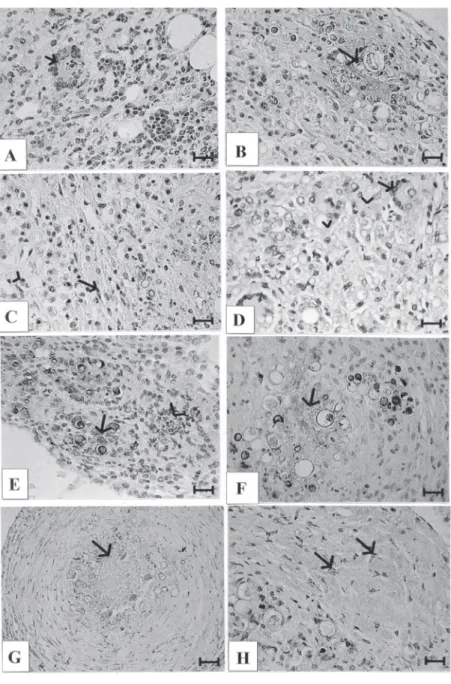
In the epiploon, TGF-ß was present on macrophages, multinucleated giant cells, lym-phocytes and fibroblasts, and absent on neu-trophils in granulomatous lesions observed in both resistant and susceptible mice in-fected with Pb18. It was also detected in areas of fibrosis and necrosis, as well as disperse in amorphous extracellular matrix, mostly in resistant mice that developed re-sidual lesions containing TGF-ß-positive pseudoxanthomatous macrophages in the later phase of infection (shown in Figure 1A-H). Decorin was present circumscribing macrophages and giant cells containing fungi, but absent on these cells. In both mouse strains, decorin was found at the periphery of the lesions and/or surrounding macro-phages and multinucleated giant cells around yeast cells, forming granulomatous struc-tures resembling fibrillar cocoons especially in concentric lesions localized in milky spot granulomas. In resistant mice, positivity was found around fibrotic and necrotic areas of encapsulated and residual lesions contain-ing lysed fungi (shown in Figure 2A-J).
TGF-ß has been associated with the anti-inflammatory response and synthesis of ECM components through activation of fibroblasts
and other cells, inducing a fibrotic reaction. The expression of TGF-ß in several cell populations, such as macrophages, giant cells and fibroblasts and/or linked to ECM ob-served here suggests an active role of this cytokine in the pattern of tissue response developed during granuloma formation and interfering with the type of lesions. By this interaction with cells and ECM, TGF-ß may influence the evolution or the control of PCM infection. In the present study, the presence of decorin forming fibrillar cocoons in the lesions suggests an effort to control P. brasiliensis dissemination by interactions of immune and inflammatory cells and ECM components against yeast cells. In an exper-imental infection of athymic and euthymic mice with P. brasiliensis, lesions with for-mation of fibrillar cocoons surrounding groups of fungal cells, surrounded or not by macrophages and giant cells were observed (14). In the literature, decorin has been shown to have a role in assembling and stabilizing the collagen fibrils; our results suggest the Figure 2. Immunohistochemical analysis of decorin in granulomatous lesions. A, Positive reaction in the ex-tracellular matrix (ECM) around small granulomatous foci (arrows) in susceptible mice at 15 days after infec-tion (bar = 31.25 µm). B, Positive loose lesion (arrows) in susceptible mice at 30 days after infection (bar = 25 µm). C, Decorin staining around small granulomas (ar-rows) in susceptible mice at 120 days after infection (bar = 31.25 µm). D, Extensive lesions with lesser deposition of decorin (arrows) around various granulo-matous foci in susceptible mice at 150 days after infec-tion (bar = 50 µm). E, Milky spot (arrow) stained in resistant mice at 15 days after infection (bar = 31.25 µm). F, Macrophages (arrowhead) and giant cells (ar-row) surrounded by positively stained ECM in resistant mice at 30 days after infection (bar = 25 µm). G, Nega-tive reaction in the cellular components (arrows) in resistant mice at 30 days after infection (bar = 25 µm).
H, Necrotic area (arrows) in resistant mice at 120 days after infection (bar = 125 µm). I, Encapsulated lesion with a fibrotic area (arrows) enclosed by decorin in resistant mice at 150 days after infection (bar = 50 µm).
participation of decorin in cocoon formation in an attempt to enclose P. brasiliensis cells, and a lesser deposition of decorin was corre-lated with extense loose granulomatous le-sions observed mainly in susceptible mice.
The present investigation of paracocci-dioidal granulomas focusing on the expres-sion of TGF-ß and decorin indicated differ-ences between susceptible and resistant mouse strains. In the earlier phases of infec-tion, resistant mice showed marked TGF-ß positivity in various cell populations and the presence of both nonencapsulated and en-capsulated granulomas. In the later phases, encapsulated granulomas predominated, with decorpositive thick fibers delimiting in-ner necrosis areas and TGF-ß-positivity around fibrotic areas and within necrosis areas, associated with amorphous ECM and lysed P. brasiliensis in the center of the lesions. Polymorphonuclear neutrophils were present during the earlier stages of the infec-tion, but were negative for TGF-ß, while positive giant cells were observed until the later phases of infection. In susceptible mice, compact lesions could be observed in the initial phases of infection, but loose granulo-mas predominated in later phases. Positivity for TGF-ß was ubiquitous, being found in
cells, predominantly in giant cells, from the onset of the infection and in both amorphous and fibrillar ECM. A positive reaction for decorin was more associated with encapsu-lated lesions and with later stages of the disease. Since these lesions were less fre-quent, a weaker expression of decorin was observed.
We described here for the first time the presence of decorin as one of the ECM com-ponents of the fibrous capsule of PCM le-sions. An association of this proteoglycan with the fibrogenic process detected mainly in lesions from resistant mice is suggested. The interactions between different cellular and extracellular host components and P. brasiliensis yeast cells present in granulo-mas are extremely complex and are impor-tant in the development of control or dis-semination of PCM infection.
Acknowledgments
We are grateful to C.S. Cunha and B.P. Albe for technical assistance and thank Dr. L.W. Fisher (National Institutes of Health, Bethesda, MD) for donation of rabbit poly-clonal antibody to mouse decorin (LF-113 clone).
References
1. Xidieh CF, Lenzi HL, Calich VLG & Burger E (1999). Influence of the genetic background on the pattern of lesions developed by resis-tant and susceptible mice infected with Paracoccidioides brasilien-sis. Medical Microbiology and Immunology, 188: 41-49.
2. Appleton I, Tomlinson A, Colville-Nash PR & Willoughby DA (1993). Temporal and spatial immunolocalization of cytokines in murine chronic granulomatous tissue. Implications for their role in tissue development and repair processes. Laboratory Investigation, 69: 405-414.
3. Hernandez-Pando R, Orosco H, Arriaga K, Sampieri A, Larriva-Sahd J & Madrid-Marina V (1997). Analysis of the local kinetics and localization of interleukin-1α and transforming growth factor-ß, dur-ing the course of experimental pulmonary tuberculosis. Immunol-ogy, 90: 607-617.
4. Grande JP, Melder DC & Zinsmeister AR (1997). Modulation of collagen gene expression by cytokines: stimulatory effect of trans-forming growth factor-beta1, with divergent effects of epidermal growth factor and tumor necrosis factor-alpha on collagen type I
and collagen type IV. Journal of Laboratory and Clinical Medicine, 130: 476-486.
5. Roman J, Jeon YJ, Gal A & Perez RL (1995). Distribution of extracel-lular matrices, matrix receptors, and transforming growth factor-ß1 in human and experimental lung granulomatous inflammation.
American Journal of the Medical Sciences, 309: 124-133.
6. Limper AH, Colby TV, Sanders MS, Asakura S, Roche PC & Deremee RA (1994). Immunohistochemical localization of transforming growth factor-ß1 in the nonnecrotizing granulomas of pulmonary sarcoido-sis. American Journal of Respiratory and Critical Care Medicine, 149: 197-204.
7. Hildebrand A, Romaris M, Rasmussen LM, Heinegard D, Twardzik DR, Border WA & Ruoslahti E (1994). Interaction of the small interstitial proteoglycans biglycan, decorin and fibromodulin with transforming growth factor beta. Biochemical Journal, 302: 527-534.
281-284.
9. Imai K, Hiramatsu A, Fukushima D, Pierschbacher MD & Okada Y (1997). Degradation of decorin by metalloproteinases: identification of the cleavage sites, kinetic analyses and transforming growth factor-ß1 release. Biochemical Journal, 322: 809-814.
10. Markmann A, Hausser H, Schönherr E & Kresse H (2000). Influence of decorin expression on transforming growth factor-ß-mediated collagen gel retraction and biglycan induction. Matrix Biology, 19: 631-636.
11. Takeuchi Y, Kodama Y & Matsumoto T (1994). Bone matrix decorin binds transforming growth factor-ß and enhances its bioactivity.
Journal of BiologicalChemistry, 269: 32634-32638.
12. Calich VLG, Singer-Vermes LM, Siqueira AM & Burger E (1985). Susceptibility and resistance of inbred mice to Paracoccidioides brasiliensis. Brazilian Journal of ExperimentalPathology, 66: 585-594.
13. Kashino SS, Calich VLG, Burger E & Singer-Vermes LM (1985). In vivo and in vitro characteristics of six Paracoccidioides brasiliensis
strains. Mycopathologia, 92: 173-178.