Source of Support: Nil
Effect of Repeated use on Topographical Features of ProTaper Next Endodontic Rotary File
Ranya Faraj Elemam1, José A Capelas2, Manuel F Vieira3 Contributors:
1PhD Candidate, Department of Endodontics, Faculty of Dental
Medicine, University of Porto, Rua Dr. Manuel Pereira da Silva, 4200-393 Porto, Portugal; 2Associate Professor, Department of
Endodontics, Faculty of Dental Medicine, University of Porto, Rua Dr. Manuel Pereira da Silva, 4200-393 Porto, Portugal;
3Associate Professor, Department of Metallurgical and Materials
Engineering, Centre for Mechanical Engineering, University of Porto, R. Dr. Roberto Frias, 4200-465 Porto, Portugal.
Correspondence:
Elemam RF. 39 4ESQ.TRAZ, Rua Manuel Pacheco Miranda, 4200-804 Porto, Portugal. Tel.: +00351934733866. Email: Ranya_Elemam@yahoo.co.uk
How to cite the article:
Elemam RF, Capelas JA, Vieira MF. Effect of repeated use on topographical features of ProTaper next endodontic rotary file. J Int Oral Health 2016;8(4):445-450.
Abstract:
Background: The purpose of this study was to evaluate the
morphological alterations of the ProTaper next rotary file (PTN) under scanning electron microscopy (SEM).
Materials and Methods: A total of 18 simulated root canals were
allocated to three groups. Six new sets of PTN instruments were used 3 times. A #10 K-file was inserted into the working length, followed by ProGlider to create a glide path. Ensuring the manufacturer’s instructions with 99% ethyl alcohol for irrigation, all canals were prepared. Files were photographed in the same position before and after three canals preparations using a high-resolution SEM.
Result: A metal strip appeared on one X1 instrument surface
preoperatively. Microcrack defects were observed on two X2 files postoperatively, and the blunt cutting edge was observed on three X1 files before and after use and one file fractured.
Conclusion: Small number of changes appeared on PTN surfaces,
yet same PTN file can be used safely 3 times to prepare multi-rooted teeth within the same patient.
Key Words: ProTaper next, rotary endodontics files, scanning electron microscopy, surface changes
Introduction
NiTi files are prone to fracture without any visible defects. Their fatigability occurs as a result of stress during instrumentation of the root canal.1 In practice, the potential difficulty in removing
fractured pieces of the broken files may compromise the outcome of endodontic treatment. This difficulty makes it imperative to know the in-service use of these instruments and identify the fracture mechanisms.
Fatigability that leads to failure of NiTi endodontic instruments can be caused by flexural fatigue or torsional
fatigue. Flexural fatigue, also called cyclic fatigue,2 occurs
when the file is used in a curved canal,3 which is the most
destructive form of stress.4 On the other hand, torsional
fatigue occurs in two forms: Dynamic fatigue, which result from frictional forces that are caused by resistance of dentin to the file’s cutting;5 and static fatigue, which occurs during
root canal preparation when the tip or any other part of the file is locked in, but the shaft continues to rotate.5 Torsional
fatigue normally shows plastic deformation followed by fracture6 and can be evaluated microscopically. Therefore,
examining the surface of endodontic files would have an important impact on the identification of fracture initiation and file failure.7
New endodontic instruments have been subjected to new strategies to improve flexibility, resistance to fracture, fatigability, and cross-sectional designs compared to conventional NiTi instruments. Manufacturers have also proposed different thermomechanical treatments. The ProTaper next (PTN) file system was produced from an M-wire alloy that has greater flexibility and fatigue resistance than conventional NiTi instruments.8
Previous studies have reported that PTN instruments have superior cyclic fatigue resistance compared with other NiTi rotary instruments.9-14 However, to date, no evaluation of the
surface characteristics of PTN files have been performed. In addition, no studies have assessed the impact of the repeated use of PTN files to simulate multi-rooted teeth. The link between fatigability of the metal surface changes and file failure with repetitive usage could possibly lead to more understanding about the characteristics of PTN files. Thus, the purpose of this study was to observe the morphological alterations of PTN files before and after continuous use by scanning electron microscopy (SEM).
Materials and Methods
Sample preparation
A total of 18 simulated root canals (16 mm in length and 60°C in curvature) in clear resin blokes (Dentsply-Maillefer Ballaigues, Switzerland) were selected for this study. The blocks were given numbers from 1 to 18 and then assembled into three groups consisting six blocks each. Six kits of a PTN (Dentsply-Maillefer Ballaigues, Switzerland) file system were also given numbers from 1 to 6; each kit was used to prepare the corresponding block group. The blocks and the file groups are detailed in Table 1.
Canal instrumentation
All canals were treated by a single operator. For each canal, the working length (WL) was estimated at the level of the apical foramen using a #10 hand K-file (Dentsply-Maillefer, Ballaigues, Switzerland), and a glide path was established using ProGlider (Dentsply-Maillefer, Ballaigues, Switzerland). PTN files were used in the canals with the crown-down technique, driven by the Wave One Endodontic Motor (Dentsply-Maillefer, Ballaigues, and Switzerland) in rotational motion. The rotational speed was set as recommended, at 350 rpm with a torque of 2.5 N/cm. The preparation was performed using two files (X1 and X2) through a gentle in-and-out motion until the full WL was reached. The instrumentation sequence was as follows: X1 file (size 17, 0.04 taper) followed by the X2 file (size 26, 0.06 taper). All canals were frequently irrigated with 99% ethyl alcohol using a 30-G side-vented needle and repeatedly recapitulated with a K-file ISO 10 to keep the glide path open.
Topographic evaluation
Before the canal preparation started, all files were examined by SEM. A high-resolution FEI QUANTA 400 FEG SEM (FEI, Hillsboro, Oregon 97124, USA) was used for this purpose. All instruments were photographed in the same position before and after each of three uses to observe any surface changes. Three views were chosen: Apical, middle, and critical point area, the latter of which is 3-5 mm away from the apical point of the file. The files were cleaned after use by ultrasonic sterilization (Biosonic UC125 Coltene Whaledent, Langenau/ Germany) for 30 min and then reexamined.
Examination criteria
The criteria used for checking the instruments’ surface defects were adopted by Eggert et al.,15 and were as follows: No visible
defects, pitting, corrosion, fretting, microcracks, fractures, metal strips, spiral distortion, blunt cutting edges, disruption of cutting edges or fatigue cracks (Table 2).
Results
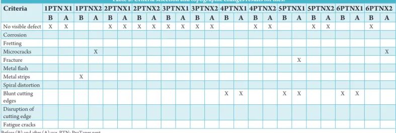
PTNX1 and PTNX2 of each of the six systems were examined two times; before (B) and after use (A) (a total of 24 files). The files showed no pitting, corrosion, fretting, spiral distortion, disruption of cutting edges or fatigue cracks. However, other changes were observed, which listed in Table 2 and shown in Figures 1-7.
Among these, 15 files showed no visible defects, and 9 files showed some topographic surface defects. One X2 file had a metal strip on the apical portion (from the top view) before its use (1 PTNX2) (Figure 1). Three X1 files had blunt cutting edges of their apical and middle surfaces before use (4 PTNX1, 5 PTNX1, and 6 PTNX1) (Figures 2 and 3); such defects persisted after usage (Figures 4-5). Small microcracks were observed on the critical point area under ×1000 magnification in two X2 files after use (1 PTNX2 and 6 PTNX2) (Figures 6a-b). Moreover, only one file had fractures after its third use (5 PTNX1) (Figure 7a and b).
Discussion
The quality of both the cutting surfaces and head surfaces of rotary NiTi files after repeated use is of clinical interest.16 Therefore,
examining the file surfaces before and after instrumentation becomes essential to understand the changes that occur before the instruments fail. This will help in the selection and application of NiTi rotary instruments during root canal treatment17 and can
provide insights for clinical use in an attempt to reduce the risk of instrument breakage within root canals.18
Our qualitative evaluation was done by SEM observation. Endodontic literature shows that SEM offers high-resolution Table 1: Sample distribution.
File system no. Group 1 Group 2 Group 3
F.S.1=1 PTNX1-X2 B1 B7 B13 F.S.2=2 PTNX1-X2 B2 B8 B14 F.S.3=3 PTNX1-X2 B3 B9 B15 F.S.4=4 PTNX1-X2 B4 B10 B16 F.S.5=5 PTNX1-X2 B5 B11 B17 F.S.6=6 PTNX1-X2 B6 B12 B18
F.S.1: File system No. 1-6, B: Endodontic block No. 1-18, PTN: ProTaper next
Table 2: Criteria selection and topographic changes results for files.
Criteria 1PTN X1 1PTNX2 2PTNX1 2PTNX2 3PTNX1 3PTNX2 4PTNX1 4PTNX2 5PTNX1 5PTNX2 6PTNX1 6PTNX2 B A B A B A B A B A B A B A B A B A B A B A B A No visible defect X X X X X X X X X X X X X X X Corrosion Fretting Microcracks X X Fracture X Metal flash Metal strips X Spiral distortion Blunt cutting edges X X X X X X Disruption of cutting edge Fatigue cracks
images and allows characterization of the topographic features that appear on the file surfaces and the fractured surfaces of broken instruments.10,15,19 SEM has also been used to observe
the dentinal debris and explore patent dentinal tubules.20 In
addition, SEM was proven to be useful in repeatedly evaluating
surface defects without affecting the physical properties of endodontic files.21
Three areas were selected for evaluating the surface changes: The middle, apical third, and critical area of the file. Those locations were in accordance with Sattapan et al.,22 who
indicated that these three areas are the most critical because the files tend to fracture close to their tip. Moreover, the tapering of the file increased toward the handle, making the bulk of the file much stronger than its tip.18
Figure 1: Metal strip - observed on the tip of 1 ProTaper next
X2 file.
Figure 2: Apical view of 4 ProTaper next X1 files before usage.
Figure 3: Middle view of 4 ProTaper next X1 files before usage.
Figure 4: Apical view of 4 ProTaper next X1 files after usage.
Figure 5: Middle view of 4 ProTaper next X1 files after usage.
Figure 6: Small microcracks observed on the critical point area
in 1 ProTaper next (PTN)X2 file (a) and 6 PTNX2 files (b). b
Before use, the files had a highly smooth surface except for one metal strip observed on the tip of one PTNX2 file (Figure 1). This finding was mentioned in previous reports15,23 as a result
of the manufacturing process. However, most defects in these reports were in the form of milling grooves, multiple cracks, and pits.
In the current study, blunt cutting edges were observed in three files before use and stayed unchanged after the third use. Similarly, in testing the light speed file, Eggert et al.,z15 described
a remaining blunt cutting edge after use and explained that it meant that the sharp cutting edges were not overused. Kumar analyzed the ProTaper Universal file (PTU) and found the same defects but only after use. The authors explained these defects as a result of the safe cutting tip and the anti-screwing design, which require less pressure to avoid such defects.23
Microcracks were also detected in this study, similarly to other work that showed that the ProTaper rotary files also exhibited microfractures after the third and fourth use compared with K3, PTU hand files.24 Microcracks or micro fractures are the
results of a rotational bending of the file within the canal due to shear forces on the blades, which later combine to become the fatigue cracks.25
Our result also revealed a fracture on one file, X1 (number five) that happened immediately after the third use. Similarly, in a study that examined the separation incidence of the PTN, the author revealed that most fractured PTN files were X1 files. Those instruments were the first used to penetrate and shape the full WL of the canal, and thus, they were more likely to suffer from fatigue.26 We used ProGlider to create glide paths before
using the PTNX1 files, which could have made our X1 files less subject to fatigue, thus reducing the number of fractures.27
SEM at larger magnifications revealed a strong plastic deformation near the fracture zone, resembling a previous study on PTN that presented typical dimpling near the center of the fracture surface.28 Such a defect indicates that the fracture
occurred by torsional stress rather than by cyclic fatigue.14,18
The same files that broke (5 PTNX1 file) had shown few changes before use (blunted cutting edge), and such changes could act as a stress concentration area and lead to fracture; the
elastic limit of the material was exceeded, which led to plastic deformation followed by fracture.29,30 The fracture occurred
4 mm from the apical point. This is comparable to previous works.2,31 that indicated that instruments did not fracture at
the tip of the file but rather at the point of supreme flexure of the shaft or the midpoint.10,32
The repeated use of NiTi files may cause the plastic deformation of the material.33 That if happened, could result
in inadequate preparation, insufficient cleansing, and shaping of the root canal system and lead ultimately to the fracture of the files.34 The present in vitro study evaluated the surface
changes of PTN rotary instruments before and after the third use under an SEM.
The results showed the low occurrence of topographic changes, low fracture incidence, and high resistance to cyclic fatigue compared to previous studies. This difference could be due to many factors; alloys such as NiTi have been proven to increase the cyclic fatigue resistance of the instruments.35 The M-wire
technology, which provides greater flexibility for the files along with the off-centered rectangular cross-sectional design, also improves the file’s strength and gives the system high resistance to cyclic fatigue.36,37 The unequal contact between the PTN
instrument and the root canal wall could also be a feature that enhanced the fracture resistance of the PTN file.10
Although PTN is able to resist high torsional stress, the incidence of fracture occurred was a result of torsional failure. Therefore, clinicians should consider these findings, especially when preparing curved root canals with the same file that have already been used to shape narrow root canals. It is recommended that the clinician use light insertion and avoid forcing the instrument apically during instrumentation.
Conclusion
In this study, the morphological alterations of PTN rotary files after preparation of three simulated root canals were examined by SEM. The changes in file surfaces were few and had no influence on the file’s stability. Thus, PTN rotary instruments were used multiple times and safely in curved simulated root canals that mimicked the clinical situation of multi-rooted canals without fear of fracture when avoiding incorrect file use.
References
1. Van der Vyver P. Creating a glide path for rotary NiTi instruments: Part two. Endod Pract 2011;17:46-53. 2. Pruett JP, Clement DJ, Carnes DL Jr. Cyclic fatigue testing
of nickel-titanium endodontic instruments. J Endod 1997;23(2):77-85.
3. Sotokawa T. An analysis of clinical breakage of root canal instruments. J Endod 1988;14(2):75-82.
4. Chakka NV, Ratnakar P, Das S, Bagchi A, Sudhir S, Anumula L. Do NiTi instruments show defects before separation? Defects caused by torsional fatigue in hand
Figure 7: Fracture view - of 5 ProTaper next X1 files (critical
area) (a), magnification of showing the plastic deformation (b). b
and rotary nickel-titanium (NiTi) instruments which lead to failure during clinical use. J Contemp Dent Pract 2012;13(6):867-72.
5. Yao JH, Schwartz SA, Beeson TJ. Cyclic fatigue of three types of rotary nickel-titanium files in a dynamic model. J Endod 2006;32(1):55-7.
6. Askeland DR, Phulé PP. The Science and Engineering of Materials, 4th ed. California: Brooks/Cole-Thomson
Learning; 2003.
7. Lee JK, Kim ES, Kang MW, Kum KY. The effect of surface defects on the cyclic fatigue fracture of HEROShaper Ni-Ti rotary files in a dynamic model: A fractographic analysis. JKACD 2007;32(2):130-7.
8. Gambarini G, Gergi R, Naaman A, Osta N, Al Sudani D. Cyclic fatigue analysis of twisted file rotary NiTi instruments used in reciprocating motion. Int Endod J 2012;45(9):802-6.
9. Capar ID, Ertas H, Arslan H. Comparison of cyclic fatigue resistance of novel nickel-titanium rotary instruments. Aust Endod J 2015;41(1):24-8.
10. Elnaghy AM. Cyclic fatigue resistance of ProTaper Next nickel-titanium rotary files. Int Endod J 2014;47(11):1034-9. 11. Ertas H, Capar ID, Arslan H, Akan E. Comparison of
cyclic fatigue resistance of original and counterfeit rotary instruments. Biomed Eng Online 2014;13:67.
12. Pérez-Higueras JJ, Arias A, de la Macorra JC, Peters OA. Differences in cyclic fatigue resistance between ProTaper next and ProTaper Universal instruments at different levels. J Endod 2014;40(9):1477-81.
13. Nguyen HH, Fong H, Paranjpe A, Flake NM, Johnson JD, Peters OA. Evaluation of the resistance to cyclic fatigue among ProTaper next, ProTaper Universal, and vortex blue rotary instruments. J Endod 2014;40(8):1190-3. 14. Pedullà E, Lo Savio F, Boninelli S, Plotino G, Grande NM,
Rapisarda E, et al. Influence of cyclic torsional preloading on
cyclic fatigue resistance of nickel - Titanium instruments. Int Endod J 2015;48(11):1043-50.
15. Eggert C, Peters O, Barbakow F. Wear of nickel-titanium lightspeed instruments evaluated by scanning electron microscopy. J Endod 1999;25(7):494-7.
16. Hanan AR, Meireles DA, Sponchiado Júnior EC, Hanan S, Kuga MC, Bonetti Filho I. Surface characteristics of reciprocating instruments before and after use – a SEM analysis. Braz Dent J 2015;26(2):121-7.
17. Pessoa OF, da Silva JM, Gavini G. Cyclic fatigue resistance of rotary NiTi instruments after simulated clinical use in curved root canals. Braz Dent J 2013;24(2):117-20. 18. Sattapan B, Nervo GJ, Palamara JE, Messer HH. Defects
in rotary nickel-titanium files after clinical use. J Endod 2000;26(3):161-5.
19. Can Saglam B, Görgül G. Evaluation of surface alterations in different retreatment nickel-titanium files: AFM and SEM study. Microsc Res Tech 2015;78(5):356-62. 20. Zmener O, Pameijer CH, Banegas G. Effectiveness
in cleaning oval-shaped root canals using Anatomic Endodontic Technology, ProFile and manual
instrumentation: A scanning electron microscopic study. Int Endod J 2005;38(6):356-63.
21. Tripi TR, Bonaccorso A, Tripi V, Condorelli GG, Rapisarda E. Defects in GT rotary instruments after use: An SEM study. J Endod 2001;27(12):782-5.
22. Sattapan B, Palamara JE, Messer HH. Torque during canal instrumentation using rotary nickel-titanium files. J Endod 2000;26(3):156-60.
23. Kumar J. A scanning electron microscopic evaluation of surface changes of new and used greater taper nickel titanium hand and rotary instruments - An in vitro study.
Endodontology 2013;25(1):37-50.
24. Subha N, Sikri VK. Comparative evaluation of surface changes in four Ni-Ti instruments with successive uses - An SEM study. J Conserv Dent 2011;14:282-6.
25. Cheung GS, Peng B, Bian Z, Shen Y, Darvell BW. Defects in ProTaper S1 instruments after clinical use: Fractographic examination. Int Endod J 2005;38(11):802-9.
26. Ertas H, Capar ID. An in vitro analysis of separation
of multi-use ProTaper Universal and ProTaper Next instruments in extracted mandibular molar teeth. Scanning 2015;37(4):270-6.
27. Berutti E, Alovisi M, Pastorelli MA, Chiandussi G, Scotti N, Pasqualini D. Energy consumption of ProTaper Next X1 after glide path with PathFiles and ProGlider. J Endod 2014;40(12):2015-8.
28. Elnaghy AM, Elsaka SE. Assessment of the mechanical properties of ProTaper Next Nickel-titanium rotary files. J Endod 2014;40(11):1830-4.
29. Blum JY, Machtou P, Micallef JP. Location of contact areas on rotary Profile instruments in relationship to the forces developed during mechanical preparation on extracted teeth. Int Endod J 1999;32(2):108-14.
30. Gambarini G. Cyclic fatigue of ProFile rotary instruments after prolonged clinical use. Int Endod J 2001;34(5):386-9. 31. Mandel E, Adib-Yazdi M, Benhamou LM, Lachkar T,
Mesgouez C, Sobel M. Rotary Ni-Ti profile systems for preparing curved canals in resin blocks: Influence of operator on instrument breakage. Int Endod J 1999;32(6):436-43.
32. Pirani C, Cirulli PP, Chersoni S, Micele L, Ruggeri O, Prati C. Cyclic fatigue testing and metallographic analysis of nickel-titanium rotary instruments. J Endod 2011;37(7):1013-6.
33. Troian CH, Só MV, Figueiredo JA, Oliveira EP. Deformation and fracture of RaCe and K3 endodontic instruments according to the number of uses. Int Endod J 2006;39(8):616-25.
34. Kuhn G, Tavernier B, Jordan L. Influence of structure on nickel-titanium endodontic instruments failure. J Endod 2001;27(8):516-20.
35. Bulem ÜK, Kececi AD, Guldas HE. Experimental evaluation of cyclic fatigue resistance of four different nickel-titanium instruments after immersion in sodium hypochlorite and/or sterilization. J Appl Oral Sci 2013;21(6):505-10.
36. Maillefer D. The ProTaper Next Brochure, 2013. Available from: http://www.dentsply.com.au/secure/downl. 37. Shen Y, Zhou HM, Zheng YF, Peng B, Haapasalo M.
Current challenges and concepts of the thermomechanical treatment of nickel-titanium instruments. J Endod 2013;39(2):163-72.