Effects of caffeic acid alkyl esters in
the control of planktonic and
sessile cells
Dissertation for Master Degree in Bioengineering – Specialization in Biological
Engineering
Mafalda Sousa Andrade
Supervisor: Prof. Manuel Simões
Co-supervisor: Prof. Fernanda Borges
“Nothing in life is to be feared, it is only to be understood. Now is the time to understand more, so that we may fear less.”
i
A
CKNOWLEDGMENTSI would like to express my gratitude towards all the people that made this Master’s Dissertation possible.
First of all, I am most grateful to my Supervisor, Professor Manuel Simões, for introducing me to this topic and for all the kind guidance and support he gave me throughout this period. In his person, I also thank Faculdade de Engenharia da Universidade do Porto, especially the Laboratory for Process Engineering, Environment, Biotechnology and Energy (LEPABE), for granting me the funds and working conditions necessary to develop this project.
I am also most thankful to my Secondary Supervisor, Professor Fernanda Borges for all the enthusiasm, sympathy and support shown during the length of this project, as well as for supplying the chemical compounds tested. In this context, I would also like to acknowledge Faculdade de Ciências da Universidade do Porto, for the use of its resources, and Sofia Benfeito and Pedro Soares, for kindly synthetizing the abovementioned compounds.
My utmost gratitude and esteem goes to Joana Malheiro, whose teaching and support were of huge importance to my project. Most of what I did and learned during the last months were only possible due to her and, in that view, I deeply appreciate all her efforts and readiness in helping me at all times and answering all of my doubts. My appreciation also goes to all the other researchers at laboratory E007, especially Anabela Borges, Catarina Meireles, Inês Gomes, Ana Luísa Gonçalves and Rita Fulgêncio who kindly helped me whenever I needed. Additionally, I also acknowledge the technicians from laboratory E-103, Paula and Sílvia, whose kindness and help were vital in several moments.
Moreover, I would like to express my gratitude to Professor Maria do Carmo Pereira for allowing the use her equipment for zeta potential measurement and Joana Loureiro for her availability and assistance in the process.
My gratefulness also goes to all of my friends: to the girls from the Biological Engineering class of 2014 for the amazing company during the last five years, but particularly during the last months by making my working days a little bit more happy; to my friends Té, Nuno, Catarina and Filipa for their long-time friendship, support and for all the fun over these years. Also, to my lab mates at laboratory E-101, Xiló and Bruno, my special thanks for making my everyday tasks much more entertaining.
And last, but definitely not the least, to my Mother and Daniel for their gigantic patience, support and encouragement at all times. I love you both and I wouldn’t have made it so far without you backing me.
ii
A
BSTRACTBacterial multidrug resistance to the commonly used antibiotics is a global concern, especially when coupled with biofilm formation, a phenomenon that causes increased resistance. In fact, biofilms are much more resistant to the action of current antimicrobial agents than planktonic cells and, thus, harder to eliminate. This issue led to a quest for the development of potential new active products and new improved alternative strategies for biofilm control. Some phytochemical products, which are produced by plants as part of their chemical strategies for stress response, including microbial attacks, are regarded as new alternative antimicrobial agents that are not as vulnerable as current drugs to bacterial resistance mechanisms.
In this view, the effects of a phytochemical (caffeic acid) and a series of alkyl ester derivatives in the control of planktonic bacterial growth and in biofilm inhibition of Staphylococcus aureus and
Escherichia coli have been studied, with the intention of analyzing the influence of the alkyl ester side
chain length in their activity. The overall results are a contribution to the rational design and development of new effective antimicrobial agents.
Caffeic acid esters were found to be effective antimicrobial agents for both the planktonic and sessile states in both bacteria. Their activity was directly dependent on their lipophilicity, i.e., on the length of their alkyl side chain, which affected bacterial susceptibility, the physicochemical properties of the bacteria and their ability to adhere to different surface materials. The compounds did not have any apparent quorum sensing inhibition activity. negative bacteria were less susceptible than Gram-positive bacteria to the action of these compounds in both planktonic and sessile forms, rendering lower susceptibilities, minor effects upon their physicochemical surface properties and higher adhesion levels. The compounds were proposed to act as membrane permeabilizers, inducing membrane alterations and causing membrane rupture and consequent cell death.
However, the influence of the alkyl side chain length is not yet fully understood, once no obvious pattern was observed apart from the fact that longer alkyl side chain compounds had better results in inhibiting bacterial growth and bacterial adhesion for the Gram-positive bacterium, while medium length alkyl side chain compounds were more effective for the Gram-negative bacterium.
Keywords: adhesion; antimicrobial activity; bacterial resistance; biofilm; biofilm prevention; phytochemical; phenolics; caffeic acid; caffeic acid alkyl esters; quorum sensing.
iii
R
ESUMOAs multirresistências bacterianas aos antibióticos comummente utilizados são uma preocupação global, especialmente quando acopladas à formação de biofilmes e ao subsequente aumento da resistência. De facto, os biofilmes são bastante mais resistentes à ação dos agentes antimicrobianos atualmente utilizados do que as células em estado planctónico e, desta forma, muito mais difíceis de eliminar. Esta problemática desencadeou a procura pelo desenvolvimento de potenciais novos produtos e estratégias melhoradas para o controlo de biofilmes. Alguns produtos fitoquímicos, os quais são produzidos por plantas como parte integrante das suas estratégias de defesa contra ataques microbianos, são considerados como potenciais e alternativos agentes antimicrobianos, não sendo tão vulneráveis aos mecanismos de resistência bacterianos como os agentes atualmente em uso.
Desta forma, os efeitos de um fitoquímico (ácido cafeico) e de uma série de derivados alquil éster no controlo do crescimento bacteriano no estado planctónico, bem como na inibição da formação de biofilmes de Staphylococcus aureus e Escherichia coli foram estudados, com a intenção de analisar a influência do comprimento da cadeia éster na atividade dos compostos. Os resultados obtidos contribuem para o design e desenvolvimento de novos e eficazes agentes antimicrobianos.
Os ésteres de ácido cafeico revelaram-se eficazes como agentes antimicrobianos, tanto para o estado planctónico como séssil, para ambas as bactérias. A sua atividade é diretamente dependente da lipofilicidade dos compostos, isto é, do comprimento da cadeia éster, a qual afetou a suscetibilidade bacteriana, as propriedades físico-químicas das bactérias e a sua capacidade de aderir a superfícies de diferentes materiais. Os compostos não apresentaram qualquer inibição aparente do quorum sensing. As bactérias Gram-negativas demonstraram ser menos suscetíveis do que as bactérias Gram-positivas à ação destes compostos (tanto no estado planctónico como séssil), apresentando suscetibilidades mais baixas. Estas também apresentam efeitos menos notórios nas suas propriedades físico-químicas e maiores níveis de adesão. Desta forma propõe-se que o mecanismo de ação destes compostos passe pela permeabilização, induzindo alterações nas membranas das bactérias e provocando rotura das mesmas e, consequentemente, morte celular.
No entanto, a influência da cadeia éster não ficou completamente clara. Nenhum padrão óbvio foi observado, à exceção do facto de que compostos com cadeias éster mais longas apresentaram melhores resultados na inibição do crescimento bacteriano e da adesão para as bactérias Gram-positivas, enquanto compostos com cadeias éster médias se revelaram mais eficazes para as bactérias Gram-negativas.
Palavras-chave: ácido cafeico; adesão; alquil ésteres de ácido cafeico; atividade antimicrobiana; biofilme; compostos fenólicos; prevenção da formação de biofilmes; fitoquímicos; quorum sensing; resistência bacteriana.
iv
T
ABLE OFC
ONTENTSLIST OF FIGURES ...vi
LIST OF TABLES ... vii
GLOSSARY AND NOMENCLATURE ... viii
1. WORK OUTLINE ... 1
1.1. Background and Project Presentation ... 1
1.2. Main Objectives and Contribution ... 2
1.3. Thesis Organization ... 2
2. LITERATURE REVIEW ... 4
2.1. Multidrug resistant bacteria and biofilms ... 4
2.1.1. Biofilms as a source of additional resistance ... 6
2.1.2. Need for new antimicrobial approaches ... 8
2.2. Phytochemicals ... 10
2.2.1. Phytochemicals in the control of planktonic and sessile bacteria ... 13
3. ANTIMICROBIAL ACTION OF CAFFEIC ACID ALKYL ESTERS IN ESCHERICHIA COLI AND STAPHYLOCOCCUS AUREUS ... 19
3.1. Introduction ... 19
3.2. Materials and Methods ... 21
3.2.1. Test microorganisms ... 21
3.2.2. Caffeic acid and alkyl ester derivatives ... 21
3.2.3. Minimum inhibitory concentration (MIC) determination ... 21
3.2.4. Minimum bactericidal concentration (MBC) determination ... 22
3.2.5. Surface hydrophobicity and its components ... 22
3.2.6. Zeta potential measurement... 24
3.2.7. Statistical analysis ... 24
3.3. Results and Discussion ... 24
3.3.1. Antibacterial activity ... 26
3.3.2. Bacterial surface charge, surface hydrophobicity and its components ... 27
3.4. Conclusions ... 34
4. THE EFFECTS OF CAFFEIC ACID ALKYL ESTERS IN THE EARLY STEPS OF BIOFILM FORMATION ... 36
4.1. Introduction ... 36
4.2. Materials and Methods ... 38
v
4.2.2. Adhesion assay ... 38
4.2.3. Quorum sensing inhibition ... 39
4.2.4. Statistical analysis ... 40
4.3. Results and Discussion ... 40
4.3.1. Bacterial adhesion ... 40
4.3.2. Quorum sensing inhibition ... 46
4.4. Conclusions ... 48
5. CONCLUDING REMARKS AND RESEARCH NEEDS ... 50
5.1. General Conclusions ... 50
5.2. Future Work ... 51
REFERENCES ... 53 APPENDIX ... I A. Caffeic alkyl esters (general data and synthesis) ... I B. MIC and MBC determination: range of concentrations tested ... VII
vi
L
IST OFF
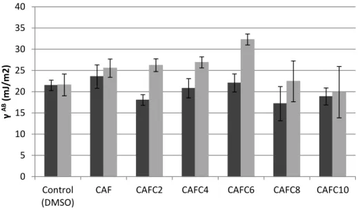
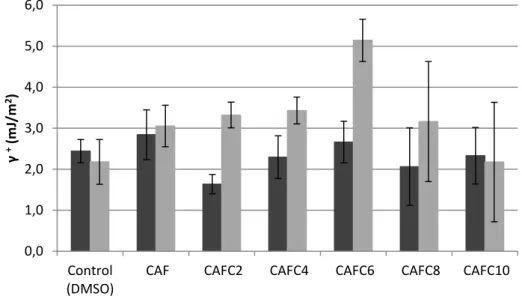
IGURESFig. 1 - Representation of the process leading to biofilm formation. ... 6 Fig. 2 – Chemical structure of a) caffeic acid and b) caffeic acid ester (R = alkyl chain). ... 13 Fig. 3 - Apolar component of the surface tension for S. aureus (■) and E. coli (■) cells after treatment with the selected compounds at 0.1 mM, for 1 h. The means ± SD (for at least three replicates) are shown. ... 29 Fig. 4 - Polar component of the surface tension for S. aureus (■) and E. coli (■) cells after treatment with the selected compounds at 0.1 mM, for 1 h. The means ± SD (for at least three replicates) are shown. ... 29 Fig. 5 – Electron acceptor parameter for S. aureus (■) and E. coli (■) cells after treatment with the selected compounds at 0.1 mM, for 1 h. The means ± SD (for at least three replicates) are shown. ... 30 Fig. 6 - Electron donor parameter for S. aureus (■) and E. coli (■) cells after treatment with the selected compounds at 0.1 mM, for 1 h. The means ± SD (for at least three replicates) are shown. ... 30 Fig. 7 – Hydrophobicity for S. aureus (■) and E. coli (■) cells after treatment with the selected compounds at 0.1 mM, for 1 h. ... 31 Fig. 8 – Adhesion of E. coli (■) and S. aureus (■) cells to PS coupons, after exposure to the selected compounds at 0.1 mM, for 1 h. The means ± SD of cell logs (i.e., log[CFU/cm2]) are shown. ... 43 Fig. 9 - Adhesion of E. coli (■) and S. aureus (■) cells to silicone coupons, after exposure to the selected compounds at 0.1 mM, for 1 h. The means ± SD of cell logs (i.e., log[CFU/cm2]) are shown. ... 44 Fig. 10 - Adhesion of E. coli (■) and S. aureus (■) cells to SS coupons, after exposure to the selected compounds at 0.1 mM, for 1 h. The means ± SD of cell logs (i.e., log[CFU/cm2]) are shown. ... 44 Fig. 11 - Example of a standard disk diffusion assay for QSI determination in C. violaceum, with CAFC4 at concentrations of a) 0.1 mM and b) 1 mM. ... 46 Fig. 12 - Growth inhibition halos observed for C. violaceum subjected to 1 mM (■) and 0.1 mM (■) of the compounds tested. ... 47
Fig. A. 1 – 1H NMR spectra for CAFC2... II Fig. A. 2 – 1H NMR spetra for CAFC4. ... III Fig. A. 3 – 1H NMR spectra for CAFC6... IV Fig. A. 4 – 1H NMR spectra for CAFC8... V Fig. A. 5 – 1H NMR spectra for CAFC10... VI
vii
L
IST OFT
ABLESTable 1 - Examples of phytochemicals with proven antimicrobial activity. ... 14 Table 2 - Examples of phytochemicals' effects in biofilm formation. ... 17 Table 3 - Minimum inhibitory concentration and minimum bactericidal concentration of caffeic acid and its alkyl ester derivatives against S. aureus and E. coli. ... 25 Table 4 - Effects of caffeic acid alkyl esters in the surface charge of S. aureus and E. coli cells. The means ± SD of the surface charge are shown, for both bacteria, after treatment with the compounds. ... 28 Table 5 - Free energy of adhesion between S. aureus and E. coli (untreated and treated
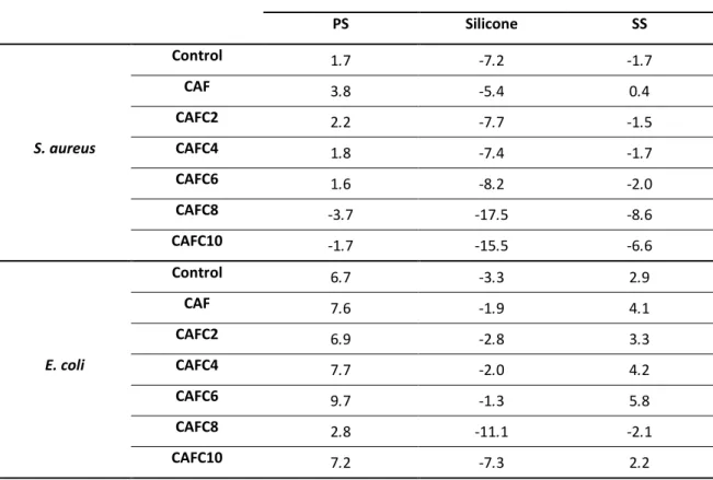
bacteria) and the different material surfaces (when immersed in water). ... 41 Table 6 - Comparison between the percentage reduction of bacterial cell adhesion in E. coli for the different materials tested. ... 45
Table A. 1 - Chemical structure and molecular weight of the caffeic acid esters tested. ... I Table A. 2 – Range of concentrations tested for MIB and MBC determination in E. coli and S.aureus. ... VII
viii
G
LOSSARY ANDN
OMENCLATURE– Electron donor parameter of the Lewis acid-base component of the surface free energy (mJ/m2)
– Electron acceptor parameter of the Lewis acid-base component of the surface free energy (mJ/m2)
– Lewis acid-base component of the surface free energy (mJ/m2
)
– Lifshitz-van der Waals component of the surface free energy (mJ/m2
) – Total free energy of interaction between two surfaces and water (mJ/m2
) – Free energy of interaction between two entities of a surface and water (mJ/m2) – Contact angle (°)
CAF – Caffeic acid
CAFCn – Caffeic acid ester with a n-carbon alkyl side chain CFU – Colony-forming units
DLVO – Derjaguin-Landau-Verwey-Overbeek theoretical approach to adhesion DMSO – Dimethyl sulfoxide
EPS – Extracellular polymeric substances LB – Luria-Bertani broth
LBA – Luria-Bertani agar
MBC – Minimum Bactericidal Concentration (mM) MHB – Mueller-Hinton broth
MIC – Minimum Inhibitory Concentration (mM) O.D.XXX nm – Optical density at XXX nm
PCA – Plate count agar PS – Polystyrene QS – Quorum sensing
QSI – Quorum sensing inhibition RMAs – Resistance-modifying agents SAR – Structure-activity relationship SS – AISI316 Stainless steel
1
C
hapter 1
1. WORK OUTLINE
1.1. Background and Project Presentation
Antibiotics have proven to be powerful drugs for the control of bacterial growth. However, their extensive and unrestricted use has imposed, over the years, a continued selective pressure upon bacteria by different drugs, which led to the development of antimicrobial resistance and, even, multidrug resistance (Abreu et al. 2012; Alekshun & Levy 2007). This is, nowadays, one of the major challenges for the industrial, environmental and biomedical sectors, where bacterial growth (especially in the sessile form of biofilms) causes several economic and public health inconveniences (Budzyoska et al. 2011; Gilbert et al. 2002). Biofilms constitute an extra source of bacterial resistance, which due to their nature, are much more resistant to the action of current antimicrobial agents than planktonic cells and, thus, harder to eliminate (Borges et al. 2014a; Gilbert et al. 2002).
For this reason, a demand for the screening and development of potential new active products and new improved alternative strategies for biofilm control began, especially regarding new classes of antimicrobials that may not be as vulnerable as current drugs to bacterial resistance mechanisms (Borges et al. 2013; Trentin et al. 2011). In this context, there is a new interest in antibacterial products that restrict the ability of bacteria to adhere to surfaces, communicate and, consequently, inhibit biofilm formation (Borges et al. 2014a; 2014b). Plant secondary metabolites (phytochemicals) have thus been implicated as potentially active and new alternative antimicrobial agents (Saavedra et al. 2010), and because they are derived from natural sources, they are considered to present a green and safe status (Borges et al. 2014a; Budzyoska et al. 2011).
Hence, this project is based upon the action of a phytochemical (caffeic acid) and some of its alkyl ester derivatives on planktonic growth control and biofilm inhibition of two selected bacteria: a Gram-positive, S. aureus, and a Gram-negative, E. coli. To my knowledge, it is the first time caffeic acid alkyl esters are tested as antimicrobials and as agents for biofilm formation prevention and microbial adhesion control.
2
1.2. Main Objectives and Contribution
The main objectives of this Dissertation are related to the testing of caffeic acid and a homologous series of caffeic acid alkyl esters with increasingly longer alkyl ester chains on planktonic growth control (i.e. for their antimicrobial activity) and on their ability to affect the bacterial physicochemical properties and act in biofilm prevention and quorum sensing inhibition. Furthermore, it is expected that this work will contribute to a better understanding of the effects of the modification of the alkyl ester side chain length in the proposed antimicrobial and anti-biofilm activities of the compounds tested, by means of a structure-activity relationship.
In this view, the final goal and contribution of this Dissertation would be the rational development of new effective antimicrobial agents, based on molecules of natural origin, particularly from plant sources, which are considered both green and not likely to trigger further bacterial resistance responses, due to their different action mechanisms from conventional antimicrobials.
1.3. Thesis Organization
This Master’s Dissertation is divided into five different chapters. In this first chapter (Work Outline), the scientific challenges that triggered the elaboration of this project are analyzed, along with the main objectives to be achieved and the general framework of the document.
A second chapter, regarding the Literature Review, focus on the relevant theoretical topics that allow a more insightful perspective into the issues being studied, as well as presenting an overall view on the main scientific breakthroughs and conclusions that have been described so far in the literature concerning the subject studied or related areas of studies.
In their turn, chapters three and four consist of the actual practical work developed in this project, including a theoretical understanding of the matters at hand, methodologies used and presentation and discussion of the results obtained and of their relevance for the topics. Particularly, in chapter 3, the antimicrobial activity and the mode of action of a phytochemical (caffeic acid) and a series of derivatives (alkyl esters) against two different bacteria are investigated. On the other hand, in chapter 4, the action of the abovementioned compounds is
3 analyzed regarding their proposed ability to prevent biofilm formation by reducing the extent of the first cell adhesion steps and by hypothetically inhibiting quorum-sensing.
Lastly, a fifth chapter is presented as a compilation of the leading conclusions withdrawn from this project and the subsequent future perspectives for the topic studied.
4
C
hapter 2
2. LITERATURE REVIEW
2.1. Multidrug resistant bacteria and biofilms
Control of microbial growth is required in many microbiologically sensitive environments, especially when the conditions for microbial proliferation are favorable (Ferreira et al. 2011). In a bacterial growth control context, the aim is to maximize bacterial inactivation or killing during the period in which active levels of chemicals are present and to minimize negative effects (such as re-growth, induction of resistance, cytotoxicity, interaction with non-target microorganisms or adverse effects on the environment), when in the presence of sub-inhibitory concentrations (Simões et al. 2009; Saavedra et al. 2010). For this purpose, antibacterial agents, routinely divided into biocides and antibiotics, are employed. These have been traditionally regarded as distinct groups of antibacterial agents by the extent of their pharmacological specificity and their degree of mammalian toxicity, being that the ideal antibiotic has a single biochemical target (i.e., a selective toxicity), whereas biocides generally possess several distinct targets, with diverse susceptibilities (i.e., a broad spectrum of usage) (Ferreira et al. 2011; Gilbert et al. 2002).
To survive in a specific environment, bacteria must respond to several stresses that lead to ill-fated growth conditions, one of which is the exposure to antimicrobial products, such as antibiotics (Simões et al. 2009). Antibiotics have proven to be powerful drugs for the control of infectious diseases and remain one of the most significant discoveries in modern medicine (Abreu et al. 2012). However, these have also been widely recognized as being used in an indiscriminate and often inappropriate fashion, by constantly being subjected to overuse, underuse and general misuse over the years, which can act as a selective pressure for the development of resistance to these compounds (Abreu et al. 2012; Bisht et al. 2009; Gilbert et al. 2002; Simões et al. 2009). The emergence of antibiotic resistance in pathogenic bacteria is a problematic and persistent concern in the medical field, with more than 70% of the bacteria that cause infections in hospitals being resistant to at least one of the most commonly used antibiotic agents (Bisht et al. 2009; Elmasri et al. 2014). Furthermore, resistant microorganisms are responsible for a decrease in the efficiency of disinfection procedures and, as a result, of
5 severe contamination in industrial (especially in the food industry), environmental and biomedical settings (Abreu et al. 2014; Borges et al. 2014a).
The resistance of pathogenic microorganisms to individual antibiotics is, by itself, a serious problem (Abreu et al. 2012). Nevertheless, over the years, continued selective pressure by different drugs has resulted in organisms bearing additional kinds of resistance mechanisms that led to multidrug resistance (Alekshun & Levy, 2007). We are now faced with a long list of microorganisms that have found ways to evade different structural classes of drugs and are no longer susceptible to most therapeutic treatments currently used (Alekshun & Levy 2007). The most problematic multidrug resistant strains include human pathogens like vancomycin-resistant enterococci, bacteria producing extended-spectrum β-lactamases (such as Pseudomonas aeruginosa, Helicobacter pylori, Acinetobacter baumannii, Escherichia coli and Klebsiella pneumonia), vancomycin-resistant and/or methicillin-resistant Staphylococcus aureus and extensively drug-resistant Mycobacterium tuberculosis (Abreu et al. 2012; Alekshun & Levy, 2007).
Antibiotic resistance occurs when bacteria change in any way that might reduce or even eliminate the effectiveness of antimicrobial agents (Bisht et al. 2009). In fact, microbial susceptibility is a continuum that reflects phenotypic and genotypic variations in natural microbial populations (Abreu et al. 2012). Therefore, bacterial resistance may be attained through intrinsic or acquired mechanisms (i.e., bacteria may acquire resistance by de novo mutation or via the acquisition of resistance genes from other microorganisms). Intrinsic resistance to antimicrobials is a natural property of the bacteria and its mechanisms are those specified by naturally occurring genes found on the host’s chromosome (even if pre-existing but previously unexpressed). These are frequently associated with cellular impermeability conveyed by the outer layers and thus, limiting the uptake of antimicrobial products. On the other hand, acquisition of new genetic material by antimicrobial susceptible bacteria from resistant ones may occur through gene transfer, either by conjugation (via plasmids and transposons), by transformation (via bacteriophages) or by transduction. The acquired resistance genes become a relatively stable part of the bacterial genome and additional resistance elements may join those already prevailing, extending the multidrug resistance phenotype. The acquired genes may enable the bacteria to produce enzymes that inactivate the antibacterial product, to modify the target site, to produce alternative metabolic pathways that bypass the antimicrobial action or to express efflux mechanisms that prevent the antimicrobial from reaching its intercellular target (Abreu et al. 2012; Alekshun & Levy, 2007; Simões et al. 2009).
6
2.1.1. Biofilms as a source of additional resistance
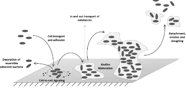
Microorganisms tendentiously grow and survive in a sessile form, i.e, as multicellular surface-attached communities called biofilms (Gilbert et al. 2002; Plyuta et al. 2013), being this the prevalent mode of microbial life in natural habitats, industrial processes and even in many infections (Borges et al. 2014a). Hence, biofilms are structured and functional consortiums of microbial cells embedded in a complex slimy matrix of extracellular polymeric substances (EPS), irreversibly attached to a surface (either biotic or abiotic) (Borges et al. 2012; 2014a; Gilbert et al. 2002; Neyret et al. 2014).
Biofilm formation is a dynamic and sequential process that, overall, includes the transport of microorganisms to surfaces, initial reversible/irreversible adhesion, cell-cell communication, microcolony formation, EPS production and biofilm maturation (Madigan et al. 2009; Simões et al.2010a).
Fig. 1 - Representation of the process leading to biofilm formation.
The process leading to the formation of biofilms (Fig. 1) is believed to start with a pre-conditioning of the adhesion surface, either by macromolecules present in the bulk environment or intentionally coated onto the surface. Then, planktonic bacteria from the surrounding medium are transported to the surface and adhere by either a nonspecific or a specific binding reaction, meaning that, if the bond is weak (reversible), bacteria may desorb from the surface into the liquid, a phenomenon that takes place simultaneously with irreversible adsorption of bacterial cells to the surface. Once bacteria are firmly attached to the surface, they start cell-to-cell signal communications, by producing specific signaling
7 molecules that take a part in growth control, replication, plasmid conjugation and secretion of various virulence factors and exopolymers. At this stage, convective and diffusive transport of substrates to and within the biofilm occurs, alongside with substrate metabolism, excretion of metabolic products, cell growth, replication and extensive EPS production (Breyers & Ratner 2004), comprising a maturation stage of the biofilm. EPS are responsible for biofilm cohesion (i.e., binding cells and other particulate materials together) and adhesion to the surface. The EPS matrix is generally composed of polysaccharides, proteins, nucleic acids, lipids and phospholipids, even though proteins and polysaccharides by themselves account for 75-89% of the biofilms EPS composition (Simões et al.2010b). Ultimately, the biofilm may experience removal of sections by detachment, erosion or sloughing (Breyers & Ratner 2004).
Biofilms are the leading example of physiological adaptation, being one of the most important sources of bacterial resistance to antimicrobial products (Borges et al. 2014a; Madigan et al. 2009). In fact, our ability to eradicate biofilms is substantially lower than that for equivalent populations of planktonic (dispersed) bacteria, which typically present susceptibilities 10 to 1000 times higher than biofilm embedded cells (Gilbert et al. 2002; Neyret et al. 2014).
However, the conventional mechanisms of antibiotic resistance found in planktonic cells (efflux pumps, modifying enzymes and target mutations) do not seem to be responsible for the protection of bacteria in a biofilm and might rather be credited to several mechanisms that can act together (Borges et al. 2012). One possible example is the poor penetration or inactivation of antimicrobials in the EPS matrix, which acts as a physical barrier in which diffusive transport prevails over convective transport, thus limiting antimicrobial penetration and preventing them from reaching their target microorganisms within the biofilm. To this purpose, a number of features are implicated, such as the binding capacity of the polymeric matrix towards the antimicrobial agent, the distribution of biomass and local hydrodynamics, the rate of turnover of the microcolony relative to the molecule diffusion rate and the production and retention of extracellular products. Other examples of biofilm resistance mechanisms comprise altered (dormant) bacterial metabolic state, presence of persister cells, resistance induced by the antimicrobial itself following the use of sub-lethal concentrations and the up-regulation of efflux pumps or potential of damaged bacterial cells to undergo apoptosis or programmed cell death (feeding the community and allowing the remaining cells to survive and proliferate in the post-treatment phase) (Borges et al. 2012; 2014a; Gilbert et al. 2002).
8
2.1.2. Need for new antimicrobial approaches
The increased bacterial resistance against the classical antimicrobial treatments leads to pathogen biofilm elimination being a major challenge with serious economic and health repercussions. Biofilms have been implicated in medicine, as the cause of many chronic and biomaterial-associated infections, and in the industrial and environmental sceneries, as the cause for biofouling, biocorrosion and biodeterioration, especially in food processing and water distribution systems (Budzyoska et al. 2011; Gilbert et al. 2002; Madigan et al. 2009). The current concerns over bacterial multi-resistance, along with biofilm resistance to, not only the conventional treatments, but also the newest generation of drugs, and the toxicity of some of the current employed antimicrobials, has led to a demand for the screening and development of potential new active products and new improved alternative strategies for biofilm control, especially regarding new classes of antimicrobials that may not be as vulnerable as current drugs to bacterial resistance mechanisms (Borges et al. 2013; Trentin et al. 2011).
The development of the so-called resistance-modifying agents (RMAs) represents an attractive strategy to mitigate the spread of bacterial drug resistance, since it could facilitate the recycling of well-established antibiotics that are often cheaper and less toxic than new candidate antimicrobials (Borges et al. 2013). The proposed RMAs are capable of partly or completely suppressing bacterial resistance mechanisms (Budzyoska et al. 2011) by combining new or established antimicrobials with the currently used antibiotics, extending the latest useful life due to a synergism that may cause improved solubility or resorption rate, enhanced bioavailability and ability to modify or even inhibit bacterial resistance mechanisms (Abreu et al. 2012).
The best option so far is to discover and develop new anti-biofilm drugs, i.e., biofilm inhibitors, whose aim, unlike antibiotics, is not to inhibit cell growth, which may thus reduce the risk of drug resistance (Lee et al. 2014). In this context, there is a new interest in antibacterial products that restrict the ability of bacteria to adhere, communicate and, consequently, form complex biofilms or, in other words, prevent the development of biofilms. In fact, to inhibit the growth of an already established biofilm (i.e., biofilm control) is far more difficult to achieve than to impair or inhibit the initial stages of biofilm formation, namely bacterial adhesion (i.e., biofilm prevention) (Borges et al. 2014a; 2014b). This new approach maintains the cells in a planktonic state, switching off the virulence expression typical of biofilms and making the microorganisms more susceptible to the action of other antimicrobials
9 (Trentin et al. 2011). In this view, understanding the relationship between adhesion and biofilm formation is crucial.
Bacterial adhesion is a complex process that is affected by many factors such as the biological properties of the bacteria (presence of fimbriae or flagella, production of EPS, etc.), the physicochemical characteristics of the bacteria (hydrophobicity, surface charge, etc.), the material’s surface properties (chemical composition, surface charge, hydrophobicity, roughness or texture) and environmental factors (temperature, pH, time of exposure, bacterial concentration, presence of chemical or antimicrobial treatment and flow conditions) (Simões et al.2010a). During biofilm formation, adhesion occurs in two different phases: in a first stage planktonic bacteria move or are moved to a surface by physical forces, such as Brownian motion, van der Waals attraction forces, gravitational forces, surface electrostatic charge or hydrophobic interactions, depending on the distance between them; on a second phase, molecular relations between bacterial surface polymeric structures and substratum surfaces become increasingly more significant (Simões et al.2010a). As for now, considerable resources have been directed towards technologies designed to inhibit microbial attachment. Prospects have included surface material coatings that prevent adhesion, responsive surfaces that phase change upon command or controlled orientation of surface-tethered adhesion molecules (Breyers & Ratner 2004). However, nowadays, the most popular approaches to prevent bacterial adhesion are through quorum sensing (QS) and motility inhibition, which are both important steps of biofilm formation and development (Simões et al.2010b).
Bacterial motility and, in particular, swimming and swarming are dependent on flagella and contribute to cell adhesion to biotic and abiotic surfaces. Swarming has long been recognized to be important for both the initial interactions with surfaces and for the movement along these and, therefore, for the early steps of biofilm formation (Lee et al. 2014; Wojnicz et al. 2012). Biofilm formation is invariably preceded by attachment mediated by the abovementioned flagellar motilities, while later stages are due to twitching motility (implicated in cell recruitment from adjacent monolayers and cell aggregate formation), which is related to type IV pili (Borges et al. 2012; Kumar et al. 2013). All considered, the importance of flagella in biofilm formation makes it an attractive target for the development of alternative biofilm control strategies (Vikram et al. 2013).
On the other hand, QS can regulate several bacterial activities, such as bioluminescence, virulence factor expression, swarming motility, sporulation and biofilm production. As a matter of fact, QS is an intercellular signaling system that allows bacteria to monitor their population density and, accordingly, control a variety of physiological processes by releasing and receiving small signaling molecules named autoinducers (Nazzaro et al. 2013;
10 Zhang et al. 2014). This cell-to-cell communication mechanism influences both initiation and maturation of bacterial biofilms and has been showed to be an important regulatory mechanism in biofilm formation/differentiation, rendering the interference with QS systems as a highly attractive and promising target to tackle biofilm control. The discovery that several products with antibiofilm activity were QS inhibitors is proof of the importance of this signaling system in biofilm prevention and began a quest for new agents that might act as nontoxic inhibitors of QS, able to control bacterial adhesion without encouraging the appearance of resistant bacterial strains (Borges et al. 2014b; Nazzaro et al. 2013; Zhang et al. 2014).
2.2. Phytochemicals
Bacteria and fungi are the leading sources of the currently available antibiotics (Cowan 1999). However, due to the already mentioned issues of toxicity and resistance, and in the quest for active products for biofilm control, an interest in products from other sources arose, particularly phytochemicals, plant secondary metabolites (Abreu et al. 2012). In fact, plants produce an enormous array of secondary metabolites, a number of which are commonly believed to be involved in chemical strategies to protect themselves against pathogen microbial attacks from an extensive range of microorganisms including fungi, yeasts and bacteria (Saavedra et al. 2010; Tegos et al. 2002). In order to be classified as antimicrobials, phytochemical products must normally present a minimum inhibitory concentration (MIC) in the range 100-1000 µg/mL in in vitro susceptibility tests (Tegos et al. 2002).
Phytochemicals are commonly classified either as phytoanticipins (molecules that are present constitutively in an inactive form, as is the case of glucosides, cyanogenic glucosides, and saponin glycosides, and are a part of the plant passive resistance mechanisms) or as phytoalexins (molecules that are produced de novo in response to tissue disruption and pathogen attack) (Abreu et al. 2012; Gibbons 2004; Vermerris & Nicholson 2008). However, at a chemical level, there is no boundary between phytoalexins and phytoanticipins, and in one plant species a certain chemical can function as a phytoalexin, whereas it has the function of a phytoanticipin in another species (Cowan 1999). Nevertheless, phytoalexins are typically low weight molecules that may include chemical classes such as polyphenols, alkaloids, glycosteroids, flavonoids, isoflavonoids, various sulfur products, di- and tri-terpenes, polyketides, lactones and naphthoquinones, all of them being common classes of phytochemicals (Tegos et al. 2002). Apart from their potential function against pathogen
11 invaders, it is believed that phytochemicals play other functions in plant physiology and functionality, as attraction pigments for pollination purposes or as protection mechanisms against UV damage or oxidative stress (Simões et al. 2009).
One of the major advantages of using phytochemicals as antimicrobial agents is the fact that, in general, their mechanisms of action differ significantly from the antibiotic ones (Saavedra et al. 2010), allowing for the discovery of new alternative and effective antimicrobial compounds. These, not only have broad-spectrum microbicidal and antibiofilm activities (posing a low risk for the development of resistance), but are also derived from natural sources, present a green and safe status (Borges et al. 2014a; Budzyoska et al. 2011).
Yet, pharmaceutical companies still prefer to pursue microbial derived products, of which there are many first class drug examples which can be readily fermented with few re-supply issues (Gibbons 2004). The chemical complexity of plant extracts, often undocumented toxicity, poor water solubility and the lack of standardization may be responsible for the apparent lack of industrial interest in phytochemicals. Other additional limitations are concerned with the access and supply, the inherent slowness of working with natural products and the costs of collection, extraction and isolation (Abreu et al. 2012).
Polyphenols are the most important and abundant group of phytochemicals (Saavedra et al. 2010). These compounds can be found in diverse dietary products like vegetables, fruits, chocolates and beverages (as coffee, tea or wine) and are involved in various functions in plants, such as growth and development regulation, interaction between plants, pathogenic (defense) and symbiotic microorganisms or ultraviolet radiation defense (Borges et al. 2014b; Manach et al. 2004; Plyuta et al. 2013). They exhibit a wide variety of biological effects including antimicrobial, antioxidant, anti-inflammatory, antiallergic, hepato- and cardioprotective, antithrombotic, antiviral, anticarcinogenic and vasodilatory actions (Merkl et al. 2010; Saavedra et al. 2010). The possible mechanisms believed to be responsible for their antimicrobial activity might include destabilization and permeabilization of cytoplasmatic membranes, efflux pump inhibition, bacterial type II fatty acid synthesis inhibition, enzyme inhibition by the oxidized products (possibly through reaction with sulfhydryl groups or through more non-specific interactions with proteins, causing a disruption of energy production) and nucleic acids synthesis inhibition of both Gram-negative and Gram-positive bacteria (Borges et al. 2013; Saavedra et al. 2010). On the other hand, regarding their antioxidant effects, polyphenols are known to delay or prevent oxidative damage by reactive oxygen species (ROS), being their abilities strongly associated with hydrogen donating and/or electron donating ability to ROS (Lee et al. 2014).
12 The polyphenol class includes simple phenolics and their derivatives: phenolic acids, such as cinnamic acids and derivatives, coumarins and derivatives, flavonoids, stilbenes, lignans and tannins. The common denominator between them is the existence of a polyphenol structure, i.e., hydroxyl groups on aromatic (benzene) rings, being classified into the different groups as a function of the number of phenol rings that they contain and of the structural elements that bind these rings to one another. Additionally, polyphenols may be associated with various carbohydrates and organic acids and with one another (Cowan 1999; Manach et al. 2004; Vermerris & Nicholson 2008). Moreover, phenolic compounds are usually found in plants as esters or glycosides rather than as free compounds (Vermerris & Nicholson 2008).
In particular, in the phenolic acid sub-categories, one can distinguish between two types of molecules: hydroxybenzoic acids and hydroxycinnamic acids (Manach et al. 2004). The numbers and positions of the hydroxyl groups on the aromatic ring causes significant changes in the properties of these molecules. In fact, the site(s) and number of hydroxyl groups on the aromatic ring are allegedly related to their relative toxicity to microorganisms, with increasing hydroxylation resulting in increased toxicity (Borges et al. 2012). Hydroxycinnamic acids have a C6-C3 skeleton (Vermerris & Nicholson 2008; Lee et al. 2014). Depending on the group substitution of the aromatic ring, different types of hydroxycinnamic acids can be found, being the most common the cinnamic, the p-coumaric, the ferulic, the caffeic and the sinaptic acids. However, as previously stated, these acids are rarely found in their free form (Vermerris & Nicholson 2008; Manach et al. 2004).
In plants, hydroxycinnamic acids are intermediates of the general phenylpropanoid pathway, the shikimate pathway and the lignin specific pathway (Barber et al. 2000; Sharma 2011). In the past, the phenylpropanoid pathway included the biosynthesis of the hydroxycinnamic acids: ferulic acid, caffeic acid and sinaptic acid from p-coumaric acid. However, nowadays, it is known that hydroxylation and methoxylation reactions do not occur at the level of the acid, but instead at more reduced forms. So, it appears that the hydroxycinnamic acids are, at least in part, synthesized through the oxidation of aldehydes, rather than via ring substitutions of the free acids. Nevertheless, this is not the exclusive route towards the substitution pattern of the phenyl ring or at least not in all plant species (Vermerris & Nicholson 2008).
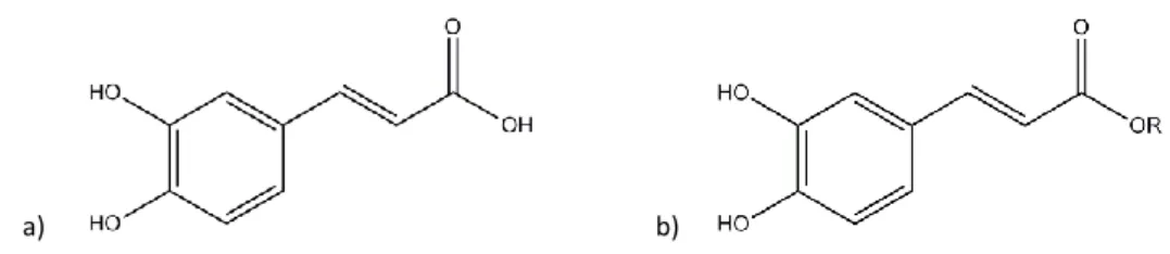
More specifically, caffeic acid (3,4-dihydroxycinnamic acid), both free and esterified (Fig. 2), is generally the most abundant phenolic acid (representing between 75% and 100% of the total hydroxycinnamic acid content of most fruit) (Manach et al. 2004; Yu et al. 2013; Sharma 2011).
13
a) b)
Fig. 2 – Chemical structure of a) caffeic acid and b) caffeic acid ester (R = alkyl chain).
Caffeic acid presents an excellent antibacterial and antioxidant activity (Manach et al. 2004; Yu et al. 2013; Sharma 2011). Furthermore, caffeic acid esters are natural components of propolis and their molecular structure contains a catechol group with an α,β-unsaturated carboxylic acid chain, which can efficiently interact with reactive oxygen species, justifying some of its activity (Uwai et al. 2008; Yu et al. 2013).
2.2.1. Phytochemicals in the control of planktonic and sessile bacteria
Phytochemicals have recently been receiving a lot of attention concerning their antimicrobial activity, as proven by the increasing amount of studies on this topic in the last few years. Furthermore, not only are phytochemicals tested for their potential antimicrobial activity and mode of action against several kinds of bacteria in the planktonic state, but also for their potential ability to act as biofilm formation inhibitors or even biofilm controllers.
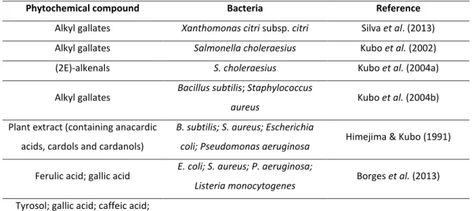
Susceptibilities of different kinds of bacteria to phytochemicals, especially phenolics, were studied by several authors (Table 1) and, generally, results demonstrated that all compounds had antimicrobial activities against the microorganisms tested. It is interesting to notice that several studies state different susceptibilities to the phytochemicals tested for Gram-positive and Gram-negative bacteria. However, there seems to be some controversy amongst authors relating to this topic (Saavedra et al. 2010). Some stated that Gram-positive bacteria are more susceptible to the action of the compounds than Gram-negative ones, while others stated the exact opposite. Merkl et al. (2010) observed that the sensitivity of Gram-positive bacteria to the hydroxycinnamic acid esters tested was higher than in Gram-negative bacteria. Other studies also proposed that phenolics were inhibitory to Gram-positive bacteria but not to Gram-negative bacteria (Himejima & Kubo 19991; Saavedra et al. 2010). Borges et al. (2012; 2013) stated that Gram-positive bacteria were less susceptible to the phenolic acids tested than Gram-negative bacteria. These results might propose that antimicrobial action of phytochemicals is not Gram-specific.
14
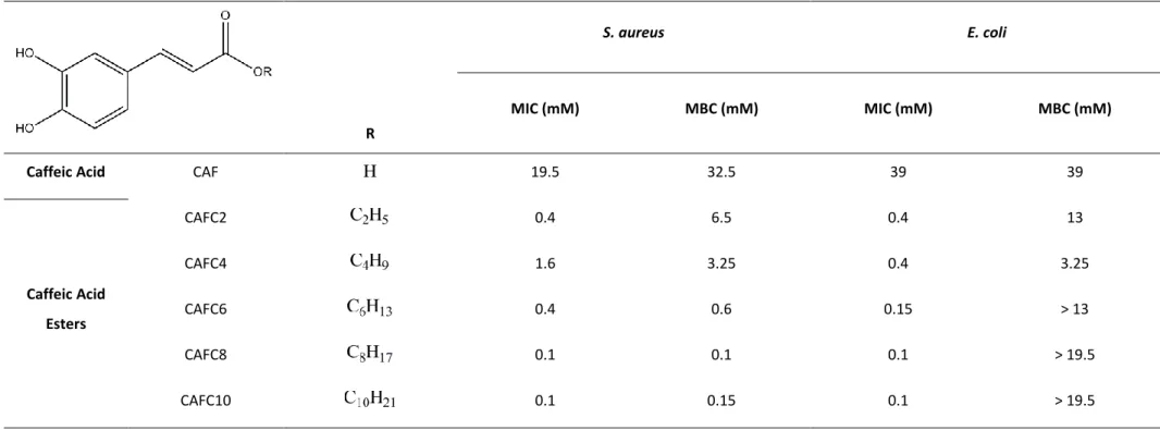
Table 1 - Examples of phytochemicals with proven antimicrobial activity.
Phytochemical compound Bacteria Reference Alkyl gallates Xanthomonas citri subsp. citri Silva et al. (2013) Alkyl gallates Salmonella choleraesius Kubo et al. (2002)
(2E)-alkenals S. choleraesius Kubo et al. (2004a)
Alkyl gallates Bacillus subtilis; Staphylococcus
aureus Kubo et al. (2004b)
Plant extract (containing anacardic acids, cardols and cardanols)
B. subtilis; S. aureus; Escherichia
coli; Pseudomonas aeruginosa Himejima & Kubo (1991)
Ferulic acid; gallic acid E. coli; S. aureus; P. aeruginosa;
Listeria monocytogenes Borges et al. (2013)
Tyrosol; gallic acid; caffeic acid; ferulic acid; chlorogenic acid;oleuropein glucoside;
epicatechin; phloridzin; allylisothiocyanate; benzylisothiocyanate
E. coli; S. aureus; P. aeruginosa;
L. monocytogenes Saavedra et al. (2010)
3,4-hydroxybenzoic acid alkyl esters
E. coli; Bacillus cereus;
L. monocytogenes Merkl et al. (2010)
p-coumaric acid; caffeic acid;
ferulic acid; coniferaldehyde; p-coumaraldehyde; sinapaldehyde
B. subtilis; E. coli; Pseudomonas
syringe Barber et al. (2000)
Taxodione;
7-(2-oxohexyl)-taxodione S. aureus Kuźma et al. (2012)
It was also established that the antimicrobial effect of hydroxycinnamic acid derivatives increases with the increasing length of the ester alkyl chain (Merkl et al. 2010; Uwai et al. 2008; Nihei et al. 2004). For instance, Merkl et al. (2010) proved that the toxicity of hydroxycinnamic acid esters increases with the increasing size of the alkyl chain, as shown by the decrease in MIC values of caffeic and ferulic acids and their correspondent alkyl esters against Escherichia coli and Bacillus cereus. However, other authors, such as Silva et al. (2013) and Kubo et al. (2002; 2004a; 2004b) demonstrated that the antimicrobial activity of alkyl esters of phenolic acids was a parabolic function of their lipophilicity, i.e., was related to the length of the hydrophobic alkyl side chain, with maximum antimicrobial activity being held by C8 to C12 alkyl esters. This means that the biological behavior of these alkyl esters could be correlated with the cutoff phenomenon, which has frequently been attributed to their
15 amphiphilic properties, which are structurally associated with the presence of two groups: phenolic groups (hydrophilic moiety) and an alkyl side chain (lipophilic tail). Therefore, the length of the carbon side chain must determine the release of the bioactive portion of these compounds (the phenolic acid) inside the cells (Silva et al. 2013).
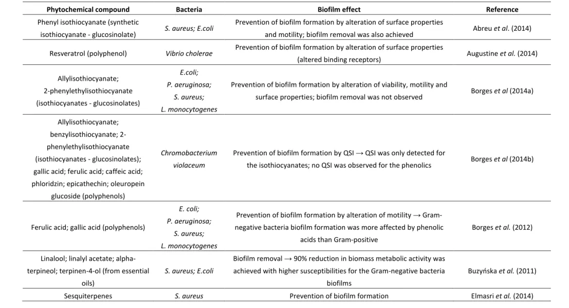
Regarding the control of biofilms, phytochemicals have been employed and studied for a variety of purposes, mainly for inhibition of bacterial adhesion through interference of these compounds in QS systems, bacterial motility or bacterial surface properties (i.e. membrane destabilization) (Table 2).
Apart from the phytochemicals described in Table 2, several others, such as flavanones, flavonoids, flavonols, furanones, hydroxycinnamic acids, rutin, epicatechin, gamma aminobutyric acid (GABA), pyrogallol, curcumin, cunnamaldehyde, furocoumarins, ursolic acid, rosmarinic acid, salycilic acid, epigallocatechin gallate, ellagic acid, tannic acid, urolithin A/B, chlorogenic acid, vanillic acid or proanthocyanidins have also been described as phytochemicals with proven anti-quorum sensing activity, for either or both Gram-negative and Gram-positive bacteria (Nazzaro et al. 2013).
Special emphasis must be given to phenolic compounds, which have been definitely receiving some attention concerning their ability to prevent biofilm formation, either by altering motility or surface properties of bacteria (hydrophobicity) or by inhibiting QS. Interestingly, some authors describe no quorum sensing inhibition (QSI) activity for phenolic acids (Borges et al. 2014b).
Overall, the results presented in the literature regarding in vitro studies of the use of phytochemicals as antimicrobials and biofilm inhibitors are very enthusiastic, especially taking into account the green and natural character of these molecules (many of which are found in human dietary products, thus likely rendering a safe cytotoxic status) and their diverse and broad spectrum modes of action, which would not further inflate the bacterial multi-resistance problematic (Borges et al. 2013; Manach et al. 2004). The therapeutic potential of phytochemical products as alternatives or even potentiators of classic antibiotics is consequently established (Abreu et al. 2012). An interesting consideration to be made is whether or not antimicrobial phytochemical compounds are naturally present in plants in enough concentration to cease potential microbial pathogen attacks. Unluckily, there is a lack of reliable and complete information regarding their levels in plants (Barber et al. 2000).
Nevertheless, besides the potential practical utility of phytochemicals as antimicrobials and RMAs, the bioprospecting results and the vast knowledge of phytochemicals diversity and functionality provides new concepts with potential application for a joint action between combinatorial chemistry and computational design for the swift discovery and synthesis of
16 new and more effective antibacterial products. In fact, some of these compounds may provide important structural scaffolds for the rational and systematic development of new drugs, based on modifications of a known antimicrobial compound (Borges et al. 2013; Kubo et al. 2002; Madigan et al. 2009; Saavedra et al. 2010; Simões et al. 2008).
In this context, structure-activity relationship studies (i.e. studies in which the relationship between the molecular structure of compounds and their biological activity) may be of use, by providing a more insightful perspective into the antimicrobial modes of action of some compounds, allowing for the screening of new activity-influencing features, and thus for the designing of drugs with the greatest potency and the least side effects.
17
Table 2 - Examples of phytochemical effects in biofilm formation.
Phytochemical compound Bacteria Biofilm effect Reference Phenyl isothiocyanate (synthetic
isothiocyanate - glucosinolate) S. aureus; E.coli
Prevention of biofilm formation by alteration of surface properties
and motility; biofilm removal was also achieved Abreu et al. (2014)
Resveratrol (polyphenol) Vibrio cholerae Prevention of biofilm formation by alteration of surface properties
(altered binding receptors) Augustine et al. (2014)
Allylisothiocyanate; 2-phenylethylisothiocyanate (isothiocyanates - glucosinolates) E.coli; P. aeruginosa; S. aureus; L. monocytogenes
Prevention of biofilm formation by alteration of viability, motility and
surface properties; biofilm removal was not observed Borges et al (2014a)
Allylisothiocyanate; benzylisothiocyanate; 2-phenylethylisothiocyanate (isothiocyanates - glucosinolates); gallic acid; ferulic acid; caffeic acid; phloridzin; epicathechin; oleuropein
glucoside (polyphenols)
Chromobacterium violaceum
Prevention of biofilm formation by QSI → QSI was only detected for
the isothiocyanates; no QSI was observed for the phenolics Borges et al (2014b)
Ferulic acid; gallic acid (polyphenols)
E. coli; P. aeruginosa;
S. aureus; L. monocytogenes
Prevention of biofilm formation by alteration of motility → Gram-negative bacteria biofilm formation was more affected by phenolic
acids than Gram-positive
Borges et al. (2012)
Linalool; linalyl acetate; alpha-terpineol; terpinen-4-ol (from essential
oils)
S. aureus; E.coli
Biofilm removal → 90% reduction in biomass metabolic activity was achieved with higher susceptibilities for the Gram-negative bacteria
biofilms
Buzyoska et al. (2011)
18 Zingerone (polyphenol) P. aeruginosa
Prevention of biofilm formation by alteration of motility; biofilm removal was ineffective, unless when used in synergism with an
antibiotic
Kumar et al. (2013)
7-(2-oxohexyl)-taxodione (terpenes
group) S. aureus
Prevention of biofilm formation; biofilm partial removal was also
achieved Kuźma et al. (2012)
Ginkgolic acids C15:1 and C17:1 S. aureus; E. coli
Prevention of biofilm formation by alteration of motility → Gram-positive bacteria biofilms were less susceptible than Gram-negative
ones
Lee et al. (2014)
Thymol; carvacrol; eugenol (phenolics) multimicrobial biofilm
Biofilm removal → 4-5 log reductions were observed, especially when
phenolics were used in synergism between them Neyret et al. (2014) 4-hydroxybenzoic acid, vanillin, gallic
acid; ferulic acid, sinaptic acid, cinnamic acid, epicathechin; chlorogenic acid (phenolics)
P. aeruginosa
Prevention of biofilm formation by alteration of motility and QSI → no motility alterations were verified and QS was promoted at sub-MIC,
which lead to an increase in biofilm formation (at higher concentrations biofilm formation was, however, prevented)
Plyuta et al. (2013)
β-sitosterol glucoside E. coli Prevention of biofilm formation by alteration of motility and QSI Vikram et al. (2013) Paeonidin-3-O-galactoside,
paeonidin-3-O-arabinoside, cyanidin-3-O-galactoside and cyanidin-3-O-glucoside
(anthocyanins)
E. coli Prevention of biofilm formation by alteration of motility and surface
properties Wojnicz et al. (2012)
Phenolic extract containing: gallic acid, catechin, epicatechin, epigallocatechin
gallate, benzoic acid, quercetin, tannins and kaempferol
C. violaceum; E.
19
C
hapter 3
3. ANTIMICROBIAL ACTION OF CAFFEIC ACID ALKYL ESTERS IN
ESCHERICHIA
COLI AND STAPHYLOCOCCUS AUREUS3.1. Introduction
Apart from the widespread bacterial resistance, susceptibility to antimicrobials intrinsically depends on the type of bacteria, with different cellular permeabilities imparted by bacterial outer layers in Gram-negative and Gram-positive bacteria being frequently associated with different susceptibility to antimicrobial products. In fact, the Gram-negative cell wall is a complex multilayered structure whereas Gram-positive cell walls are typically much thicker and consist almost entirely of a single type of molecule (Madigan et al. 2009). Gram-positive and Gram-negative bacteria do share the inclusion of peptidoglycan in their cell walls. Peptidoglycan is a polymer composed of N-acetyl glucosamine, N-acetyl muramic acid and amino acids. In Gram-positive bacteria, peptidoglycan accounts for nearly half of the entire cell and most of the cell wall (about 90%), whilst In Gram-negative bacteria, peptidoglycan only accounts for about 10% of the cell wall (Madigan et al. 2009; Maisuria 2009).
Gram-negative bacteria, such as E. coli, surround themselves in a double membrane, where the inner or cytoplasmatic membrane is mainly composed of phospholipids and the outer membrane is a second lip bilayer containing lipopolysaccharides (LPS). The surface of the cell wall of Gram-negative bacteria is considered hydrophilic because of the LPS, which together with the outer bilayer membrane protect Gram-negative bacteria against membrane destabilizers that weaken the inner cytoplasmic membrane. The outer membrane is nevertheless a permeable barrier to hydrophilic low-molecular-weight substances due to the existence of narrow porin channels. In that view, the outer membrane acts as an efficient permeability barrier against macromolecules and hydrophobic (or even amphipathic) substances. Moreover, the low fluidity of the LPS leaflet slows down the inward diffusion of lipophilic products (Kubo et al. 2004a; Madigan et al. 2009; Maisuria 2009; Simões et al. 2009).
By contrast, Gram-positive bacteria, such as S. aureus, present a single membrane surrounding the cell, mainly composed of peptidoglycan, being two-fold to eight-fold larger than the cell wall of Gram-negative bacteria and not containing LPS (Madigan et al. 2009;
20 Maisuria 2009). As a result, Gram-negative bacteria are generally less susceptible to antimicrobial action, as their cell walls present a more significant barrier to surpass (Kubo et al. 2002; Simões et al. 2009; Tegos et al. 2002).
In the sense that cell walls differ in their hydrophilicity/lipophilicity, surface hydrophobicity is an important factor to be taken into account when studying the antimicrobial action of a molecule upon bacterial cells. In fact, alterations in the bacterial cells surface physicochemical properties upon antimicrobial treatment may be a useful indicator of its mode of action and potential as an inhibitor of biofilm formation, since hydrophobicity has been considered the most important short-range interaction in bacterial adhesion (Simões et al. 2007).
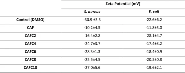
Other surface properties, such as surface charge, may also contribute to analyze the interaction between an antimicrobial and the bacterial surface. The surface charge of cells is frequently determined based on their zeta potential, which is calculated from the electrophoretic motility of cells in the presence of an electrical field, under defined pH and salt concentrations (Borges et al. 2013). When applying an electric field across a bacterial suspension, bacteria with non-zero zeta potential migrate towards the electrode of the opposite charge, with a velocity proportional to the magnitude of their zeta potential (Ferreira et al. 2011). Bacterial cells normally present a negative surface charge, due to the presence of anionic groups in their membranes, such as carboxyl and phosphate groups (Borges et al. 2013).
In particular, for the series of alkyl caffeates used in this study, it is relevant to note that these molecules possess a head-and-tail structure, similar to an amphiphile. Amphiphiles are molecules with two parts: they have a water-loving part (hydrophilic) and a water-hating part (hydrophobic), usually a long-chain alkyl group (Maisuria 2009). In this case, the amphiphile of caffeic acid esters is associated with the presence of the two groups: the phenolic groups (hydrophilic moiety) and the alkyl side chain (lipophilic tail) (Silva et al. 2013). This is actually the case for several phenolic acid esters, which, as already mentioned, display a parabolic antimicrobial activity as function of their lipophilicity. This biological behavior could thereby be correlated with the cutoff phenomenon, very distinctive of amphiphilic substances: antimicrobial properties of amphiphiles tend to increase with increasing alkyl chain length; however, increasing their length decreases the solubility of amphiphiles in aqueous media, lowering their biological activity levels. This phenomenon is known as the cutoff effect due to insolubility (Maisuria 2009) and is believed to explain, in part, the parabolic antimicrobial activity of such compounds.
21 All of the caffeic acid alkyl esters in the homologous series of compounds studied present the same hydrophilic portion, but increasingly long alkyl side chains, thus distinguishing the role of the hydrophobic alkyl portion in their antimicrobial action and allowing to perform a SAR study (Kubo et al. 2002).
Therefore, the main objectives of this work were to assess the antimicrobial activity and the mode of action of caffeic acid alkyl esters against Gram-positive and Gram-negative bacteria.
3.2. Materials and Methods
3.2.1. Test microorganismsThe Gram-negative bacterium Escherichia coli CECT 434 and the Gram-positive bacterium Staphylococcus aureus CECT 976 were used in this study. These microorganisms have previously been employed as model microorganisms for antimicrobial tests with phytochemical products (Saavedra et al. 2010; Borges et al. 2012). The bacteria were cryopreserved at -80⁰C, in a mixture of Mueller-Hinton broth (MHB, Merck) and 30% (v/v) glycerol, and subcultured in Plate Count Agar (PCA, Merck) at 30⁰C for 24 h, before testing.
3.2.2. Caffeic acid and alkyl ester derivatives
The compounds tested included caffeic acid and some of its ester derivatives. Caffeic acid (CAF) was obtained from Sigma-Aldrich, while its esters were kindly synthetized and provided by Prof. Fernanda Borges and her team, from Faculdade de Ciências da Universidade do Porto. The collection includes C2, C4, C6, C8 and C10 alkyl esters of caffeic acid (CAFC2, CAFC4, CAFC6, CAFC8 and CAFC10, respectively) (Appendix A).
Stock solutions of all tested compounds were prepared in dimethyl sulfoxide (DMSO, Fisher), under sterile conditions, and kept in the dark, at room temperature, for a maximum of two weeks. Serial dilutions of the stock solutions were prepared in DMSO, whenever needed.
3.2.3. Minimum inhibitory concentration (MIC) determination
The antimicrobial activity of the tested compounds was measured by means of their minimum inhibitory concentrations.
22 For each bacteria, overnight cultures were prepared in 250 mL sterile flasks containing around 50 mL of previously autoclaved (at 121⁰C for 15 min) MHB, and incubated at 30⁰C, under agitation at 120 rpm (in an incubation-shaking cabinet Sartorius Certomat® BS-1), after inoculation. The optical density at 600 nm (OD600 nm) of the overnight cultures was set to 0.1.
MIC values were determined in sterile 96-well flat-bottomed polystyrene tissue culture microtiter plates (Orange Scientific). In each well, a volume of 20 μL of compound’s solution was added to 180 μL of cell culture. All test compounds were tested in a range of different concentrations (Appendix B), in duplicates. Positive and negative controls were established as follows: 200 µL of sterile distilled water; 180 µL of sterile MHB + 20 µL of the highest concentration solution tested; 180 µL of cell culture + 20 µL of DMSO; 200 µL of cell culture.
The OD600 was measured at t=0 h in a microtiter plate absorbance reader (Biotek
Synergy HT) and at t=24 h, after incubation at 30⁰C and 120 rpm (in an incubation-shaking cabinet Sartorius Certomat®
BS-1). The MIC was defined as the lowest concentration of test compound which would inhibit the visible growth of microorganisms after the 24 h incubation (Merkl et al. 2010; Nihei et al. 2004).
3.2.4. Minimum bactericidal concentration (MBC) determination
After MIC determination, 3 x 10 µL from each MIC experiment were plated out on PCA. Plates were incubated at 30⁰C for 24 h and growth was visually inspected. The MBC was determined as the lowest concentration of compound in which total inhibition of growth was observed and, consequently, no CFU were detected on the solid medium (Ferreira et al. 2011).
3.2.5. Surface hydrophobicity and its components
The influence of treatment with the caffeic acid derivatites on the physicochemical surface properties of both bacteria tested was assessed by contact angle measurement.
For each bacterium, overnight cultures were prepared in 1000 mL sterile flasks containing around 250 mL of previously autoclaved MHB, and incubated at 30⁰C, under agitation at 120 rpm, after inoculation. The cells were centrifuged twice at 3202 g for 10 min (at 25⁰C) and washed with sterile saline solution (0.85% (w/v) NaCl, BDH Prolabo). The optical density at 640 nm (OD640 nm) of the cell suspensions was set to 0.44. A volume of 45 mL of this
culture was added to 5 mL of test compound (to a final concentration of 0.1 mM), in 250 mL sterile shake flasks, and incubated for 1 h at 30⁰C and 120 rpm. A negative control was prepared with DMSO. Bacterial lawns (i.e. homogeneous layers of cells) were then prepared