*Correspondence: Xixiang Ying. School of Pharmacy, Liaoning University of Traditional Chinese Medicine. No.77 Shengming 1 Road, DD port, Dalian, 116600, P.R. China. E-mail: [email protected]
A
vol. 48, n. 1, jan./mar., 2012
HPLC method for the simultaneous determination of four
compounds in rat plasma after intravenous administration of
Portulaca oleracea
L. extract
Zhongzhe Cheng, Ming Xie, Wenjie Zhang, Lan Cheng, Yang Du, Yunjiao Wang, Xixiang Ying
*,
Tingguo Kang
Department of Pharmaceutical Analysis, School of Pharmacy, Liaoning University of Traditional Chinese Medicine, China
The objective of the present study was to develop a simple and selective HPLC method for the simultaneous determination of hesperidin (HP), caffeic acid (CA), ferulic acid (FA) and p-coumaric acid (p-CA) in rat plasma after intravenous administration of Portulaca oleracea L. extract (POE). With the hyperoside as the internal standard, the sample pretreatment procedure involved simple single-step extraction with methanol of 0.2 mL plasma. The mobile phase consisted of methanol-acetonitrile-tetrahydrofuran-0.5% glacial acetic acid (5:3:18:74, v/v/v/v). The calibration curves were linear over the range of 0.1-25 μg mL-1, 0.1-25 μg mL-1, 0.1-25 μg mL-1and 0.015-3 μg mL-1 for HP, CA, FA and
p-CA, respectively. The method developed was suitable for the pharmacokinetic study of HP, CA, FA and p-CA in rats after intravenous administration of POE.
Uniterms:Portulaca oleracea L./pharmacognosy. High Performance Liquid Chromatography/rat plasma. Vegetal extract/pharmacokinetic study. Medicinal plants.
O objetivo do estudo foi desenvolver um método simples e especíico de HPLC para a determinação simultânea de hesperidina (HP), ácido caféico (CA), ácido ferúlico (FA) e ácido p-cumárico (p-CA) em plasma de rato após a administração intravenosa de extrato Portulaca oleracea L. (POE) empregando hyperosídeo como padrão interno de referência. Metanol foi empregado para os analitos em plasma (0,2 mL). A fase móvel isocrática foi composta por metanol-acetonitrila-tetraidrofurano-0,5% ácido acético glacial (5:3:18:74, v/v/v/v). Curvas de calibração foram lineares na faixa de concentração de 0,1-25 μg mL-1, 0,1-25 μg mL-1, 0,1-25 μg mL-1 e 0,015-3 μg mL-1 para HP, CA, FA e p-CA, respectivamente. O método desenvolvido foi adequado para estudo farmacocinético de HP, CA, FA e
p-CA em ratos após a administração intravenosa de POE.
Unitermos: Portulaca oleracea L./farmacognosia. Cromatograia Líquida de Alta Eiciência/plasma de rato. Extrato vegetal/estudo farmacocinético. Plantas medicinais.
INTRODUCTION
Portulaca oleracea L., a well-known traditional
Chi-nese medicine, is used as a treatment for removing heat, counteracting toxicity, cooling blood, hemostasia and anti-dysentery (PRC, 2010). As a widespread and abundant plant in the world, Portulaca oleracea L. is also used as a folk medicine in many countries. Recently, a number of
pharmacological studies showed that Portulaca oleracea
L. had potential activities of relaxing skeletal muscle in rat (Parry et al., 1993), analgesic and anti-inlammatory
(Chan et al., 2000), wood healing effect (Rashed et al., 2003), meliorating abnormal uterine bleeding (Shobeiri et al., 2009), neuropharmacological effect (Radhakrishnan
et al., 2001) and so forth. In addition, phenolic acids and
lavone glycosides, being important chemical constituents
of this plant, have also been reported, such as hesperidin (HP), caffeic acid (CA) (Yang et al., 2007), ferulic acid (FA) and p-coumaric acid (p-CA) (Xiang et al., 2005).
health for the antioxidant effect of FA and CA (Maurya et al., 2010), anticancer activity of HP (Lee et al., 2010) and by the property of p-CA for inhibiting growth of bacteria (Aziz et al., 1998). Thereby, these bioactive constituents can be used as marker compounds to characterize the
Portulaca oleracea L. extract (POE). More recently, some
in vivo investigations have focused on the
pharmacokinet-ics of individual HP (Jin et al., 2010), FA (Zhang et al., 2009) and CA (Uang et al., 1997) by the HPLC, LC/MS and LC/MS/MS methods, respectively. However, little attention has been devoted to the pharmacokinetic study of bioactive components in POE. Therefore, the present
study is the irst to report a HPLC method developed and
validated for simultaneously determination of HP, FA, CA and p-CA in rat plasmaand its pharmacokinetic study after intravenous administration of POE.
MATERIALS AND METHODS
Plant material
Portulaca oleracea L. were collected from Shenyang
(Liaoning, China) in September 2010, and identiied by
Professor Yanjun Zhai. Voucher specimens (No. 2010090) were deposited at the School of Pharmacy, Liaoning Uni-versity of Traditional Chinese Medicine.
Reagents and chemicals
HP, CA, FA and the internal standard (IS), hypero-side, were obtained from the National Institute for the Con-trol of Pharmaceutical and Biological Products (Beijing, China). P-CA was provided by Nanchang Beta Biotech Co., Ltd. (Nangchang, China). All the chemical structures of the four compounds and IS are shown in Figure 1. Water
was puriied with a Milli-Q® Biocel Ultrapure Water
Sys-tem (Millipore, Bedford, MA, USA. Methanol, acetonitrile and tetrahydrofuran were all of HPLC grade and provided by Damao (Chemical Reagent Plant, Tianjin, China). All other reagents were of analytical grade (Jinfeng Chemical Factory, Tianjin, China).
Apparatus and chromatographic conditions
The Agilent 1100 series HPLC system (Agilent technology, Palo Alto, CA, USA) consisted of a quaternary Pump (G1310A), a vacuum degasser (G1322A), a UV/VIS spectrophotometric detector (G1314A) and Chemstation software (Agilent). An analytical Kromasil C18 column (5
µm, 150 × 4.6 mm, Sanjie Science and Technologies, Da-lian, China) connected with a KR C18 guard column (5 μm,
35 × 8.0 mm, Dalian Create Science and Technology Co., Ltd., China) was employed to determine the analytes at ambient temperature. The mobile phase consisted of a mixture of methanol-acetonitrile-tetrahydrofuran-0.5%
glacial acetic acid (5:3:18:74, v/v/v/v), which was iltered and degassed under reduced pressure before use. The low
rate was constantly kept at 1 mL min-1 and chromatograms
were recorded at 283 nm for HP, 322 nm for FA, CA, IS and p-CA, respectively.
Preparation of POE
The dried whole plant of Portulaca oleracea L. (5 kg) was extracted with 60% ethanol (50 L) twice by
re-luxing 2 h, then iltrates were collected and combined, and
ethanol evaporated under reduced pressure. Subsequently, the residue was passed through an AB-8 macroporous resin column (10 × 120 cm, Shanghai, China). To eliminate impurity, the column was eluted with 20 L water, and with 50 L of 60% ethanol. After removing the ethanol of 60% ethanol fraction in vacuo, the supernatant was dried in a water bath. Subsequently, the dried powders (68 g) were
extracted with methanol by reluxing 1 h, and the solvent
was recovered under reduced pressure. Following this, the
residue was extracted with 50 mL of a mixture of ethanol-water (5:95, v/v) in an ultrasonic bath for 45 min. The
supernatants were iltered with a 0.45 μm membrane ilter
and stored at 4 °C prior to use. The prepared solution was analyzed by HPLC-UV/VIS. The contents corresponding to HP, CA, FA and p-CA were 7.2, 6.7, 24 and 17 μg mL-1,
respectively.
Preparation of standards and quality control samples
Stock solutions of HP, CA, p-CA and IS were pre-pared by dissolving in methanol to yield concentrations
of 200, 200, 170, 124 μg mL-1 while FA in 70% methanol
was 200 μg mL-1, respectively. The combined working
solutions were prepared with diluted stock solution in
series to concentrations of 0.4-100 μg mL-1 for HP, CA,
and FA and also, 0.06-12 μg mL-1 for p-CA. The working
solution of IS was 12.4 μg mL-1 and obtained by diluting
stock solution with methanol. All the working solutions were stored at 4ºC. Calibrators of HP, CA and FA (0.1, 0.2,
0.4, 1.0, 2.0, 5.0, 10.0 and 25.0 μg mL-1) and p-CA (0.015,
0.03, 0.075, 0.3, 0.75 and 3.0 μg mL-1) were prepared by
adding standard mixture working solutions (50 μL) and working solution IS (50 μL), respectively, to 200 μL drug-free rat plasma. The quality control (QC) samples were
prepared at low, middle and high concentrations (0.25,
12.5 and 20 μg mL-1 for HP, 0.25, 12.5 and 20 μg mL-1 for
CA, 0.25, 12.5 and 20 μg mL-1for FA and 0.04, 1.5 and
2.4 μg mL-1 for p-CA) in bulk, and aliquots were stored at
-20 °C until analysis.
Plasma extraction
Before deproteinating, 20 μL of acetic acid, 50 μL of IS were added to 200 μL plasma. The proteins of plasma
were precipitated with 1 mL methanol, followed by vortex mixing for 1 min and then centrifuged at 890 g for 15 min. The supernatant was collected and evaporated to dryness at 40 ºC under a stream of nitrogen. The residue was then
dissolved in 200 μL of mobile phase, and centrifuged at
15,092 g for 10 min. The injection volume of supernatant
was 20 μL for analysis.
METHOD VALIDATION
Selectivity
The selectivity was determined by comparing chromatograms of different blank plasma obtained from rat with those of corresponding standard plasma samples
spiked with HP, FA, CA, p-CA and IS and plasma sample after intravenous administration of POE.
Linearity, LOD and LOQ
Linearities were evaluated over the concentration
range of 0.1-25, 0.1-25, 0.1-25 and 0.015-3 μg mL-1 for
HP, CA, FA and p-CA, respectively. The calibration curves were prepared by exact concentrations of working solu-tions added to blank plasma and established by weighted (1/c2) least square linear regression analysis based on the peak area ratio of each analyte to the internal standard. The
limit of detection (LOD) and the limit of quantiication (LOQ) were deined as the lowest concentration of each
analyte and determined by a signal-to-noise ratio of 3 and 10 with an acceptable accuracy (RE %) within ±20% and a precision (RSD %) that did not exceed 20%.
Precision and accuracy
The validation of intra-day precision and accuracy
were evaluated with replicates of QC plasma samples at
the three concentration levels on the same day as well as inter-day precision on each day for 3 separate days. The
intra-day and inter-day precision was deined as the rela
-tive standard deviation (RSD) and accuracy was deter-mined by calculating the relative error (RE).
Extraction recovery
The extraction recoveries of HP, CA, FA and p-CA were determined by comparing the peak area of each
compound from QC samples that were at the three con
-centrations low, middle and high, to that of the unextracted standard solutions containing the equivalent amount of analytes (n = 6).
Stability
Stability was investigated at low, middle and high
concentrations of QC samples. A short-term stability ex
-periment was carried out at ambient temperature (25 °C) for 24 h whereas long-term stability was tested by storage
at -20 °C for 1 month, respectively. QC samples were sub
-jected to three freeze (-20 °C)-thaw (room temperature) cycles to determine freeze-thaw stability.
Animals and pharmacokinetic study
University of Traditional Chinese Medicine (Shenyang, China). Before the experiments, all rats were kept in a controlled environment for 1 week and had free access to standard laboratory food and water intake. The rats were fasted 12-16 h prior to administration of the POE. Animal experiments were carried out in accordance with the Guidelines for Animal Experimentation of Liaoning University of Traditional Chinese Medicine, and the pro-cedure was approved by the Animal Ethics Committee of the institution.
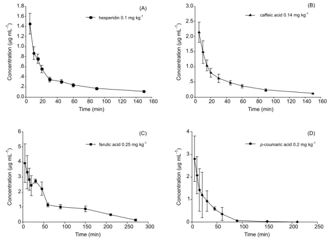
Five rats were administered POE solution via tail vein injection at a dose of 11 mL kg-1 (approximately
equivalent to 0.1 mg kg-1 of HP, 0.14 mg kg-1 of CA,
0.25 mg kg-1 of FA and 0.2 mg kg-1 of p-CA, respectively).
Blood samples (0.4 mL) from orbital sinus were taken into heparinized tubes at times of 5, 10, 15, 20, 25, 30, 45, 60, 90, 150, 210, 270 min after dosing, and then centrifuged at 890 g for 15 min, immediately. Plasma was then trans-ferred into clean test tubes and stored at -20 ºC until analysis.
RESULTS AND DISCUSSION
Method development
Good separations and suitable retention time of four analytes were obtained in isocratic elution using an analytical Kromasil C18 column (5 µm, 150 × 4.6 mm, Sanjie Science and Technologies, Dalian, China) with mobile phase consisting of methanol-acetonitrile-tetrahydrofuran-0.5% glacial acetic acid (5:3:18:74, v/v/v/v) after various mixtures of methanol, acetonitrile, tetrahydrofuran water solution solutions were used. The addition of 0.5% glacial acetic acid in the solvent system led to the sharp peak shapes. Moreover, to simultaneously determine four analytes, use of wavelengths ranging from 280 to 330 nm was attempted. Stronger signals were only observed when the wavelength was set at 283 nm for HP and 322 nm for CA, FA and p-CA, respectively. Vitexin-4”-O-glucoside, vitexin-2”-O-rhamnoside and baicalin had been considered as IS, the retention time of these, however, proved unsuitable due to the presence of interference by endogenous materials. Ultimately, hyperoside, given its similar chromatographic behavior to the analytes, was chosen as the internal standard for the assay. To improve the solution of analytes, a mixture of ethanol-water (5:95, v/v) 50 mL was used to extract residue in the ultrasonic bath during the preparation of POE. The sample pretreatment was also optimized by precipitation of protein with methanol and addition of
20 μL acetic acid to the plasma.
Method validation
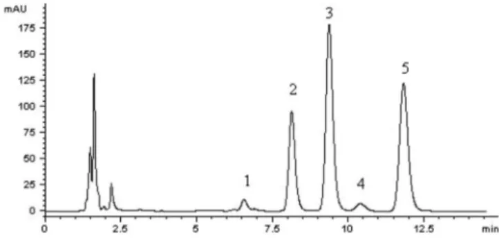
The typical chromatograms of blank plasma, plasma spiked with analytes and IS are shown in Figure 2A and 2B, respectively. The chromatogram obtained from plasma samples after intravenous administration is shown in Figure 2C and the result indicated the absence of interfering endogenous peaks observed at the retention time of analytes. The retention times of HP, CA, FA, IS and p-CA were approximately 7.31, 8.68, 10.04, 11.72 and 12.72 min, respectively. The total run time was 14.0 min.
Eight-point calibration curves over the
concentra-tion range of 0.1-25 μg mL-1for HP, CA and FA, and
six-point 0.015-3 μg mL-1 for p-CA, respectively, were used
to evaluate the linearity. The weighted (1/c2) least squares
FIGURE 2A - Representative chromatogram of blank plasma.
FIGURE 2B - Representative chromatogram of plasma spiked
with hesperidin, caffeic acid, ferulic acid, p-coumaric acid and IS.
FIGURE 2C - Representative chromatogram of plasma sample
linear regression was employed to obtain the regression equations: typically y = 0.6192x + 0.038 (r = 0.9985, HP), y = 0.0482x -0.0193 (r = 0.9979, CA), y = 0.1026x - 0.0374 (r = 0.9981, FA), y = 5.1443x - 0.0664 (r = 0.9968, p-CA).
The limits of quantification (LOQ, S/N = 10)
for HP, CA, FA and p-CA were 0.11, 0.105, 0.12 and
0.014 μg mL-1, respectively, whereas the limits of
de-tection (LOD, S/N = 3) were 0.035, 0.031, 0.036 and
0.003 μg mL-1 for HP, CA, FA and p-CA, respectively.
The data for intra- and inter-day precision at the three concentrations ranged from 0.5 to 12.5%, and RE were -4.7 to 2.0%, which conform to the criteria for the analysis of biological sample according to guidance of FDA (USFDA. 2001).
Mean extraction recoveries at three concentration levels for HP, CA, FA and p-CA were 90.87%, 92.64%, 96.75%, and 93.72%, respectively, and the RSD had a range of 6.4-8.5%, suggesting negligible loss during extraction.
The short-term, long-term and freeze-thaw stabil-ity of analytes in plasma ranged from 93.49 to 99.95%, indicating no significant degradation occurred during
chromatography, extraction and sample storage processes.
Application to pharmacokinetic study
Pharmacokinetic analysis was performed with 3p97 (Practical Pharmacokinetic Program, 1997, China) to calculate the relevant parameters by both compartment and non-compartment approaches. Moreover, terminal half-life (t1/2) was deined as t1/2 = 0.693/λz and the terminal
elimination rate, λz, was calculated by natural logarithms of linear regression on the last ive points.
According to AIC and R2 (Table I), a
two-compart-ment open model (Weight = 1) yielded the best it to the
plasma concentration-time curves for HP, CA and p-CA, and one-compartment open model (Weight = 1/c2) was optimal for FA, respectively. The plasma concentration
proiles and pharmacokinetic parameters of HP, CA, FA
and p-CA in rat plasma after intravenous administration of POE are illustrated in Figure 3 and Table II. The results showed that the apparent volume of distribution (Vc) of FA was larger than other analytes, suggesting a wider distribution of FA in the body. Furthermore, all half-lives
TABLE I - AIC and R2 data of hesperidin, caffeic acid, ferulic acid and p-coumaric acid
model weight HP CA FA p-CA
AIC R2 AIC R2 AIC R2 AIC R2
1 1 -24.6 0.91 -19.6 0.93 -9.16* 0.94 -23.8 0.97
2 1 -39.3* 0.99 -45.9* 0.99 -9.13 0.96 -34.6* 0.99
3 1 -35.4 0.99 -41.5 0.99 -5.13 0.96 -30.1 0.99
1 1/c -12.3 0.60 -11.1 0.79 1.52 0.84 -18.5 0.95
2 1/c -30.5 0.97 -40.3 0.99 -5.68 0.94 -20.8 0.97
3 1/c -26.9 0.97 -36.8 0.99 -1.48 0.94 -17.0 0.97
1 1/c2 -2.20 0.40 -5.60 0.58 8.90 0.67 1.3 0.62
2 1/c2 -21.4 0.92 -37.0 0.99 -2.04 0.92 -2.8 0.85
3 1/c2 -17.3 0.92 -30.8 0.99 1.97 0.92 -1.2 0.32
1 1 -14.2 0.91 -9.5 0.99 3.86 0.93 -14.1 0.97
2 1 -31.5 0.99 -42.1 0.99 3.72 0.95 -26.5 0.99
3 1 -28.7 0.99 -38.1 0.88 7.86 0.95 -22.3 0.99
1 1/c -4.40 0.73 -4.6 0.99 -0.17 0.95 -13.7 0.97
2 1/c -26.4 0.98 -39.6 0.99 -0.83 0.97 -15.4 0.98
3 1/c -24.5 0.98 -33.3 0.99 2.82 0.97 -14.3 0.99
1 1/c2 2.90 0.39 -2.5 0.86 -2.45 0.96 8.8 0.79
2 1/c2 -17.6 0.96 -32.2 0.99 1.66 0.97 -3.8 0.96
3 1/c2 -19.8 0.98 -33.9 0.99 3.32 0.97 0.56 0.96
* Representation of best it result of AIC for each group, based on least value. AIC denotes Akaike’s Information Criterion, AIC=NlnRe+P (N represents data of point, Re is residual sum of squares, P refers to parameter member of model), and R2 is square
TABLE II - Pharmacokinetic parameters of hesperidin, caffeic acid, ferulic acid and p-coumaric acid in rat plasma (mean ± SD,
n = 5) after intravenous administration of POE
Parameter (Units) HP CA FA p-CA
Dose (mg/kg) 0.1 0.14 0.25 0.2
Vc (L/kg) 0.0442 ± 0.0063 0.0410 ± 0.0040 0.0778 ± 0.0021 0.0442 ± 0.023
T1/2ke (min) - - 62.90 ± 0.53
-T1/2α (min) 5.69 ± 1.94 5.50 ± 0.60 - 3.97 ± 0.93
T1/2β (min) 62.66 ± 1.57 51.32 ± 2.08 - 23.38 ± 2.79
aAUC
0→t (mmol·min /L) 0.11 ± 0.02 0.45 ± 0.49 1.51 ±0.71 0.52 ± 0.67
CL (kg·L/ min) 0.0014 ± 0.007 0.0017 ± 0.0007 0.0008 ± 0.0006 0.0023 ± 0.0007
MRT0→t
(min) 43.95 ± 0.93 38.69 ± 1.68 81.25 ± 0.11 28.46 ± 0.17
bAUC
0→t (mmol·min /L) 0.08 ± 0.12 0.40 ±0.33 1.58 ± 0.87 0.50±0.05
T1/2z (min) 99.606 ± 0.013 40.7435 ± 1.34 65.5490 ± 6.07 39.906 ±14.20
adata calculated by compartmental approach and b non-compartmental approach. V
c is apparent volume of distribution; T1/2ke,
T1/2α,T1/2β, are all half-life parameters, representing half-life of central compartment, absorption and distribution phase, respectively,
T1/2z is terminal half-life; AUC is area of under curve; MRT is mean residence time.
of HP, CA and p-CA were shorter than their respective
β half-lives, which indicated that HP, CA and p-CA
pre-sented a rapid distribution phase and a more prolonged
elimination phase. A shorter T1/2z of HP was obtained in our
investigation, compared with values in earlier literature, indicating that HP suffered more rapid elimination than its
individual form (Li, et al., 2008). Moreover, both α half-life and β half-half-life of CA in this study were longer than
previously reported values after intravenous administra-tion of individual drug at higher dose (Uang, et al., 1997). This phenomenon may be ascribed to the coexisting effect of multiple components in POE. In addition, the plasma concentration profiles of FA exhibited a second peak after intravenous administration of POE at 0.5 h, which presented a different pharmacokinetic process compared with individual FA (Zhang, et al., 2009). CA was able to be enzymatically converted to FA in hepatocytes, and FA being an O-methylated product of CA was reabsorbed in blood (Moridani et al., 2002), which accounted for the second peak. Possibly, the coexisting constituents in POE impacts on the pharmacokinetic behavior of FA. Given the diverse groups of compounds present in POE, the pharmacokinetic character of our target compounds may be interactively affected by other coexisting components, either due to competition of drug carrier in transportation between them, or because the activity of enzymes, such as CYP450s, was prompted or inhibited by other coexisting constituents during metabolism.
CONCLUSIONS
The present article described the development and validation of an HPLC method for the simultaneous de-termination of HP, CA, FA and p-CA in rat plasma, which can play an important role in clarifying the mechanism of
action and eficacy of POE via the pharmacokinetic study
of the four bioactive constituents.
ACKNOWLEDGMENTS
This study was supported by the Foundation for Excellent Youth Scholars in pharmacy of the Liaoning University of Traditional Chinese Medicine (Grant no. yxrc0906).
REFERENCES
AZIZ, N.; FARAG, S.; MOUSA, L.; ABO-ZAID, M. Comparative antibacterial and antifungal effects of some phenolic compounds. Microbios, v.93, n.374, p.43-54, 1998.
CHAN, K.; ISLAM, M.W.; KAMIL, M.; RADHAKRISHNAN, R.; ZAKARIA, M.N.; HABIBULLAH, M.; ATTAS A. The analgesic and anti-inlammatory effects of Portulaca oleracea L. subsp. Sativa (Haw.) Celak. J. Ethnopharmacol., v.73, n.3, p.445-451, 2000.
GUIDANCE for industry: bioanalytical method validation. In: U.S. Department of Health and Human Services. Food and Drug Administration, Center for Drug Evaluation and Research (CDER), Center for Biologics Evaluation and Research (CBER). Available at: <www.fda.gov/downloads/ Drugs/GuidanceComplianceRegulatoryInformation/ Guidances/UCM070107.pdf>. Accessed on: 05 may 2011.
JIN, M.J.; KIM, U.; KIM, I.S.; KIM, Y.; KIM, D.H.; HAN, S.B.; KWON, O.S.; YOO, H.H. Effects of gut microlora on pharmacokinetics of hesperidin: a study on non-antibiotic and pseudo-germ-free rats. J. Toxicol. Environ. Health A., v.73, n.21-22, p.1441-1450, 2010.
LEE, C.J.; WILSON, L.; JORDAN, M.A.; NGUYEN, V.; TANG, J.; SMIYUN, G. Hesperidin suppressed proliferations of both human breast cancer and androgen-dependent prostate cancer cells. Phytother. Res., v.24, suppl.1, p.15-19, 2010.
LI, Y.M.; LI, X.M.; LI, G.M.; DU, W.C.; ZHANG, J.; LI W.X.; XU, J.; HU, M.; ZHU, Z. In vivo pharmacokinetics of hesperidin are affected by treatment with glucosidase-like BglA protein isolated from yeasts. J. Agric. Food Chem., v.56, n.14, p.5550-5557, 2008.
MAURYA, D.K.; DEVASAGAYAM, T.P. Antioxidant and prooxidant nature of hydroxycinnamic acid derivatives ferulic and caffeic acids. Food Chem. Toxicol., v.48, n.12, p.3369-3373, 2010.
MORIDANI, M.Y.; SCOBIE, H.; O’BRIEN, P.J. Metabolism of caffeic acid by isolated rat hepatocytes and subcellular fractions. Toxicol. Lett., v.133, n.2-3, p.141-151, 2002.
PARRY, O.; MARKS, J.A.; OKWUASABA, F.K. The skeletal muscle relaxant action of Portulaca oleracea: role of potassium ions. J. Ethnopharmacol., v.40, n.3, p.187-194, 1993.
THE PHARMACOPOEIA OF THE PEOPLE’S REPUBLIC OF CHINA. Beijing: China Medical Science And Technology Press, 2010. v.1, p.46.
RASHED, A.N.; AFIFI, F.U.; DISI, A.M. Simple evaluation of the wound healing activity of a crude extract of Portulaca oleracea L. (growing in Jordan) in Mus musculus JVI-1. J. Ethnopharmacol., v.88, n.2-3, p.131-136, 2003.
SHOBEIRI, S.F.; SHAREI, S.; HEIDARI, A.; KIANBAKHT, S. Portulaca oleracea L. in the treatment of patients with abnormal uterine bleeding: a pilot clinical trial. Phytother. Res., v.23, n.10, p.1411-1414, 2009.
UANG, Y.S.; HSU, K.Y. A dose-dependent pharmacokinetic study on caffeic acid in rabbits after intravenous administration. Biopharm. Drug Dispos., v.18, n.8, p.727-736, 1997.
XIANG, L.; XING, D.; WANG, W.; WANG, R.; DING, Y.; DU, L. Alkaloids from Portulaca oleracea L. Phytochemistry, v.66, n.21, p.2595-2601, 2005.
YANG, Z.J.; ZHENG, Y.N.; XIANG, L. Study on chemical constituents of Portulaca oleracea. Zhong Yao Cai, v.30, n.10, p.1248-1250, 2007.
ZHANG, T.; YANG, X.; ZHANG, P.; ZHU, M.; HE, Z.; BI, K. Determination of ferulic acid in rat plasma by liquid chromatography-tandem mass spectrometry method: application to a pharmacokinetic study. Anal. Lett., v.42, n.14, p.2157-2169, 2009.
Received for publication on 14th July 2011