BIOINSPIRED DEGRADABLE SUBSTRATES WITH
EXTREME WETTABILITY PROPERTIES – in vivo
study of standard and superhydrophobic poly(L-lactic
acid) films
DISSERTAÇÃO DE MESTRADO EM MEDICINA VETERINÁRIA
NICOLE LOUISE LÂNGARO AMARAL
Orientadora:
Professora Doutora Maria Isabel Ribeiro Dias
Universidade de Trás-Os-Montes e Alto Douro
Coorientador:
Professor Doutor Carlos Alberto Antunes Viegas
Universidade de Trás-Os-Montes e Alto Douro
Composição do júri:
Professor Doutor Luis Maltez da Costa Professora Doutora Maria dos Anjos Pires
ii NOME: NICOLE LOUISE LÂNGARO AMARAL TELEMÓVEL: (+351) 934704698
CORREIO ELECTRÓNICO: [email protected]
DESIGNAÇÃO DO MESTRADO: MESTRADO INTEGRADO EM MEDICINA VETERINÁRIA
TÍTULO DA DISSERTAÇÃO DE MESTRADO EM MEDICINA VETERINÁRIA: BIOINSPIRED DEGRADABLE SUBSTRATES WITH EXTREME WETTABILITY PROPERTIES – in vivo study of standard and superhydrophobic poly(l-lactic acid) films ORIENTADORES:
PROFESSORA DOUTORA MARIA ISABEL RIBEIRO DIAS PROFESSOR DOUTOR CARLOS ALBERTO ANTUNES VIEGAS ANO DE CONCLUSÃO: 2017
DECLARO QUE ESTA DISSERTAÇÃO DE MESTRADO É RESULTADO DA MINHA PESQUISA E TRABALHO PESSOAL E DAS ORIENTAÇÕES DOS MEUS SUPERVISORES. O SEU CONTEÚDO É ORIGINAL E TODAS AS FONTES CONSULTADAS ESTÃO DEVIDAMENTE MENCIONADAS NO TEXTO E NA BIBLIOGRAFIA FINAL. DECLARO AINDA QUE ESTE TRABALHO NÃO FOI APRESENTADO EM NENHUMA OUTRA INSTITUIÇÃO.
VILA REAL, 06 DE DEZEMBRO DE 2017
iii
Dissertação apresentada à Escola de Ciências Agrárias e Veterinárias da Universidade de Trás-os-Montes e Alto Douro, como requisito para a obtenção do título de Mestre em Medicina Veterinária.
iv ACKNOWLEDGMENTS
First of all, I want to acknowledge all my family for teachings and inspiration to follow this amazing profession. Thanks for the example of ethics, persistence, and courage, for encouraging me to follow pathways that make me competent to practice veterinary medicine, for the love, strength, patience and for understanding my absence during this time far away.
I also thank my friend at heart and colleague, Shayra Bonatelli, who always listened to me, supported me and encouraged me, even living far away so many years.
My special thanks are directed to my professor, Maria Isabel Dias, for the opportunity to carry out this work, for the companionship and support during the difficult moments when I was alone in Portugal, besides her teachings during the period, helping in my professional qualification, and for presenting me to exceptional people in the research areas.
Professor Carlos Viegas, thank you for the guidance in the preparation of the master's dissertation, for the dedication and concern to always provide quality on teaching.
Thanks also to the animals that, unintentionally, provided me the learning during the course and realization of this research.
To the Department of Veterinary Sciences of the University of Trás-os-Montes and Alto Douro, for the global support that made feasible the experimentation. To Professor António Manuel Silvério Cabrita of the Faculty of Medicine of the University of Coimbra, for making time and space available for histopathological study. To Professor João Mano and PhD Mariana B. Oliveira for performing the preparation of the biomaterials and for the orientation about the research carried out with them.
v ABSTRACT
The biomaterials stimulate inflammatory responses after in vivo implantation and their surface wettability is known to have great influence on this aspect. It has been shown that PLLA is widely used for medical devices with rare cases of complications. However, the impact of extreme wettability surfaces on the cells behavior remain not completely understood. We evaluated the inflammatory response in vivo of PLLA with different wettabilities after subcutaneous implantation in rats. The materials were implanted in a total of 18 rats divided into two groups: control group (PLLA standard/hydrophobic) and experimental group (PLLA superhydrophobic). For each group, three animals (n=3) were euthanized on day 7, 14 or 60 and histological cuts of the surrounding tissue of the implants were analyzed with hematoxylin and eosin (HE) and Masson’s trichrome (TM). A minimal to moderate inflammatory response was observed for PLLA superhydrophobic surface along the time and a mild to moderate for PLLA standard. At the day-7, the inflammatory reaction was classified as moderate reactive for both biomaterials and at the day-14 and -60, there were only scant inflammatory cells surrounding the implant. A reduction of the inflammatory process was verified after 60 days in comparison to 7 days for both groups, better seen in the PLLA superhydrophobic group. The TM staining showed the formation of a fibrous capsule surrounding both materials at all the intervals. The fibrous capsule at the day-7 was not well-organized, with a minimal and loose arrangement of collagen and many inflammatory cells between fibroblasts. At day-60 the capsule was well-organized containing densely packed collagen fibers, several fibroblasts and few inflammatory cells. The capsule thickness measurement revealed statistically difference along the time only in the PLLA standard group. At day-60, the capsules were thicker, with more densely arranged collagenous tissue compared with those at day-7. No difference was found for the capsule thickness related with the type of biomaterial implanted. We demonstrated good biocompatibility for hydrophobic and superhydrophobic PLLA, with no signs of severe inflammation. There was a well-ordered host response with wound healing signs and the inflammatory response decreased along the time.
Keywords: tissue engineering, superhydrophobic, biomaterial, biocompatibility, inflammatory response, wettability.
vi RESUMO
Quando implantados in vivo, os biomateriais estimulam respostas inflamatórias e a sua molhabilidade da superfície pode ter grande influência sobre este aspecto. O poli(L-ácido láctico) (PLLA) é amplamente utilizado para dispositivos médicos com raros casos de complicações. Porém, o impacto dos valores extremos de molhabilidade da superfície no comportamento das células não está completamente elucidado. Avaliamos a resposta inflamatória in vivo do PLLA com diferentes valores de molhabilidade após implantação subcutânea em ratos. Os materiais foram implantados em 18 ratos divididos em dois grupos: grupo controle (PLLA padrão/hidrofóbico) e grupo experimental (PLLA superhidrofóbico). Para cada grupo, três animais (n = 3) foram eutanasiados no dia 7, 14 ou 60 e os cortes histológicos do tecido circundante aos implantes foram analisados com as colorações Hematoxilina e Eosina (HE) e Tricrômio de Masson (TM). Observou-se uma resposta inflamatória de mínima a moderada para o PLLA superhidrofóbico e leve a moderada para o PLLA padrão ao longo do tempo. No dia 7, foi classificada como moderadamente reativa para ambos os biomateriais e nos dias 14 e 60 notou-se apenas poucas células inflamatórias rodeando o implante. Houve redução do processo inflamatório após 60 dias de implantação em comparação com o dia-7 para ambos os grupos. A coloração de TM mostrou uma cápsula fibrosa envolvendo tanto o PLLA padrão quanto o superhidrofóbico em todos os intervalos de tempo. A cápsula fibrosa no dia 7 era pouco organizada, com um arranjo mínimo e solto de colágeno, e muitas células inflamatórias entre fibroblastos. No dia 60, a cápsula estava bem organizada, contendo densas fibras de colágeno, vários fibroblastos e poucas células inflamatórias. A medida da espessura da cápsula revelou diferença estatística ao longo do tempo apenas no grupo padrão PLLA. No dia 60, as cápsulas eram mais espessas, com colágeno mais densamente disposto, em comparação com o dia 7. Não foi encontrada diferença na espessura da cápsula relacionada ao tipo de biomaterial. Demonstrou-se boa biocompatibilidade para o PLLA hidrofóbico e superhidrofóbico, sem sinais de inflamação grave, com sinais de cicatrização das feridas e diminuição da resposta inflamatória ao longo do tempo.
Palavras-chave: engenharia de tecidos, superhidrofobicidade, biomateriais, biocompatibilidade, resposta inflamatória, molhabilidade.
vii TABLE OF CONTENTS
ACKNOWLEDGMENTS ... iv
ABSTRACT ... v
RESUMO ... vi
LIST OF ABBREVIATIONS AND ACRONYMS ... viii
LIST OF FIGURES ... ix
LIST OF TABLES ... xi
CHAPTER 1. INTRODUCTION ... 1
1.1 TISSUE ENGINEERING AND REGENERATIVE MEDICINE ... 2
1.1.1 BIOMATERIALS FOR TISSUE ENGINEERING APPLICATIONS ... 4
1.1.1.1 NATURAL MATERIALS ... 8
1.1.1.2 SYNTHETIC MATERIALS ... 10
1.1.2 SUPERHYDROPHOBIC SURFACES ... 13
1.1.2.1 APPLICATION OF SUPERHYDROPHOBIC SURFACES IN THE BIOMEDICAL FIELD ... 18
1.1.3 IMUNNE RESPONSE TO BIOMATERIALS ... 20
CHAPTER 2. EXPERIMENTAL SECTION ... 25
2.1 OBJECTIVE ... 26
2.2 MATERIALS AND METHODS ... 26
2.2.1 POLY (L-LACTIC ACID) SURFACES ... 26
2.2.2 IMPLANTATION OF THE SURFACES IN RATS ... 27
2.2.3 HISTOLOGICAL ANALYSIS ... 29 2.2.4 STATISTICAL ANALYSIS ... 30 2.3 RESULTS ... 31 2.4 DISCUSSION ... 36 2.5 CONCLUSION ... 41 REFERENCES ... 42
viii LIST OF ABBREVIATIONS AND ACRONYMS CA – contact angle
ECM – extracellular matrix FBGCs – foreign body giant cells FDA - Food and Drug Administration HE – hematoxylin and eosin
IgG - immunoglobulin G Mw – molecular weight PMN – polymorphonuclear PLLA – poly(L-lactic acid) PDLA – poly(D-lactic acid) PDLLA – poly(D,L-lactic acid)
PLGA - poly(lactide-co-glycolide acid) PLA – poly(lactic acid)
PGA – Poly(glycolic acid) RM – regenerative medicine SEM – standard error mean TE – tissue engineering
TERM – tissue engineering and regenerative medicine TM – Masson’s trichome
ix LIST OF FIGURES
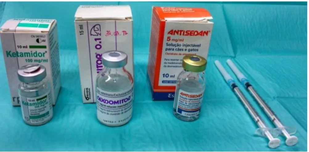
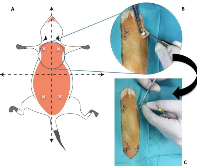
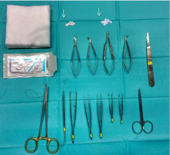
Figure 1 – TE triad of cells, chemical (bioactive molecules, e.g., growth factors) and physical (bioreactor) signals, and the scaffold which allows cells to migrate, adhere and produce new tissue. Adapted from [10]. ... 3 Figure 2 – PGA, PLA and their copolymer PLGA are different biomaterials used and widely investigated for TE purposes. PLA refers to a family of polymers: PLLA, PDLA and PDLLA. ... 12 Figure 3 – Wetting on the surfaces. (a) Surfaces with water CAs above 90° are considered hydrophobic. (b) Surfaces with water CAs above 150° are termed superhydrophobic. Roughness is also responsible for superhydrophobicity. (ɵ: angle) Adapted from [77]. ... 14 Figure 4 - The cell type temporal variation in the inflammatory response to implanted biomaterials. Adapted from [138]. ... 22 Figure 5 - Schematic representation of the experimental process to produce superhydrophobic surfaces. Adapted from [149]. ... 26 Figure 6 - Drugs utilized for intraperitoneal anesthesia (Ketamidor® and Dexdomitor®) and reversal of anesthesia (Antisedan®) in the Rattus norvegicus rats. ... 27 Figure 7 – Schematic representation of the process of implantation of PLLA surfaces. A) The animals were immobilized and placed in a ventral position. The dorsal skin of the animals was shaved, washed, and disinfected. Two paravertebral incisions were made at the level of scapula and two at the level of pelvis (rosy). B) A subcutaneous pocket was created and the implant was inserted. C) After implant insertion, the skin was closed using skin non-absorbable suture thread. ... 28 Figure 8 – Surgical instruments utilized in the implantation procedure (lint, scissor, scalpel, needle holder, tweezers, and suture thread) and the implant disks (white arrows). ... 29 Figure 9 – Abscess formation close to the surgical incision – 7 days PLLA standard surface (scale bar 50µm). ... 31 Figure 10 – Representative histological section (HE staining) of tissue reaction to the PLLA standard surface at day-7. Mononuclear cells (blue arrowheads), PMNs (red arrows), vessels (black star), endothelial cell (black arrow). ... 32 Figure 11 – Representative histological section (HE staining) of tissue reaction to the PLLA standard surface at day-60 close to the implant site. Mononuclear cells (blue arrowheads), fibroblast (yellow arrows), vessels (black star), endothelial cell (black arrow), collagen (C). 32
x
Figure 12 – TM staining of fibrous capsule at day-7 and day-60 of PLLA standard and superhydrophobic group. (*: implant site). ... 33 Figure 13 – Capsule thickness (µm) along the time considering the measurement of PLLA standard and superhydrophobic. Mean ± SEM. ... 34 Figure 14 – Fibrous capsule thickness around PLLA standard and superhydrophobic 7, 14 and 60 days after implantation. Mean ± SEM. ... 34 Figure 15 – Comparison between PLLA standard and superhydrophobic capsule thickness (µm). Mean +/- SEM. ... 35
xi LIST OF TABLES
Table 1 – List of important aspects to be considered in developing biomaterials for clinical and commercial use. Adapted from [17]. ... 4 Table 2 - Representative list of some polymers used in TERM. Adapted from [40]. ... 7 Table 3 - Summary of main properties and applications of some natural polymeric biomaterials. Adapted from [33]. ... 9 Table 4 - A summary of the main properties and applications of some synthetic polymeric biomaterials. Adapted from [33]. ... 10 Table 5 - Sequence/continuum of host responses following implantation of biomaterials. Adapted from [118, 123]. ... 21 Table 6 – Inflammation score based on the number of inflammatory cells for PLLA standard and PLLA superhydrophobic in the day-7, day-14 and day-60. ... 33 Table 7 – Measurement of fibrous capsule thickness (µm) surround PLLA standard and superhydrophobic at day-7, day-14 and day-60. Data not connected by same letter are significantly different (p<0.05). Data are expressed as mean ± SEM. ... 35
1
2
1.1 TISSUE ENGINEERING AND REGENERATIVE MEDICINE
Over the past years, the Regenerative Medicine (RM) approaches have been studied for many reasons. The injury or failure of an organ or tissue is one of the most frequent, devastating and costly problem in human health care [1]. Reviewing the lack of consensus about a clear and precise definition of RM, Daar and Greenwood (2007) defined it as “an interdisciplinary field of research and clinical applications focused on the repair, replacement or regeneration of cells, tissues or organs to restore impaired function resulting from any cause, including congenital defects, disease, trauma and ageing” [2].
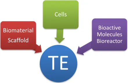
Traditionally, RM uses a combination of several technologies that stimulates and supports the body’s own self-healing ability. It may include, but is not limited to, the use of soluble molecules, gene therapy, stem and progenitor cells therapy [2], immunomodulation, nanomedicine and Tissue Engineering (TE) [3]. The TE strategies stand on three main pillars: cells, scaffolds and bioactive molecules, often combined into complex systems (Figure 1) where scaffolds are typically seeded with cells and/or growth factors and give the mechanical support to cell growth and proliferation, acting as a temporal template for tissue formation [3, 4]. These mainstream components can be cultured in bioreactor systems [5] that provide biochemical and physical regulatory signals under a closely monitored and tightly controlled environment (in
vitro) to encourage the cells to undergo differentiation and/or to produce extracellular matrix
(ECM), prior to implantation into the patient [6]. The studies around RM and TE in the last years made clear that these two subjects are extremely related because of their similar objectives. Correspondingly, these two fields have been merging in nowadays as a single research pursuit, originating the wide-ranging field of Tissue Engineering and Regenerative Medicine (TERM) [3].
The progress in the field of TERM has caused a revolution in present and future trends of medicine and surgery [1, 5] and on the development of off-the-shelf tissue-engineered products, holding the potential to manage wide range of diseases, pathological conditions and traumas in the upcoming years [4]. In fact, advanced TERM approaches bring new therapeutic options for all human population, where bioengineered materials are able to improve patient outcomes and recovery time [4, 5]. In veterinary medicine, some species have been studied with the same purpose. The therapeutic options for animals include wound healing, bone regeneration, drug and vaccine delivery [7]. For the athlete horses, for example, the RM therapies based on the use of growth factors and cells aim to improve the quality and speed of
3
healing for faster returning to the competitions [8]. Scaffolds for the repair and reconstruction of dermatologic, musculotendinous and urogenital structures are also used for veterinary applications in other species [9].
Figure 1 – TE triad of cells, chemical (bioactive molecules, e.g., growth factors) and physical (bioreactor) signals,
and the scaffold which allows cells to migrate, adhere and produce new tissue. Adapted from [10].
Langer and Vacanti (1993) describe the high economic costs and the problems with organ transplantation and reconstructive surgeries. They suggest that TE can enable future savings with human healthcare by providing alternatives cheaper than donor organs and means of intervention before patients are critically ill [1]. Moreover, the engineering of cells, tissues, and organs in an external controlled environment before surgical transplantation can significantly reduce the complications of donor site morbidity [11, 12] and immunologic rejection [11, 13], consequently decreasing health care costs related to ineffective or inadequate approaches [4].
The success of TERM technologies demands deep investigation of the biological mechanisms responsible for the repairing process against the injuries, as well as the knowledge concerning the new biomaterials under consideration [5].
TE
Biomaterial
Scaffold
Cells
Bioactive
Molecules
Bioreactor
4
1.1.1 BIOMATERIALS FOR TISSUE ENGINEERING APPLICATIONS
Biomaterials for TE purposes must follow rigorous criteria and requirements to be accepted by regulatory agencies for being manufactured for clinical use [14]. In this sense, biocompatibility of the scaffold/matrix components (e.g., source, purity, and contaminates), physical properties (e.g., mass, volume, density, and porosity), degradation kinetics and sterility are essential aspects related to the safety and performance of TERM products that should be considered [15].
Materials used in biomedical applications must be nontoxic, biocompatible, and suitable for the specific application which have been designated for. It means that biomechanical properties and physical structure must be appropriated [16]. They should not contain impurities, initiators, additives, stabilizers, emulsifiers or coloring leachable that would cause in vivo reactions [14].
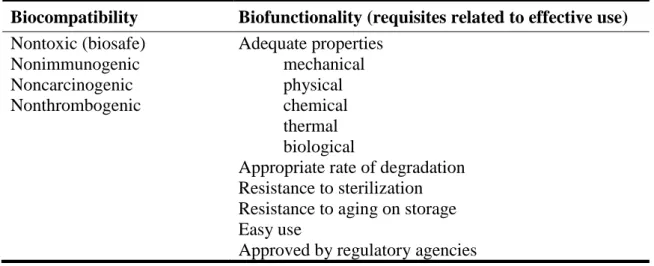
Table 1 – List of important aspects to be considered in developing biomaterials for clinical and commercial use.
Adapted from [17].
Biocompatibility Biofunctionality (requisites related to effective use) Nontoxic (biosafe) Nonimmunogenic Noncarcinogenic Nonthrombogenic Adequate properties mechanical physical chemical thermal biological
Appropriate rate of degradation Resistance to sterilization Resistance to aging on storage Easy use
Approved by regulatory agencies
To avoid graft failure, the biomaterial must maintain its biostability and biofunctionality during the expected implant life to ensure the function of the organ or tissue. In other words, the mechanical, chemical and structural properties for long-term use must not change over the time [14].
Considering their biostatibility, biomaterials can be classified as biostable/bioinert, bioabsorbable, and bioactive. The biostable materials, such as metals, ceramics and glasses, are projected to stay in a body the whole lifetime of the patient, functioning appropriately and causing minimal response in the surrounding tissues [16]. A common feature of these
5
biomaterials is that after implantation a layer of diverse unspecific proteins is adsorbed on their surface, attracting cells to grow and form fibrous tissue that will completely encapsulate the implant with time. Biodegradables are applicable to those medical devices with ability to undergo a progressive degradation while new tissue regenerates and heals [18] and ideally stay in the body only temporarily, while serving the pre-designed function, disappearing without the necessity of a second surgical intervention to remove them [19]. Another desirable feature is that they do not interfere on imaging diagnosis after being resorbed, which facilitates subsequent medical evaluations [20]. Bioactive materials, in other hand, are capable of stimulating the surrounding tissue with the aim of leading or activating the cells to specific responses and behaviors and enhance tissue growth [16, 18]. The stability features can be combined to obtain optimized materials exhibiting tailored mechanical properties and controllable degradation rates in the body [21, 22].
The choice between a material requiring long-term stability or one bioresorbable depends of its application, the organ function to be repaired, and the time of implantation that is desired [14]. Several biomaterials for TERM applications are designed to degrade or resorb
in vivo during the tissue regeneration. Synthetic polymers such as poly(glycolide/L-lactide) and
polydioxanone, or natural materials such as collagen and hyaluronic acid are commonly used for this purpose [15]. In this sense, bioabsorbable polymers are preferred candidates for developing therapeutic devices such as temporary prostheses, three-dimensional porous structures as scaffolds for TE and as controlled/sustained release drug delivery vehicles [23], since they are able to be broken down and excreted or resorbed without requiring a second surgery, reducing medical costs and other inconveniences for the patients [20, 24].
Clinically, it is desirable that materials have a predictable bioabsorption profile, because the recovery speed of the damaged tissue and the mechanical properties of the tissues may be different along the body [25]. In 2013, Willbold et al. demonstrated a correlation between the deterioration of a biodegradable metal and the site of implantation. The corrosion rate in subcutaneous was the fastest, followed by intramuscular and bony implantation of the samples. The reason for this behavior in different anatomical locations could be explained by the local blood flow that was higher in the subcutaneous site [26]. Artzi et al (2011) also analyzed materials implanted in different target sites, namely subcutaneous, intramuscular and intraperitoneal spaces. The distinctive erosion profiles in this study were correlated with the fluid volume in each site [27].
6
The first-generation biomaterials were designed initially to achieve adequate mechanical proprieties, such as strength, and a relative state of “bioinertness”, with a minimal toxic response of the host [16]. Nowadays, with the improvements in the technologies approaches, the surface design is projected to be able to direct the surrounding biological processes to attain a desired response depending on the specific application [28]. In other words, the ideal biomaterial should recapitulate the form and activity of the ECM that supports the seeded cells in vivo [11], promoting their differentiation and proliferation towards the formation of a new tissue [29]. The combination of bioactivity and biodegradability is probably the most relevant characteristics in the new biomaterials that are able to stimulate particular cellular responses at the molecular level [18]. In the past decade, it has become increasingly apparent that these behaviors of biomaterials play a fundamental role in the viability and functionality of cells, tissues, and organs [11].
Generally, the classification of biomaterials fall into one of three categories: (1) naturally derived materials such as collagen or hyaluronic acid, proteins, peptides or carbohydrates, (2) synthetic polymers such as polymeric (e.g. polyglycolic acid and polylactic acid) or inorganic materials (e.g. ceramic and metals), or (3) processed tissue derived from human or animal sources, as decellularized tissue matrices obtained via treatment with a detergent [11, 30, 31]. The most commonly materials used in clinical applications are natural and modified natural materials, but also metals, ceramics, synthetic polymers, and composites [16].
Polymers are long-chain molecules that consist of a number of small repeating units [32]. They possess significant potential since flexibility in chemistry gives rise to materials with great physical and mechanical property diversity [24]. Polymers are relatively weak and ductile compared to inorganic materials such ceramics, glasses, and metals, however, due to their versatility, easiness of processing, and biocompatibility, many of them are widely and effectively used for replacement, support, augmentation, or fixation of living tissues [16]. Moreover, polymers can be prepared in different compositions with a wide diversity of structures and properties that other materials cannot [33]. Among them, the natural ones are really attractive options, mainly due to their similarities with the ECM and good biological performance [29, 34]. Natural and synthetic polymers can be combined resulting in new materials with appropriate mechanical properties of the synthetic component and biocompatibility of the biological component [35].
7
The biodegradable polymers have basically two major applications; as biomedical polymers for health care and as environment-friendly polymers that do not exert adverse effects on animals and plants on the earth [36].
Some of the current applications of biodegradable polymeric materials in the surgery and pharmacology include: temporary prostheses, drug delivery and targeting systems, and medicated prostheses [17]. The use of biomaterials to deliver biologically active agents directly to the site of disease in a controlled manner, sparing off-target tissue toxicities, is an interesting concept to facilitate localized therapies such as tumors [37] and periodontal diseases [38], for example. Biomaterials can also be used as scaffolds for cell transplantation; as barriers at the cellular or the protein level to guide tissue regeneration; as tissue adhesives or structural supports to bear mechanical loads during healing or regeneration; and as provisional matrices [39].
The main groups of polymeric materials used in biomedical applications and some examples of each group can be summarized as follows (Table 2).
Table 2 - Representative list of some polymers used in TERM. Adapted from [40].
Classification Polymers
Natural polymers Collagen, albumin, gelatin, agarose, alginate, carrageenan, hyaluronic acid, dextran, chitosan.
Synthetic polymers
Biodegradable Poly(lactic acid), poly(glycolic acid), poly(hydroxyl butyrate), poly(ε-caprolactone), poly(dioxanones), poly(sebacic acid), polyamino acids, polyphosphates, polyurethanes, polyortho esters.
Non-biodegradable Carboxymethyl cellulose, polymethacrylate, poly(methyl methacrylate), polyvinyl pyrrolidone.
In the environmental field, polymers can be used to packaging, mulching films, agricultural staples, coatings to protect seeds, chewing gums, cigarette filters, cartridge and cartridge wax [17]. Thinking about a green environment, biodegradable polymers are very attractive, but still expensive for production [36].
8 1.1.1.1 NATURAL MATERIALS
Naturally derived polymers are available in large quantities and usually biodegradable [33]. They offer the advantage of being similar, sometimes identical, to naturally occurring substances of ECM, avoiding the stimulation of chronic inflammation or immunological reactions, often noticed with synthetic polymers [29]. Furthermore, due this similarity to biological macromolecules, natural biomaterials are able to be designed to work efficiently at molecular, rather than macroscopic level [41].
Another interesting characteristic of natural polymers is their ability of being degraded and remodeled by cell-secreted enzymes [42], a virtual assurance that the implant will be eventually metabolized and be removed by normal metabolic processes [41].
Some natural polymers have antibacterial properties and are used as coating materials for alleviating pathogenic colonization on surfaces. The coatings are noncytotoxic and exhibit a high degree of stability under expected conditions. For example, agarose works as antibacterial coating for biomedical devices and quaternized chitosan for preventing pathogen transmission in the environment [43].
For delivery systems, they offer the advantage of being usually non-toxic, even at high concentrations, so they can readily be incorporated into oral delivery or bolus matrix delivery systems [42].
The classification of natural polymers, some examples, their properties and applications are summarized in the Table 3.
The principal disadvantage of natural polymers lies in their structural complexity that makes difficult the development of reproducible production methods [33]. The natural variability in structure of substances derived from natural sources and the chemical difference from one species to another and from one tissue to the next induces batch to batch variations [41].
Other potential problems when using a natural polymer as biomaterial include: deficiency in bulk quantity and expansive, and the variability of degradation rate from patient to patient, once it depends upon enzyme quantities [44].
Nowadays, with the advances in biotechnology, natural polymers can be synthetized by the fermentation of micro-organisms [45] or produced in vitro by enzymatic polymerization [46].
9
Table 3 - Summary of main properties and applications of some natural polymeric biomaterials. Adapted from
[33].
Natural polymer Main applications and comments
Proteins and protein-based polymers
Absorbable, biocompatible, nontoxic, naturally available, typically elastic materials used as implants and in TE.
Collagen Absorbable sutures, sponge wound dressing, drug
delivery, artificial skin, coatings to improve cellular adhesion, guided tissue regeneration in dental applications, scaffold for reconstruction of blood vessels, wound closure.
Albumin Cell and drug microencapsulation.
Poly(amino acids) Nontoxic, nonantigenic and biocompatible. Used as oligomeric drug carriers.
Polysaccharides and derivatives
From vegetable sources
Carboxymethyl cellulose Non-biodegradable. Cell immobilization via a combination of ionotropic gelation and polyelectrolyte complex formation (e.g. with chitosan), in drug-delivery systems and dialysis membranes.
Cellulose sulphate Component of polyelectrolyte complexes for immunoisolation.
Agarose Largely used as supporting materials in clinical
analysis and as an immobilization matrix. Alginate (marine sources, algae) Excellent gel-formation properties; relative
biocompatibility; batch-to-batch variations. Used as immobilization matrices for cells and enzymes, controlled release of bioactive substances, injectable microcapsules for treating neurodegenerative and hormone-deficiency diseases.
Carrageenan Excellent thermoreversible properties. Used for microencapsulation.
From human and animal sources
Hyaluronic acid Excellent lubricant, potential therapeutic agent. Heparin and heparin-like
glycosaminoglycan
Antithrombotic and anticoagulant properties. Extensively used in surgery.
Microbial polysaccharides
Dextran and its derivatives Excellent rheological properties. Plasma expander. Widely used as drug carrier.
Chitosan and its derivatives Biocompatible, nontoxic, excellent gel- and film-forming ability. Widely used in controlled-delivery systems.
10
1.1.1.2 SYNTHETIC MATERIALS
Synthetic polymers offer a number of advantages for applications in TERM. Unlike natural materials, they can be easily reproduced keeping quality and purity [47] and have better mechanical and thermal stability [48]. Moreover, they can be fabricated into various shapes with desired morphologic features [49], including three-dimensional structures with a projected dimension by three-dimensional printers [50]. They are available in many compositions with readily adjusted properties by processing, copolymerization and blending, which optimize their mechanical and biological properties [33].
In the biomedicine field, synthetic polymers are often used for TE in various areas such as the cardiovascular system, orthopedics, neurology, drug delivery systems and others [51], as represented below (Table 4).
Table 4 - A summary of the main properties and applications of some synthetic polymeric biomaterials. Adapted
from [33].
Synthetic polymers Main applications and comments
Aliphatic polyesters
Poly(lactic acid), poly(glycolic acid) and copolymers
Used in sutures, drug-delivery systems, barrier membranes, guided tissue regeneration (dental applications), orthopedic applications, stents, staples and TE. Biodegradable. Often copolymerized to regulate degradation time. Poly(ε-caprolactone) and
copolymers
Biodegradable, used as a matrix for long term drug-delivery systems, cell microencapsulation. Properties can be changed by chemical modification, copolymerization and blending. Polyamides (nylons) Sutures, dressing, haemofiltration membranes. Poly(ortho esters) Surface-eroding polymers. Application in sustained
drug delivery, stents, ophthalmology.
Poly(cyano acrylates) Biodegradable. Used as surgical adhesives and glues, potentially used in drug delivery.
Polyphosphazenes Made into films and hydrogels. Applications in drug delivery, blood contacting devices, skeletal reconstruction.
Thermoplastic polyurethanes Good elastomeric properties. Used in permanently implanted medical devices (prostheses, vascular grafts), catheters and drug-delivery systems. Initial candidates for the artificial heart.
11
Poly(glycolic acid) (PGA), poly(lactic acid) (PLA) and their copolymer
[poly(lactide-co-glycolide)(PLGA)] are becoming the most commonly used [23 , 52] and the most widely
investigated for TE purposes [18, 53]. In particular, synthetic biodegradable polymers have attracted special attention because they better control their physico-chemical properties [18] and they can be metabolized by human body [23, 52]. These polymers degrade by nonenzymatic hydrolysis and their nontoxic degradation products are eliminated from the body by natural metabolic pathways [20], in the form of carbon dioxide and water [54] (e.g.: urine) [20]. The biodegradation rate of synthetic biodegradable polymers depends of their characteristics such as shape, molecular weight, composition, monomer conversion, macromolecular orientation, etc. and can be controlled by alteration of some features, such as copolymer ratio [25]. The degradation times can be achieved from several weeks to several months [54] for applying to clinical uses.
Although the degradation products have shown to be nontoxic, the concern with the use of some specific polymers still remains because they can provoke adverse effects or alter local microenvironment in vivo, reducing local pH, and consequently inducing inflammatory response and injuries in the cell health at the implant site [55]. As synthetic polymers are often associated with inflammatory reactions, except for poly(ethylene oxide) and PLGA that show good biocompatibility, their use is limited to solid, unmoving, impermeable devices [33]. Other disadvantages of the synthetic polymers, such as poor processability and loss of mechanical properties very early during degradation, are also reported [49].
Among the biopolymers used in the medical field, the polyester PLA has received significant attention, not just because it is made from renewable resources, but also because it provides excellent properties at a low cost compared to other traditional biodegradable polymers used for the same purposes [56]. PLA represents one of the most important biodegradable polymers, being the preferred alternative to its homologous PGA because of its degradation time [25]. The hydrophobic characteristic of PLA makes its degradation slower than PGA. The water absorption of thin films is limited by its hydrophobicity that slows down the backbone hydrolysis rate. According to the available data, the estimate duration of PLA degradation process is one to two years [57].
Owing to mechanical, biological and thermoplastic properties, PLA is convincingly accepted for using in biomedical applications, such as bone fixation devices. It is derived from lactic acid, a naturally occurring organic acid that can be produced by fermentation [23] of sugars obtained from renewable resources such as sugarcane [20]. Additionally, PLA is
12
approved by the Food and Drug Administration (FDA) [49], can be produced using low energy and used in an environmentally friendly cycle [58], being considered an eco-friendly biomaterial [58, 59].
Despite the advantages, some drawbacks may limit the use of PLA in certain applications. The limitations include poor toughness and lack of reactive side-chain groups [58].
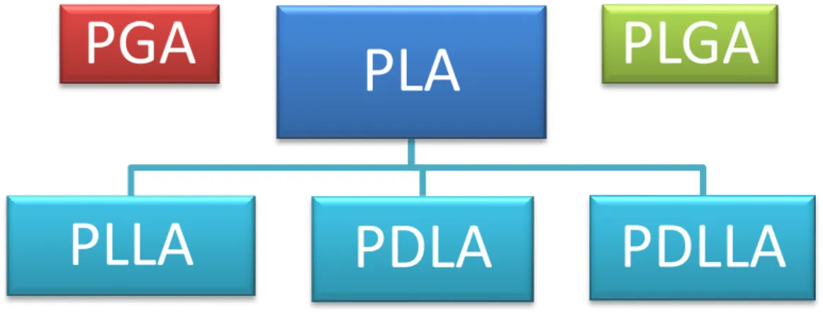
Lactic acid is a chiral molecule, existing in L and D isomers and the term “poly-lactic acid” refers to a family of polymers: pure poly(L-lactic acid) (PLLA), pure poly(D-lactic acid) (PDLA), and poly(D,L-lactic acid) (PDLLA) [52] - a racemic mixture of PLLA and PDLA (Figure 2). As far as use in biomedical research, only PLLA and PDLLA have shown promising and have been widely studied [24]. Because it is preferentially metabolized in the body, the (L) isomer of lactic acid is often chosen for most applications [49].
Figure 2 – PGA, PLA and their copolymer PLGA are different biomaterials used and widely investigated for TE
purposes. PLA refers to a family of polymers: PLLA, PDLA and PDLLA.
PLLA is a semi-crystalline polymer [60], aliphatic polyester with good biodegradability and biocompatibility [16], versatility [61], reasonable mechanical properties, and processability in forming fibers [16]. The hydrolytic degradation process of aliphatic polyesters occurs by random scissions of ester bonds within the polymer chains [62]. Of the two enantiomeric forms (PLLA and PDLA), PLLA degrades the slowest [59], because the material has no affinity with body fluids [63]. The water uptake of PLLA during the hydrolysis is one of the responsible for the process of mass loss [64]. PLLA degradation process provides a significant increase in the crystallinity with ageing time [65], which restricts the water uptake into the polymer matrix, making the hydrolytic process difficult [64]. Consequently, some portions of biomaterial
PGA
PLA
PLLA
PDLA
PDLLA
13
remain protected against the water and the sorption processes lasts longer. Therefore, crystallization appears to be effective in increasing the hydrolytic stability [57].
In order to modify the degradation time to obtain a desirable time scale for specific application, investigators have blended or copolymerized PLLA with other degradable polymers [64, 66]. It offers great promise in a wide range of commodity applications, although features such as high rigidity and hydrophobicity limit its use in some areas [63].
PLLA is widely used in compounding with other materials for sutures and bone fillings [16] and also for medical devices (e.g., screws for fixation of tendon to bone and bone to bone) with rare complications [67]. However, in 1995, Bergsma et al. reported swelling at the site of implantation in four patients three years after implantation of PLLA and associated the disintegration of PLLA with it [68]. In dermatology area, Funt and Pavicic (2013) reported dermal filler complications such as granuloma in the implant site [69], but this reaction is usually attributed to inadequate techniques and not to the implant itself [70].
In summary, the success of synthetic polymers as biomaterials mainly relies on their wide-ranging mechanical properties, transformation processes that allows a variability of shapes to be easily achieved, and low production costs. On the other hand, biological polymers present good biocompatibility but their mechanical properties are usually inferior, the necessity of preserving biological properties complicates their processability, and their production or recovery costs are very high [70, 71]. “Bioartificial polymeric materials” is a term to designate a new class of materials based on blending synthetic and natural polymers, where the final purpose is produce materials for biomedical applications that possess both good mechanical properties and biocompatibility, overcoming the poor performance of each one in these features [72].
1.1.2 SUPERHYDROPHOBIC SURFACES
To meet the specifications which biomaterial was designed for, it must exhibit expected mechanical, physical, or electrical properties [14]. The good performance clinically of a biomedical device depends on the identification and controlled modification of key intrinsic surface properties [33]. Characteristics such as charge, polarity and energy, heterogeneous distribution of functional groups, wettability, chain mobility, as well as morphological and
14
topographical aspects, including texture, smoothness and roughness, should be considered in this sense [14].
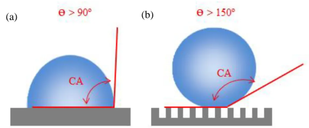
Among the methods to characterize the wettability of the surface, the water contact angle (CA) measurement is often used [73]. According to Marmur (2012), CA is defined as the angle between the solid surface and the tangent to the liquid surface (on the liquid side of it), at the three-phase contact line. In his study, a well-defined terminology that accounts for both the chemistry of a solid surface and its wetting functionality is presented [74]. In summary, surfaces with water CAs above 90° are considered hydrophobic, and those with CAs above 150° are termed superhydrophobic (Figure 3) [75]. Moreover, the combination of suitable surface roughness and low-surface-energy materials is also responsible for superhydrophobicity [73, 76].
Figure 3 – Wetting on the surfaces. (a) Surfaces with water CAs above 90° are considered hydrophobic. (b)
Surfaces with water CAs above 150° are termed superhydrophobic. Roughness is also responsible for superhydrophobicity. (ɵ: angle) Adapted from [77].
In the nature, we can find many superhydrophobic surfaces that possess water-repellent, self-cleaning and anti-icing properties [78]. In 1997, Barthlott and Neinhuis documented an almost complete self-cleaning ability by water-repellent plant surfaces, such as lotus leaf. In nature, many terrestrial plants and animals have the ability to create their superhydrophobic surfaces from a microscopic roughness over coated with specific functional groups [79]. Since then, the lotus leaf has become widely explored and has inspired many investigators to research others superhydrophobic plant surfaces [78] such the petal of red rose [80]. Compared with the lotus effect, which water droplets roll off the surfaces independent of their chemical nature or size providing a very effective anti-adhesive property against particulate contamination [79], the petal effect does not permit the water droplet roll off even when the petal is turned upside down [80].
15
Nowadays, the studies around non-wettable surfaces with high water CAs and facile sliding of drops continue to receive important attention [81] not only for academic reasons, but also for practical applications such the production of hydrogels. The drops placed onto a superhydrophobic surface almost completely surrounded by air or another desired atmosphere will maintain the spherical shape. This permits an efficient preparation of a large range of systems under mild conditions avoiding any loss of the cargo during the process and a good control over the size of the particles [82].
Hydrogels are cross-linked hydrophilic polymers of natural or synthetic origin [83] that swell significantly when placed in a polar liquid solution [84]. Hydrophilic functional groups attached to the polymer enable the hydrogels to retain high percentage water content [85], over 90% [83]. Superhydrophobic surfaces permit to encapsulate cells into hydrogels beads by gravitational dripping [82]. Hydrogels are mechanically strong and resistant to heat, wear and attack by solvents. However, they are relatively inflexible, insoluble and infusible. Most of them have applications on medical field as drug delivery and for TE [86].
Due to the limitation on fluid absorption, water-repellent devices are desirable where long-term mechanical resistance is required, as previously mentioned. For example, orthopaedic and dental implants must be water repellent to avoid any degradation or erosion processes leading to changes in toughness and loss of mechanical strength [33]. High hydrophobicity can significantly enhance the resistance against hydrolytic degradation [87] through a weak interaction between body fluid and the implanted biomaterial [63]. Others must have limited moisture penetration, as pacemakers and artificial blood vessels [33]. Sun et al. (2005) reported the effect of special nanostructures on blood compatibility. The results demonstrated excellent anti-adhesion to platelets and the relatively low platelet activation making nanostructured superhydrophobic films good candidates for utilization in artificial organ implantation, manmade blood vessels, and other blood-contacting medical devices [88]. The surface energy of an implant, indirectly measured by liquid-solid CA, also affect the biological response to the implant such as adhesion of proteins and other macromolecules onto the surface, hard and soft tissue cell interactions and bacterial adhesion and subsequent biofilm formation [89]. Two materials with similar surface energies but different water-sorption characteristics can possess different biocompatibilities [33]. However, wettability alone does not play the dominant role in determining subsequent cell behavior; the functional groups on the surfaces also affect the biomaterial performance [90]. In the study performed by Sartoretto et al. (2015), the bone healing was not affected by micro topography but by chemical changes.
16
Surfaces with less carbon had a markedly enhanced hydrophilicity and this accelerated osseointegration and increased the area of the bone-to-implant interface [91]. Depending on the objective of the biomaterial it should stimulate cell adhesion or suppress the attachment of specific proteins and cells [92], so wettability, surface energy and chemical property can determine the choice of a biomaterial.
It has been shown that the surface roughness of implants may also influence biological responses [93]; however surface wettability was recognized as a critical factor to explain the different cell behavior on biomaterials when comparing these two properties [60]. The cells can adhere and proliferate on both superhydrophobic and superhydrophilic surfaces, but constant contact to superhydrophobic surfaces is required for cell division and proliferation on it [94]. Modifying just one side of the surface to transform it in superhydrophobic can permit the use of this biomaterial for bone guided regeneration where this surface do not allow cell growth, for example [95].
According to Ishizaki, Saito & Takai (2010), cells easily adhere and proliferate immediately after seeding on superhydrophilic surfaces [94]. Oliveira et al. (2011) demonstrated higher cells proliferation in surfaces with water CA ranging from 13º to 30º, independently of being rough or smooth [95]. Sawase et al. (2008) studied the effect of photo-induced hydrophilicity on initial cell behavior and bone formation. The CA of the biomaterial irradiated with ultraviolet (UV) light decreased and the cell attachment and proliferation on this hydrophilic disk increased improving the initial cell reactions and enhancing early bone apposition to the implant [93].
Last studies highlighted the influence of roughness on cell behavior and protein adsorption demonstrating that total protein adsorption and cell viability at the rough surfaces are generally lower than at the corresponding smooth surfaces in superhydrophobic biomaterials [60]. However, the same authors concluded that chemistry and topography did not have the same importance to cell behavior as the wettability. Aqueous solutions in contact with superhydrophobic films have less actual surface area available for protein adsorption than the surface area of a flat surface [77] as it is demonstrated on Figure 3. On the other hand, Oliveira et al. (2011) concluded in their study that superhydrophilic surfaces seem to be ideal for repellence of proteins [95].
It has been demonstrated that increased surface roughness is also an important physical factor for bacterial adhesion [96]. In general, the surfaces with a higher number of attachment points will attract more cell attachment [97]. Bacterial adhesion shows a direct positive
17
correlation with the surface roughness [98-100]. It possible to take an example from dental analysis that showed an increase in plaque accumulation in rough surface above a certain threshold of roughness [101].
Analyzing the surface wettability, Tang et al. (2011) concluded that although the
Staphylococcus aureus was not totally absent on the superhydrophobic surfaces and the amount
of adhered bacteria increased with time, they were much less in quantity and more scattered than those on the hydrophilic and hydrophobic surfaces and could be easily removed. The experiment results show that superhydrophobic surfaces display high resistance to bacterial contamination and have a strong potential to reduce device-associated infection [102]. On the other hand, Sousa et al. (2011) showed that superhydrophobic and hydrophobic PLLA surfaces are able to be colonized by bacterial cells, although this effect can be due to the combined effect of the different PLLA and S. aureus specific surface morphologies, since S. aureus cells seem to fit perfectly the irregularities on the roughness of superhydrophobic surface and, thus, end up having a greater contact area than on the smooth hydrophobic surface [103].
The database of bacterial adhesion on superhydrophobic surfaces is not yet sufficiently extensive and systematic to completely understand the mechanisms of this process. With this purpose, the tests should be standardized and more bacterium types should be tested. More parameters such as surface roughness, morphology, functional group and the free energy from the superhydrophobic surfaces should also be studied to analyze their effect on bacterial adhesion [97].
It is not surprising that contradictory results have been observed in different studies, since the variability on surface evaluations and the experimental conditions applied on them make difficult to comparison about the influence on cells behavior, protein adsorption [104] and bacterial adhesion [97]. It is hard to define the true surface reactions responsible for the performance of biomaterial because several surfaces can be identical on wettability while their surface chemistries can remain quite different [90]. According to Wennerberg and Albrektsson (2009), the difficulty is occasionally attributed to the terminology assumed by the authors. In addition, many investigators falsely assume the roughness of the implant based on the surface preparation and many other studies use only qualitative techniques to define it [104].
18
1.1.2.1 APPLICATION OF SUPERHYDROPHOBIC SURFACES IN THE
BIOMEDICAL FIELD
Various different natural, synthetic and hybrid polymers are available for biomedical applications in diverse areas. Specifically, superhydrophobic surfaces have been actively studied for the use in the industries and further in the biomedical field as substrates to control protein adsorption, cellular interaction, and bacterial growth, as well as platforms for drug delivery devices and for diagnostic tools [105].
The fact that cells adhere very differently to hydrophobic and hydrophilic substrates can be used in favor of biomedicine [92]. The different ability for proteins to adsorb in these substrates has been used to produce smart surfaces for programmed adsorption and release of proteins in the context of microfluidic devices [106].
New methodologies based on the use of superhydrophobic surfaces propose the production of compartmentalized multilayered polymeric spherical particles with controlled size and layer thicknesses of distinct materials that could allow the distribution of cells or drugs by layers and the use in a wide range of applications including cosmetics, pharmacy, agriculture, food technology and biomedicine [107]. Others recent studies propose the production of smart systems incorporating responsive substances, for example magnetic responsive particles or networks, containing temperature responsive polymers. Even growth factors or other unstable as expensive non-volatile molecule could also be integrated into such particles with high levels of efficiency [108].
Additionally, superhydrophobic surfaces with controlled wettable spots can be used as platforms to produce microarray chips for multiplexing evaluation, offering the possibility to screen individually and in the same chip different combinations of biomaterials under different conditions, including different cells, culture media or solutions with diverse proteins or other molecules [109]. These systems exhibiting patterned high-contrast wettability regions act as mini-bioreactors with distinct behaviors in each spot that may be used to distinct applications needs [110] on TERM, cellular biology, diagnosis, drug discovery and drug delivery monitoring [109]. In this sense, Ishizaki, Saito & Takai (2010) also studied the cell behavior on superhydrophobic and superhydrophilic micropatterned surfaces. The results show that the method could contribute to development of cell-based technologies including biosensors for the screening of drug libraries as well as for better understanding the eukaryotic cells interactions with implantable biomaterials and the communication between them [94].
19
It was recently shown that rough superhydrophobic PLLA surfaces are colonized by bacterial cells, introducing a possible application of PLLA-based superhydrophobic materials as bacterial colonization substrata with potential to be used as carriers for biomass immobilization in bio-reactors [103]. However, other study indicated that superhydrophobic surface shows high resistance to bacterial contamination and could be used in the clinical practice as antimicrobial to reduce device-associated infections [102]. This characteristic makes these materials suitable in extracorporeal medical devices, by making the device free of contamination and easy to clean [88]. For further information, researches should focus in analyzing different bacteria, regarding their Gram-type and morphology, on superhydrophobic surfaces with distinct properties.
As previously described, the excellent anti-adhesion to platelets and the relatively low platelet activation, show the useful of these films in artificial organ implantation and blood vessels, and other blood-contacting medical devices [88].
Li et al. (2013) demonstrated the application of superhydrophobic surfaces in conducting biological assays for rapid human blood typing analysis using a liquid drop micro reactor with only a small amount of blood sample. The characteristics of superhydrophobic surfaces can help the pathological laboratories on diagnosis because they enable the blood and antibody droplets to have a spherical shape, making easier to photograph, record and analyze the haemagglutination reaction inside the droplet by software [111].
Concluding, superhydrophobic surfaces can be used to produce biomaterials with a wide potential applications including catheters, endotracheal tubes, or medical instruments with a superhydrophobic coating to reduce bacterial adhesion when in contact with blood or bodily fluids; controlled patterns of superhydrophobic and superhydrophilic regions used to construct cellular microarrays or engineered tissues; disposable microfluidic diagnostic devices, where the superhydrophobic coating supports droplets or facilitates fluid flow; and coated medical devices for drug delivery [105].
Regarding the efficiency of the therapy, each application designed for a biomaterial demands appropriated characteristics such as physical, chemical, biological, biomechanical and degradation properties. For this purpose, investigators keep focus in studying a wide range of natural or synthetic polymers [112]. Superhydrophobic surfaces made by these sources have shown great potential for such applications in the biomedical field and should be better studied to improve the efficiency in the treatment of human and veterinary patients.
20
1.1.3 IMUNNE RESPONSE TO BIOMATERIALS
Once the inflammatory response caused by biomaterials is an unavoidable event affecting the tissue regeneration, attention to this aspect should be considered when developing TE strategies [113]. Although improvements has been made concerning about biocompatibility, many materials and procedures are associated with side effects such as inflammation, infections and subsequent loss of function [114], as well as fibrosis and thrombosis [115] inducing bioincompatibility.
In general, failure of most implants results from an inadequate host response to the material because of the organism inability to predict and regulate biological phenomena, such as protein adsorption and cell interactions [116]. Polymorphonuclear leukocytes, monocytes, macrophages and foreign body giant cells (FBGCs) play a central role in the foreign body reaction and immune inflammatory responses, processes that affect the biostability, biocompatibility and effectiveness of the implant [117].
Biocompatibility is generally defined as “the ability of a biomaterial, prosthesis, or medical device to perform with an appropriate host response in a specific application” [118] and it implies the absence of adverse reactions, local or systemic, caused by material to the tissue directly or through the release of their material constituents [119].
The ability to mimic repair processes following injury and to control reactions like inflammation has been shown to dictate the efficiency of biomaterial implants [120], as mentioned above. Then, in order to design materials with a good performance to ensure a desirable cell survival, migration and adhesion, a depth understanding of the host response is required [121].
Once implanted, the biomaterials surfaces interact with the surrounding tissues [116, 121], producing some degree of tissue damage, which will initiate two principal reactions, inflammation and the related response of repair – the wound healing [120]. In this interaction, host reactions incorporate a combination of many processes [122], summarized in the Table 5. The response to tissue injury is dependent on multiple factors including the extent of injury, blood-material interactions and the extent of the inflammatory response that consequently will affect later events [118]. Microvascular injury, protein exudation and accumulation, and activation of the humoral and cellular defense systems are some key points occurring during the early inflammatory response [124].
21
Table 5 - Sequence/continuum of host responses following implantation of
biomaterials. Adapted from [118, 123].
Injury post implantation Blood/material interactions Acute inflammation
Chronic inflammation Granulation tissue Foreign body reaction
Fibrosis/fibrous capsule formation
The contact between the blood and the implant is an inevitable and early occurrence after almost all implantation procedures in biological tissue [124], including soft and hard tissues (e.g. dental implants, prostheses). This initial contact with the blood influences the inflammatory reaction against the material [114] and triggers a complex series of interlinked events including protein adsorption, platelet and leukocyte activation/adhesion, and the activation of coagulation [125] and an immediate complement-mediated inflammatory response [125, 126].
Blood-biomaterial contact induce the quick adsorption of proteins onto a biomaterial surface [127], regarded as important determinant of the acute inflammatory response [128] and the first major step in the integration of an implanted device with implications for nanotechnology, biomaterials and biotechnological processes [129].
Among these host proteins that spontaneously associate with implant surfaces, albumin, immunoglobulin G (IgG) and fibrinogen usually predominate [130]. Albumin is known to decrease platelet adhesion to polymer surfaces and improve the biocompatibility, preventing the formation of thrombus [131]. IgG involves the activation of the complement system and subsequent stimulation of adherent macrophages by complement products [132]. Adsorbed fibrinogen has been shown to be the primary component of plasma responsible for acute inflammatory responses, mainly by facilitating phagocyte recruitment at implant surfaces [133]. Also, fibrinogen enhances platelet adhesion [134, 135] stimulating thrombosis [125, 136], and bacterial colonization [137].
Blood-interactions and protein adsorption on the surface of biomaterial is followed by acute inflammation, with attraction of polymorphonuclear leukocytes (PMN). Sequentially,
22
chronic inflammation characterized by the presence of monocytes and lymphocytes usually resolve within the first 2-3 weeks following implantation [123].
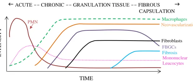
The predominant cell type present in the inflammatory response varies along the time (Figure 4) [118, 138]; however the components of the reaction within the implant site may also vary depending of the surface properties [138]. In general, neutrophils predominate during the first several days and disappear after 24 to 48 hours following injury and then are replaced by monocytes that after differentiate into macrophages which are very long-lived (up to months) [118] and the principal responsible for wound healing [138].
Implanted biomaterials usually provoke a persistence of an inflammatory stimulus leading to chronic inflammation characterized by the presence of macrophages, monocytes and particularly lymphocytes and plasma cells with the proliferation of blood vessels and connective tissue [139]. Implant failure may be caused by the fragments of implanted biomaterials that lead to chronic inflammation [128], the main reason for this undesirable outcome.
The granulation tissue process involves proliferation, maturation, and organization of endothelial cells into capillary tubes, fibroblasts proliferation and subsequently synthesis collagen and proteoglycans [140]. The sequence ultimately ends in the formation of foreign body giant cells at the tissue/material interface [121].
Figure 4 - The cell type temporal variation in the inflammatory response to implanted biomaterials. Adapted from
[138].
The following inflammatory and wound healing response after biomaterial implantation is characterized by the foreign body reaction [121], composed of FBGCs and the components of granulation tissue described above (e.g. macrophages, fibroblasts, collagen and capillaries)
TIME
ACUTE -- CHRONIC -- GRANULATION TISSUE -- FIBROUS
CAPSULATION IN TEN S IT Y PMN Macrophages Neovascularization FBGCs Fibroblasts Fibrosis Mononuclear Leucocytes23
[138, 140]. Monocytes adhered to the biomaterial surface differentiate into macrophages that fuse to form the FBGCs [123] and remain at biomaterial-tissue interfaces for the lifetime of the device [122]. Maluf-Meiken et al. (2006) showed that the number of multinucleated giant cells increased significantly from the seventh day after implantation of bioabsorbable polymer, decreasing from the twenty-eighth day to the sixtieth day and increasing again from the ninetieth day [141].
The end-stage healing response to biomaterials is generally followed by fibrosis or fibrous encapsulation [140]. Mainil-Varlet, Gogolewski & Nieuwenhuis (1996) studied the tissue capsule formed around PLLA implanted in the subcutaneous tissue of sheep and noticed the capsule consisting of fibroblasts, fibrocytes, phagocytes, a few FBGCs and PMN cells. At three months post-implantation, the capsule was denser, its thickness and cellularity had slightly increased compared to one month time point and continually increased until 6 months when it showed more matured [142].
As previously described, the cell-interaction with the implant is largely dependent on the cell type and surface properties of the materials [116] such as wettability, energy, roughness, charge and chemical composition [143]. Such properties modulate the foreign body reaction in the first two to four weeks following implantation [121] and the intensity and the time variables also depend of the extent of injury created in the implantation procedure [140].
The quality of protein adhesion in the early phases of inflammation influences the cells morphology and their proliferation and differentiation ability [144]. The absence of adsorbed proteins, or interference with their function, modifies the cells behavior preventing their attachment [145]. Therefore, it is reasonable to accept that modifications in distribution of adsorbed proteins can be used in favor to engineer materials with the desired performance for a specific application [146] and with lower or no complement-activating properties [114]. Ekdahl et al. (1993) analyzed the complement activation in vitro and the results suggested enhancing the biocompatibility of polystyrene surfaces after surface modifications [147].
The biocompatibility of polymers is also defined by the degradation products and their active biocompatibility must be demonstrated over time. The chemical, physical, mechanical and biological properties of a biodegradable material will vary and these changes can cause long-term host responses to these biomaterials to be greatly different than the initial response [24]. In fact, degradation products of polymers may reduce the microenvironment pH and consequently affect the integrity of the cells [16]. The degradation products of PLLA, for example, reduce local pH, accelerate the polyester degradation rate and induce inflammatory
24
reaction [57]. Although PLA-PGA biomaterials are generally biocompatible and non-toxic, Athanasiou, Niederauer & Agrawal (1996) summarized some studies with PLA and PGA that reported inflammatory reactions usually occurring 7-20 weeks after implantation in the body [148].
Although inflammatory reaction is usually associated to implants failure, it was also reported to help the triggering of tissue regeneration (e.g. neural regeneration) [113]. Studies have been demonstrated that there is still a necessity to understand the mechanisms involved in the biocompatibility in order to improve the biomaterials and the patient’s recovery, reducing undesirable reactions, treatment time and cost. For this purpose, more detailed in vivo studies should be performed to better explaining biomaterial-host interaction.
25