Influence of Light Quality and Intensity on Adventitious Root
Formation in Microshoots of Pinus pinea L.
C. Ragonezi1,a, M.R. Castro1, K. Klimaszewska2, M. Lima1 and M.A. Zavattieri1 1
Laboratory of Plant Breeding and Biotechnology, ICAM, University of Évora, Ap. 94, 7002-554 Évora Codex, Portugal
2
Natural Resources Canada, Canadian Forest Service, Laurentian Forestry Centre, 1055 du P.E.P.S., P.O. Box 10380, Stn. Sainte-Foy, Québec, G1V 4C7, Canada
Keywords: stone pine, micropropagation, Gro-lux lamps (GL), Cool-white lamps (CW) Abstract
In the present study of Pinus pinea L., further improvement of microshoot rooting was achieved by applying Cool-white light at increased intensity from 60 to
90 µmol m-2 s-1. In contrast, light provided by Gro-lux lamps promoted rooting of
the microshoots at the same frequency regardless of its intensity. Majority of
microshoots (70.4%) grown under Cool-white lamps at the intensity of 90 µmol m-2
s-1 were also significantly taller when compared with those from other tested
treatments.
INTRODUCTION
The light-mediated changes in plant growth and development are referred to as photomorphogenesis, and plants have extraordinary versatility to perceive different light signals in different developmental contexts. Different groups of photoreceptors of the photosensory systems regulate plant development, namely cryptochromes, phototropins and phytochromes each of them monitoring different wavelengths regions of the spectrum (Quail, 2002). Plants use phytochrome to detect and respond to red and far-red wavelengths and cryptochromes were the first blue light receptors isolated and characterized. Light can also modify the efficacy of plant growth regulators (PGRs) as well as affect the endogenous hormone balance. Auxin plays a central role in the determination of rooting capacity, and light conditions are known to affect auxin metabolism and tissue receptivity (Reid et al., 1991). From an applied point of view, plant morphogenesis may be influenced by the correct choice of lamps and filters (Fuerakranz et al., 1990). For example, red-light improved rooting percentage and root numbers in shoots of two genotypes of grape propagated in vitro (Poudel et al., 2008). However, little is known about the effect of green and yellow lights, which seem to be involved in the regulation of in vitro plant development (Loreti et al., 1991).
The purpose of this work was to establish if light quality and intensity, within the visible range, influence root growth and development of Pinus pinea microshoots with the aim of enhancing the present rooting protocol achieved with Cool-white (CW) lamps.
MATERIAL AND METHODS Plant Material
Cotyledons from non-germinated embryos of stone pine (Pinus pinea L.) were used as explants. The seed coat was cracked with a nut cracker and discarded. The remainder megagametophytes were surface sterilized by immersion in 70% (v/v) ethyl alcohol for 2 minutes followed by three rinses in sterile bi-distilled water. They were then disinfected with sodium hypochlorite 10% (v/v) (commercial bleach with 5% free chlorine) for 25 minutes followed by four rinses in sterile bi -distilled water. All of the following steps were carried out under aseptic conditions. An embryo was excised from the megagametophyte by making a longitudinal incision with a scalpel and by gently pulling the edges of the cleft with two forceps. Finally, the cotyledons were excised from
a
the embryo axes with a cut at their bases.
Microshoot Induction from Embryo Cotyledons
The whole set of cotyledons of each of 20 excised embryos was cultured separately in a Petri-dish (9 x 1.5 cm) containing WPM (McCown and Lloyd, 1981) medium supplemented with 5 mg/L of benzylamino purine (BAP) for shoot organogenesis. After a month, the explants with shoot buds were transferred to a fresh PGR-free medium with 2 g/L of activated charcoal (AC) to promote shoot elongation.
Rooting of Microshoots
Seven, eleven, and six clonal shoots were tested at 60, 90, and 110 µmol m-2 s-1, respectively. In all experiments, shoots 2 cm in height were transferred to WPM with half concentration of the macronutrients, 0.65% Difco Bacto-Agar and different carbon sources (see below), and adjusted to pH 5.8 before autoclaving. For root induction, which lasted two weeks, the medium was supplemented with 10.7 μM naphthalene acetic acid (NAA) (Oliveira et al., 2003), and 0.12 M glucose (WPMRI). The cultures were kept for two weeks in a growth chamber; the first week in darkness. During the 2nd week of induction (under 16 h photoperiod and constant temperature of 19ºC) and during root expression phase, two light sources were used: Sylvania Gro-lux lamps 18W (GL) and Philips Cool-white lamps 18W (CW) at different photosynthetic photon flux (PPF – see above) depending on the experiment. In all experiments root expression medium was WPM consisting of half concentration of the macronutrients, without PGRs and with 0.058 M glucose (WPMRE) at 16 h photoperiod and 24/19ºC day/night temperatures. The root emergence was monitored for six weeks.
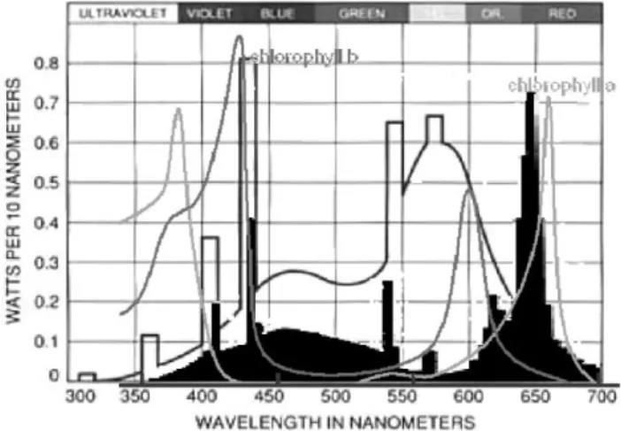
Light Sources Spectra
Gro- lux: The color tone of GL light is violet; this is the result of the combination of blue and red wave lengths. Cool-white: Broad-spectrum CW light lamps supply blue, yellow, and green light but very little far red light (Fig. 1). Light intensity was measured in the middle of the culture flask with the quantum sensor (Skye Instruments Ltd., UK SKP 200).
Evaluated Parameters and Statistical Analyses
Rooting percentages and the number of roots produced over time for each light quality and intensity were monitored. The percentages of rooted microshoots were compared by the analysis of variance (ANOVA) for clones and light sources using Statistica six Sigma. Means were compared by Duncan’s Range test.
RESULTS
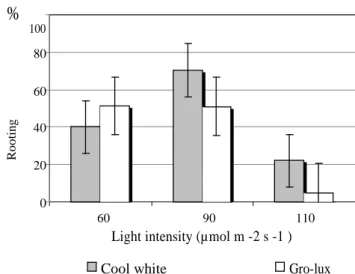
P. pinea microshoots rooted at 40% under CW lamps and at 47.1% under GL
lamps at 60 µmol m-2 s-1 (Fig. 2, Experiment 1). In order to test if increased to 90 µmol m-2 s-1 light intensity from both types of lamps would influence the rooting response; one additional lamp was added in each tissue culture chamber (Experiment 2). Since there were no statistical differences among clones in the first experiment, the data analysis in experiment 2 considered only light quality and light intensity. In the latter experiment 70.4% microshoots rooted under CW and 51% under GL lamps (Fig. 2). There was a significant difference in the rooting percentage under CW lamps at 60 versus 90 µmol m-2 s-1, but no difference was observed at the two intensities for GL lamps (Fig. 2, Table 1). This indicated that the P. pinea microshoots were not sensitive to variation in the photosynthetic photon flux (PPF) from GL lamps with respect to rooting response. When the light was increased to 110 µmol m-2 s-1 , reduction of the rooting percentages was observed for both lamp types, particularly for GL (Fig. 2).
The number of roots produced per week increased exponentially during two weeks on WPMRE medium under both types of light (Fig. 3).
DISSCUSION
During the first week of the rizhogenic process (induction) the combination of 0.12 M glucose with 10.7 μM NAA in the medium, and continuous darkness were the most favorable conditions compared with previously tested ones (unpublished results).
Rooting percentage was influenced by different light quality according to the PPF used. The fact that similar rooting percentages were obtained for GL lamps at 60 and 90 µmol m-2 s-1 indicated that an increase in light intensity did not stimulate rooting. In contrast, increased intensity of CW light increased the rooting percentage by more than 30%. This is an interesting result from the practical point of view because without any change in the tissue culture protocol, except for one additional CW lamp in the culture chamber, it was possible to obtain an increased number of rooted stone pine plantlets. However, at a higher light intensity (110 µmol m-2 s-1) ARF was inhibited under both lamps indicating that optimal conditions were exceeded.
Comparing both fluorescent lamps spectra, GL lamps emit blue and red waves and CW more yellow and green. These differences in the spectra could explain the results of this study, which are consistent with the previous observations that phytochrome was mainly involved in root formation (Tyburski and Tretyn, 1999) and that blue light inhibited photomorphogenesis (Seibert et al., 1975). The yellow component of CW lamps might have enhanced adventitious rooting in P. pinea similarly to the results of Fuerakranz et al. (1990). The authors reported that yellow light and CW lamps were superior for rooting of Prunus serotina compared with red and blue lights alone.
Maximum rooting that occurred after approximately 15 days in the expression medium and was consistent in all the treatments, indicated that the time to trigger microshoot response and growth of new roots were independent from the light quality, and that this parameter was determined by other physiological factors.
ACKNOWLEDGEMENT
This work was supported by FCT Portugal: Analysis and Mastering of Root Growth Signalling by Ectomycorrhizal Fungi on Pinus pinea L. Microshoot Cultures.
Literature Cited
Fuerakranz, H.A., Nowak, C.A. and Maynard, C.A. 1990. Light effects on in vitro adventitious root formation in axillary shoots of mature Prunus serotina. Physiol. Plant. 80:337–341.
Loreti, F., Muleo, R. and Morini, S. 1991. Effect of light quality on growth of in vitro cultured organs and tissues. Proc. Int. Plant Propagator’s Soc. 40:615–623.
Lloyd, G. and Mc Cown, B. 1981. Commercially feasible micropropagation of mountainlaurel, Kalmia latifolia, by the use of shoot tip culture. Proc. Plant Prop. Soc. 30:421–427.
Oliveira, P., Barriga, J., Cavaleiro, C., Peixe, A. and Potes. 2003. Sustained in vitro root development obtained in Pinus pinea L. inoculated with ectomycorrhizal fungi. Forestry 76:579–587.
Poudel, P.R., Kataoka, I. and Mochioka, R. 2008. Effect of red- and blue -light-emitting diodes on growth and morphogenesis of grapes. Plant Cell Tiss. Org. Cult. 92:147– 153.
Quail, P.H. 2002. Photosensory perception and signaling in plant cell: new paradigms? Current Opin. Cell Biol. 14:180–188.
Reid, D., Beall, F.D. and Pharis, R.P. 1991. Environmental cues in plant growth and development. p.65–181. In: F.C. Steward (ed.), Plant Physiology – A Treatise, Vol X. Academic Press, San Diego
Seibert, M., Wetherbee, P.J. and Job, D.D. 1975. The effects of light intensity and spectral quality on growth and shoot initiation in tobacco callus. Plant Physiol. 56:130–139. Tyburski, J. and Tretyn, A. 1999. Organogenetic response of photomorphogenic mutants
Whitelam, G.C. and Halliday, K.J. 1999. Phytochrome takes a partner! Current Biology 9:225–227.
Table
Table 1. Duncan test for variable rooting percentage.
Light Intensity GL 60, GL 90, GL 110, CW 60, CW 90, CW 110, quality 47143 45714 05000 42643 71571 16667 1 GL 60 0,8900 0,0005* 0,6830 0,0229* 0,0089* 2 GL 90 0,0007* 0,7663 0,0215* 0,0103* 3 GL 110 0,0012* 0,0000* 0,2629 4 CW 60 0,0127* 0,0161* 5 CW 90 0,0000* 6 CW 110 Figures
Fig. 1. Spectra of Sylvania Gro-lux lamps (white) and Philips Cool-white lamps (black).
% 100 80 60 R o o ti n g 40 20 0 60 90 110
Light intensity (µmol m -2 s -1 )
Cool white Gro-lux
Fig. 2. Rooting of P. pinea microshoots under different light quality and intensity. See significance in the Duncan Range test above.
20 15 ro o ts 10 of nº 5 0 0 1 2 3 4 5 6 Gro-Lux weeks Cool White
Fig. 3. Number of roots produced per week considering all microshoots for each light treatment under Cool-white and Gro-lux lamps at 60 µmol m-2 s-1.