Uncovering the role of angiogenesis in spinal cord
regeneration in zebrafish
Aluno: Benedict Paul Both
Orientadora: Doutora Ana Catarina Ribeiro
Instituto de Histologia e Biologia do Desenvolvimento
Faculdade de Medicina da Universidade de Lisboa
2015/2016
Abstract: The adult mammalian spinal cord has a very limited capacity of cellular
regeneration and functional recovery after trauma. The inefficient restoration of the vascular system following injury significantly contributes for the impaired repair of the nervous tissue. In contrast to mammals, the adult zebrafish is able to fully recover after spinal cord injury. This study aimed to explore the role of blood vessels in the successful repair of the zebrafish spinal cord. This work revealed how blood vessels in the spinal cord respond to mechanical forces imposed on the nervous tissue. After the disruption caused by the injury, the blood vessels rearrange themselves, converging to the center of the lesion. Moreover the vasculature remains considerably affected even in later time points when the zebrafish regains swimming capacity. This data suggests that even a partial repair of the vascular system is sufficient to sustain the metabolic requirements of the regenerating spinal cord. For further investigation, the role of new blood vessels in this process can be assessed by using a VEGF-R inhibitor. Put together, this knowledge might provide clues about the factors that restrict the restoration of the vasculature in the mammalian spinal cord and could be instrumental for the development of future therapies.
Abstract: A medula espinal dos mamíferos tem uma capacidade muito limitada de
regeneração celular e recuperação funcional após o trauma. A restauração ineficaz do sistema vascular após lesão contribui significativamente para a reparação limitada do tecido nervoso. Em contraste com os mamíferos, o peixe-zebra adulto é capaz de recuperar totalmente após uma lesão medular. Este estudo teve como objetivo explorar o papel dos vasos sanguíneos na reparação bem sucedida da medula espinhal do peixe-zebra. O trabalho revelou como os vasos sanguíneos da medula espinhal respondem a forças mecânicas aplicadas no tecido nervoso. Após a disrupcão causada pela lesão, os vasos sanguíneos reorganizam-se, convergindo para o centro da lesão. Além disso, a vasculatura permanece consideravelmente afectada, mesmo em tempos posteriores, quando o peixe-zebra recupera a capacidade de nadar. Estes dados sugerem que mesmo uma reparação parcial do sistema vascular é suficiente para manter os requisitos metabólicos da medula espinal em regeneração. Para uma investigação mais aprofundada, o papel de novos vasos sanguíneos no presente processo pode ser avaliada pela utilização de um inibidor de VEGF-R. Em conjunto, estes dados podem proporcionar pistas sobre os factores que impedem o
Introduction
The vertebrate spinal cord has a key role in the integration of sensory inputs and in the coordination of motor output (1). Spinal Cord Injury (SCI) in humans has a severe and permanent impact on motor and sensory function, due to the inability of the central nervous system to spontaneously regenerate and a current lack of efficient therapies.
The inefficient response of the mammalian spinal cord to injury appears to derive from two main features: the formation of a glial scar and the impaired ability to form new neurons and oligodendrocytes (2). Damage to the spinal cord results in the immediate loss of neurons and glia, and in axonal disruption. The outcome of the injury is greatly aggravated by a secondary reaction. Astrocytes accumulate at the site of the injury forming a glial scar, which constitutes a physical barrier across the spinal cord and contains axon growth inhibitors. In addition, injury triggers apoptosis of neurons and oligodendrocytes, resulting in the demyelination of spared axons.
Although a population of spinal cord cells - ependymal cells - shows stem cell properties in vitro, in vivo they generate mostly astrocytes, very few oligodendrocytes and no neurons (2). Consequently, the tissue is unable to restore all cell lineages lost with SCI and regain structural and functional integrity. An additional and critical aspect of SCI in mammals is the response of the spinal cord vascular system (3). During spinal cord injury the vascular architecture is disrupted, an event that impacts the regeneration of the spinal cord at several levels. Immediately after SCI the severed blood vessels at the injury epicenter lead to hemorrhage, exposing spared neurons to toxic substances present in the blood, thus causing more cell death. Additionally, in the vicinity of the injury blood vessels become hyperpermeable, due to compromised Blood-Brain Barrier (BBB), and allow the passage of inflammatory cells and toxic molecules that exacerbate cell death (4). Hence, the vascular system critically contributes for the secondary injury events that result in the additional loss of initially spared nervous tissue.
The disruption of the vascular system also affects other steps of spinal cord repair, namely the survival of newly formed cells. Blood vessels deliver oxygen and nutrients to cells and remove metabolic waste, therefore are crucial for the function of a highly metabolically active tissue such as the spinal cord. Following spinal cord injury the blood supply to the nervous tissue is compromised, not only due to the damaged vasculature but also to the limited regeneration of the severed blood vessels at the injury site. Although new blood vessels appear to form at different time-points post-injury in the mouse spinal cord, these vessels lack a proper BBB and may leak toxic molecules into the tissue. Moreover, as many of the new blood vessels fail to establish connection with other spinal cord cells such as
astrocytes and pericytes, the malfunctioning vessels are removed at later phases (5,6). Without the reestablishment of the spinal cord vasculature at the injury site the survival of cells in this region is compromised.
The multiple deleterious events that occur downstream of spinal cord injury entail that any successful repair therapy must comprise a combination of targets. Given the importance of the vascular system, the induction of functioning blood vessels should be one of the targets in strategies that require long-term survival of new cells, such as therapies based on the recruitment of endogenous neural stem cells towards specific lineages or cell transplantation therapies. However, it is important to bear in mind that manipulating blood vessel formation could have a dual effect, since the stimulation of angiogenesis might aid repair but the leakiness of new blood vessels could exacerbate tissue loss. In order to achieve successful functional recovery it is important to understand how this delicate balance can be maintained. Insight into this process could be obtained using zebrafish as a regeneration model.
In stark contrast with the mammalian spinal cord, whose limited regenerative attempts fail to restore function, the zebrafish spinal cord successfully regenerates. The injured spinal cord of adult zebrafish does not develop a glial scar and is able to regenerate axons and reconnect to the appropriate targets (7). In addition, the zebrafish spinal cord appears to be more permissive to the progression of neurogenesis, as new motor neurons are generated after injury from a pool of Olig2+ ependymal cells (8).
Although the cellular and molecular mechanisms underlying the successful regeneration of the zebrafish spinal cord are starting to be understood, the contribution of the vascular system to this process is still unknown. A better knowledge of the interplay between the blood vessels and the spinal cord during regeneration and how to achieve fully mature blood vessels could be instrumental for the development of viable therapies in humans. The relationship between blood vessels and the regenerating spinal cord of adult zebrafish will be addressed in this project.
The primary objective of the study is to perform a detailed characterization of the vasculature of the adult zebrafish spinal cord in control and injury conditions, using a
Tg(fli1-EGFP) transgenic line and adult zebrafish angiography to visualize blood vessels. The
analysis is performed at different times post-injury, to address different aspects: [1] early time points to reveal how the vasculature is disrupted and how the adjacent tissues are affected; [2] later time points to uncover the mechanisms involved in the formation of new blood vessels at the injury epicenter. Although some reports have described blood vessels in the zebrafish spinal cord, a thorough characterization is yet to be made.
Methods
The fli1-EGFP zebrafish transgenic line was used because it has endothelial cells labelled. The spinal cord of transgenic animals was dissected and analysed in whole-mount using confocal microscopy. The resulting data was analysed using image processing software to obtain a map of the vasculature.
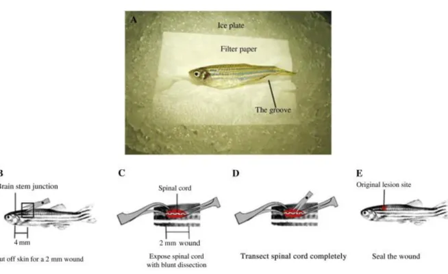
The spinal cord crush injury protocol established in the laboratory (9) (Fig. 1) was used to investigate the changes in blood vessel architecture in response to damage. To perform the injury an incision was made on the side of anesthetized fli1-EGFP zebrafish to expose the vertebral column. The spinal cord was then crushed dorsoventrally with forceps, at a level halfway between the dorsal fin and the operculum. The wound was sealed with Vetbond (3M) and the fish allowed to recover in individual tanks for up to 6 weeks following injury. As a control, a sham injury was performed in age-matched zebrafish, in which an incision was made at the side of the animal but the spinal cord will not be crushed. The vasculature of control and injured spinal cords were mapped in whole-mount.
Figure 1. Spinal cord injury protocol. A) Preparation before surgery: the fish is placed on an ice plate,
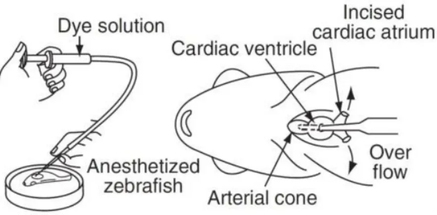
Due to a lack of sufficient fli1-EGFP transgenics, we also used wild type zebrafish to perform the spinal cord injury protocol. After the proper time of recovery we performed angiography in the wild type zebrafish injecting rhodamine dextran (10 kDa) in the cardiac ventricle (Fig. 2).
Figure 2. Angiography technique: injection of rhodamine dextran (10 kDa) in the cardiac ventricle (9).
To verify if the results obtained with angiography in wild type zebrafish are similar to the ones obtained with the transgenic line, we injected the rhodamine dextran in the cardiac ventricle of a transgenic fli1-EGFP zebrafish and saw that there is an overlap between the fluorescent endothelial cells and the rhodamine dextran. (Fig. 3)
Figure 3 Angiography in Tg(fli1:EGFP) zebrafish showing overlap between the green fluorescent endothelial cells and the rhodamine dextran (10 kDa).
The manipulated spinal cords were extracted and analysed at different time post-injury to assess the progression of regeneration: 3, 7, 10 and 21 days post-post-injury (dpi). The vasculature of control and injured spinal cords were mapped in whole mount as described above. Additional quantification of Vascular Networks was performed using a AngioTool 0.5 software (10).
Results
In the present work we wanted to study from a structural point of view the normal vascular architecture, as well as the events that occur after a spinal cord injury.
We applied the spinal cord injury protocol in two types of zebrafish: the wild type in which angiography was used to see the blood vessels and the transgenic fli1-EGFP zebrafish with labelled endothelial cells.
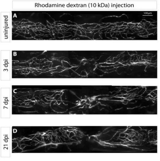
Analysing the transgenic lines, the zebrafish spinal cord in uninjured condition reveals an organized vasculature (Fig. 4A, B). In the same manner the uninjured wild type spinal cord vasculature injected with rhodamine dextran (10kDa) shows an organized pattern of blood vessels with similar density throughout the entire examined area (Fig. 5A). At the initial stage following spinal cord injury (1 dpi) there is a rupture of the blood vessels at the lesion site (Fig. 4C,D) throughout the analysed segment, both centrally and laterally. At 3 dpi the blood vessels suffer a recoil process from the injury site (Fig 4A and 5B) which is suggested by a decrease in this area of both the blood vessel area and length, compared to the anterior and posterior regions (Fig. 6A, 6B – central lesion, green bar), leaving the injured area poorly vascularized. When observing the vasculature of the spinal cord at 7 dpi the blood vessels suffer a reorganization process. They rearrange themselves converging to the injury site (Fig. 5C). Following this process the density of vessels increases at the centre of the injury (Fig. 6A-central yellow column) and decreases at the lateral sides. In contrast, the vasculature length maintains its decreased value registered at 3 dpi. At 21 dpi the vasculature still remains considerably affected (Fig. 5D) greatly differing from the uninjured organization pattern of the vessel architecture. Simultaneously, we saw that the vascular area and length did not increase from the values registered at 7 dpi (Fig. 6A, B). This data is in great contrast to the zebrafish continuous recovering swimming capabilities registered at this time point.
Figure. 4. Whole mount images of Tg(fli1:EGFP) zebrafish spinal cord at different days-post-injury. A) Central view and B) Lateral view: images of normal vasculature with organized pattern in an uninjured zebrafish spinal cord. C) Central view and D) Lateral view: images of the disrupted vasculature just after the injury (1 dpi). E) Central view and F) Lateral view: images showing recoil of the blood vessels from the injury site (3 dpi).
Figure 5. Whole-mount images of wild type zebrafish with spinal cord angiography at diferent days-post-injury. A) Normal vasculature in an uninjured wild type spinal cord. B) Disruption of the blood vessels 3 dpi. C,D) Evolution of vessels architecture at 7 and 21 dpi, respectively showing the converging blood vessels towards the centre of the lesion.
Figure 6. Quantification of the vascular network at the lesion site and at an anterior and posterior position relatively to the lesion site. A) Vascular area in the uninjured condition and at 3, 7 and 21 dpi. B) Vascular length in the uninjured condition and at 3, 7 and 21 dpi.
Discussion
This study provided the first map of the spinal cord vasculature over the course of regeneration in adult zebrafish. The analysis revealed the normal organization pattern of the vascular architecture in the uninjured condition and the changes in blood vessel caliber and density in the vicinity of the injury from very early on. The loss of motor function following spinal cord injury is accompanied by the rupture and loss of the initial organization pattern of the vessel architecture. Strikingly the poorly vascularized area remains substantially affected even at later time points when the injured fish have started to recover their swimming capabilities. Although the vessel density from later time points (7 and 21dpi) registers an increase from the 3dpi value it never approaches the initial value in the uninjured condition. There is however a reorganization process in which the blood vessels converge towards the
product of angiogenesis or merely the reorganization and anastomosis of the remaining vessels.
The image data of the compromised vessel architecture and the persistence of low structural parameters verified in later periods, despite the motor recuperation raise the hypothesis that even a partial repair of the vascular system is sufficient to sustain the metabolic requirements of the regenerating spinal cord. This raises several important questions about how does a diminished supply affect and contribute to the regeneration process and how does the cells grow despite it.
However, further work is needed to assess the functionality of these blood vessels. In the future we also plan to investigate the role of new blood vessel formation in the structural and functional recovery of the spinal cord. By using a VEGF-R inhibitor we can study how the inhibition of angiogenesis affects the functional repair of the spinal cord after an injury. Other important aspect will be uncovering the role of the vessel integrity and permeability and how does it affect the delicate balance between the deleterious inflammatory agents in the early events following injury and the delivery of mediators that stimulate cellular regeneration and differentiation.
This work has revealed new insight into the vascular system of the zebrafish spinal cord and provides a basis for further investigation aiming to understand how this organism successfully coordinates blood vessel formation and spinal cord repair.
References
(1) Jessell TM. (2000). Neuronal specification in the spinal cord: inductive signals and transcriptional codes. Nat Rev Genet 1, 20‐29.
(2) Barnabé-Heider F and Frisén J. (2008). Stem cells for spinal cord repair. Cell Stem Cell. 3(1):16-24.
(3) Oudega M. (2012). Molecular and cellular mechanisms underlying the role of blood vessels in spinal cord injury and repair. Cell Tissue Res 349:269–288.
(4) Mautes AEM, Weinzierl MR, Donovan F and Noble LJ. (2000). Vascular Events After Spinal Cord Injury: Contribution to Secondary Pathogenesis. Phys Ther 80:673-687.
(5) Casella GT, Marcillo A, Bunge MB, Wood PM. (2002). New vascular tissue rapidly replaces neural parenchyma and vessels destroyed by a contusion injury to the rat spinal cord. Exp Neurol 173:63–76.
(6) Goritz C, Dias DO, Tomilin N, Barbacid M, Shupliakov O, Frisen J. (2011). A pericyte origin of spinal cord scar tissue. Science 333:238–242.
(7) Becker T, Wullimann MF, Becker CG, Bernhardt RR and Schachner M. (1997). Axonal
regrowth after spinal cord transection in adult zebrafish. J Comp Neurol 377, 577–595. (8) Reimer MM, Sörensen I, Kuscha V, Frank RE, Liu C, Becker CG and Becker T. (2008).
Motor neuron regeneration in adult zebrafish. J. Neurosci. 28, 8510– 8516.
(9) Hui SP, Dutta A and Ghosh S. (2010). Cellular response after crush injury in adult zebrafish spinal cord. Dev. Dyn. 239(11), 2962-79.
(10) Enrique Zudaire, Laure Gambardella, Christopher Kurcz, Sonja Vermeren (2011). A Computational Tool for Quantitative Analysis of Vascular Networks. PLoS ONE | www.plosone.org 9 November 2011 | Volume 6 | Issue 11 | e27385
Special thanks
I would first like to thank professor Leonor Saude and doctor Ana Ribeiro whose help and assistance were essential to the present work. Also I would like to thank Lara, Aida and everyone in the Fish Facility for their support, friendship and availability.


