Development of an
anti-adhesive
coating using an
extracellular polymer
from a marine
cyanobacterium
Bruna Filipa Vieira Costa
Dissertação de Mestrado apresentada à
Faculdade de Ciências da Universidade do Porto em Biologia
Funcional e Biotecnologia de Plantas
2018
D ev elopmen t of an an ti-ad he siv e co ati ng us ing an ex tracell ul ar po ly mer from a mari ne cy an ob ac teri um Brun a F ilipa V iei ra Cost aMSc
FCUP ANO 2.º CICLODevelopment of an
anti-adhesive coating using an
extracellular polymer from a
marine cyanobacterium
Bruna Filipa Vieira Costa
Mestrado em Biologia Funcional e Biotecnologia de Plantas
Faculdade de Ciências da Universidade do Porto Departamento de Biologia
2017/1018
Orientador:
Fabíola Moutinho, Pós-Doutorada,
i3S-Instituto de Investigação e Inovação em Saúde, INEB- Instituto de Engenharia Biomédica,
Universidade do Porto
Coorientador:
Paula Tamagnini, Professora Associada
Faculdade de Ciências, e Líder do grupo de Bioengenharia e Microbiologia Sintética,
I3S - Instituto de Investigação e Inovação em Saúde, IBMC - Instituto de Biologia Molecular e Celular, Universidade do Porto
FCUP Development of an anti-adhesive coating using an extracellular polymer from a marine cyanobacterium
ii
Todas as correções determinadas pelo júri, e só essas, foram efetuadas. O Presidente do Júri,
FCUP Development of an anti-adhesive coating using an extracellular polymer from a marine cyanobacterium
FCUP Development of an anti-adhesive coating using an extracellular polymer from a marine cyanobacterium
iv
“
Enquanto não alcances
Não descanses.
De nenhum fruto queiras só metade.”
Miguel Torga, Diário XIII
FCUP Development of an anti-adhesive coating using an extracellular polymer from a marine cyanobacterium
FCUP Development of an anti-adhesive coating using an extracellular polymer from a marine cyanobacterium
vi
Acknowledgements
Gostaria de começar por agradecer a toda a minha família, principalmente aos meus pais e irmão, por todo o apoio, paciência e compreensão durante este último ano. Agradecer também às minhas primas Catarina, Mariana e Salomé e à minha madrinha, Flora, por todo o apoio e partilha, não só durante o desenvolvimento desta dissertação, mas também por todo o acompanhamento durante o meu crescimento, estando sempre presentes nos melhores e nos piores momentos, acreditando sempre em mim.
Além da minha família, gostaria de agradecer também às minhas colegas e companheiras de trabalho por todas as partilhas e experiências vividas, por me acompanharem todos os fins de semana e tornarem o trabalho melhor.
Às minhas amigas de sempre, em especial à Catarina Amorim, um muito obrigada por todas as conversas e por toda a motivação dada durante este percurso. Mais ainda, não poderia deixar de agradecer às minhas amigas e companheiras, Diana Moreira e Maria João Martins, por todo o percurso vivido lado a lado ao longo do mestrado, por todos os desabafos e partilha durante estes dois anos. Espero que esta amizade perdure e que os nossos futuros sejam aquilo que sempre desejamos.
Um agradecimento muito especial ao meu namorado, Frederico Ribeiro, por toda a dedicação, apoio e motivação dados ao longo de todos estes anos e principalmente nos últimos meses, um muito obrigado por toda a paciência.
Quero também agradecer à minha professora orientadora, Fabíola Moutinho, por todo o apoio e orientação prestados ao longo de toda a dissertação, assim como à professora coorientadora deste projeto, Paula Tamagnini. Agradecer também à professora Cristina Martins por toda a ajuda e por tão bem me ter recebido no grupo Bioengineered Surfaces. Agradeço também à Rita Mota por toda a ajuda prestada ao longo desta dissertação assim como por toda a troca e partilha de ideias. A todas estas pessoas, agradeço toda a partilha de conhecimento, de técnicas, supervisão, esclarecimento de dúvidas e confiança depositada, assim como por acreditarem no meu potencial para a participação em diversos projetos.
Quero também deixar uma palavra de agradecimento a todas as pessoas dos grupos Bioengineered Surfaces e Bioengineering & Synthetic Microbiology por toda a ajuda e apoio no decorrer dos trabalhos laboratoriais, por toda a partilha e por me fazerem sentir bem-vinda, sem eles não seria possível.
FCUP Development of an anti-adhesive coating using an extracellular polymer from a marine cyanobacterium
vii Agradeço também aos técnicos Dalila Pedro, Ricardo Vidal e André Maia por todas as formações, ajuda e disponibilidade ao longo do desenvolvimento desta dissertação. O meu obrigada também ao Centro de Materiais da Universidade do Porto (CEMUP), especialmente à técnica Daniela Silva.
Muito obrigado a todos. Bruna Costa
Financial acknowledgements
This work was financed by FEDER - Fundo Europeu de Desenvolvimento Regional funds through the COMPETE 2020 - Operacional Programme for Competitiveness and Internationalisation (POCI), Portugal 2020, and by Portuguese funds through FCT - Fundação para a Ciência e a Tecnologia/Ministério da Ciência, Tecnologia e Ensino Superior in the framework of the project "Institute for Research and Innovation in Health Sciences" (POCI-01-0145-007274), by the project NORTE-01-0145- FEDER-000012, Structured Programme on Bioengineering Therapies for Infectious Diseases and Tissue Regeneration, supported by Norte Portugal Regional Operational Programme (NORTE 2020), under the PORTUGAL 2020 Partnership Agreement, through the European Regional Development Fund (ERDF), POCI-01-0145-FEDER-028879 and the AntiBioCoat project (Anti-adhesive Biopolymer Coating) - RESOLVE 2016: Technology Transfer Ignition Program in the Health Sector, Portugal (NORTE2020) (NORTE-01-0246- FEDER-000018-RESOLVE-55536).
FCUP Development of an anti-adhesive coating using an extracellular polymer from a marine cyanobacterium
viii
Resumo
Os dispositivos médicos são amplamente utilizados na medicina moderna para melhorar a saúde geral do doente bem como a sua qualidade de vida, evitando muitas vezes uma morte precoce. A aplicação de dispositivos médicos induz, contudo, uma reação ao corpo estranho, o que prejudica a resposta imunológica normal, afeta o fluxo sanguíneo, altera o microambiente local, possibilitando a contaminação bacteriana, que pode originar uma infecção grave. A maioria das infeções relacionadas com dispositivos médicos é causada por biofilmes bacterianos que são difíceis de erradicar. Os tratamentos atuais provaram ser insuficientes, para além de serem tóxicos e de poderem promover resistência bacteriana. Logo, são necessárias novas estratégias para prevenir a infeção, nomeadamente evitando os primeiros passos da colonização bacteriana e consequente formação de biofilme.
Neste trabalho, um polímero extracelular produzido por uma cianobactéria unicelular marinha, Cyanothece sp. CCY 0110, foi utilizado para o desenvolvimento de um revestimento antiaderente, vocacionado para materiais usados na produção de cateteres. Neste âmbito, foram utilizados diferentes parâmetros para avaliar o crescimento da cianobactéria e foi otimizado o método de extração do polímero que foi posteriormente utilizado como revestimento. Este revestimento (revestimento RPS) foi caracterizado por diferentes técnicas, nomeadamente goniometria (molhabilidade), elipsometria (espessura) e espectroscopia de fotoeletrões excitados por raios X (composição elementar). O revestimento RPS foi avaliado em termos de sua eficácia antiaderente contra agentes etiológicos relevantes, nomeadamente Staphylococcus
aureus, S. epidermidis, Pseudomonas aeruginosa e Escherichia coli usando normas
internacionais (ISO) (ISO 22196). Foram também avaliadas a degradabilidade (ISO 10993-13), biocompatibilidade (ISO 10993-5) e trombogenicidade (ISO 10993-4) deste revestimento. O revestimento RPS possui uma espessura reduzida e características hidrofílicas. Este revestimento manteve-se estável durante 30 dias sob condições de armazenamento padrão, e preveniu a adesão bacteriana em mais de 90%. Após os ensaios de degradação foram observadas algumas alterações nas características da superfície do revestimento, mas a sua eficácia antiaderente não sofreu alterações significativas. Este revestimento parece não ter um efeito citotóxico em fibroblastos L929. Para além disso, também preveniu a adesão e ativação de plaquetas, sendo por isso um bom candidato para aplicações em que seja necessário o contato com sangue.
O desenvolvimento deste revestimento antiaderente é um passo importante para o estabelecimento de uma nova tecnologia capaz de prevenir infeções associadas a dispositivos médicos.
FCUP Development of an anti-adhesive coating using an extracellular polymer from a marine cyanobacterium
ix
Palavras-chave: Revestimento antiaderente, Cianobactérias, Cyanothece sp., Polímero extracelular, Infeções associadas a dispositivos médicos.
FCUP Development of an anti-adhesive coating using an extracellular polymer from a marine cyanobacterium
x
Abstract
Medical devices are widely used in modern medicine improving overall health and quality of patient’s life and often acting as lifesavers. However, medical device application induces a foreign body reaction that impairs normal immunological response, affecting blood flow, altering local microenvironment and enabling bacterial contamination originating infection. Most device-related infections are caused by biofilms that are very difficult to eradicate. Current treatments have proven to be insufficient, often beingtoxic and sometimes even promoting bacterial resistance. Therefore, new strategies are needed to prevent infection, namely the control of the first step of bacterial colonization and consequent biofilm formation.
In this work, an extracellular polymer, produced by the marine unicellular cyanobacterium Cyanothece sp. CCY 0110 was explored for the development of an anti-adhesive coating, specificallymeant for catheter-related materials. For this purpose, different parameters were used to evaluate the cyanobacterium growth, and its polymer was extracted to subsequently be applied as a coating. This coating (RPS-coating) was characterized by different surface techniques, namely goniometry (wettability), ellipsometry (thickness) and X-ray photoelectron spectroscopy (elemental composition before and after degradation assays). The RPS-coating was evaluated using international ISO standards in terms of its bacterial anti-adhesive efficiency against relevant etiological agents, namely Staphylococcus aureus, S. epidermidis,
Pseudomonas aeruginosa and Escherichia coli (ISO 22196). The coating degradability
(ISO 10993-13), biocompatibility (ISO 10993-5) and thrombogenicity (ISO 10993-4) were also evaluated. The RPS-coating revealed to have a low thickness (nanorange) and higher hydrophilic properties. Moreover, the coating was stable during 30 days under standard storage conditions Importantly, RPS-coating prevented bacterial adhesion in more than 90%, for all tested bacteria. The degradation assay promoted some changes in the surface characteristics parameters, but the coating anti-adhesive efficiency was not substantially altered. In addition, our results showed a non-cytotoxic effect of the coating towards L929 fibroblast cells. Furthermore, the RPS-coating also prevented platelet adhesion and activation, being therefore a candidate for blood-contacting applications.
The development of this anti-adhesive coating is an important step towards the establishment of a new platform technology capable of preventing medical device associated-infections.
FCUP Development of an anti-adhesive coating using an extracellular polymer from a marine cyanobacterium
xi
Keywords: Anti-adhesive coating, Cyanobacteria, Cyanothece sp., Extracellular
FCUP Development of an anti-adhesive coating using an extracellular polymer from a marine cyanobacterium
xii
Table of Contents
Acknowledgments vi Resumo viii Abstract x
Table of Contents xii
List of figures xiv
List of abbreviations xviii
1. Introduction 1
1.1. Medical devices associated-infections 1
1.2. Biofilm formation 2
1.3. Preventive care guidelines, treatments and new solutions 3
1.4. Cyanobacteria and its extracellular polymeric substances 6
1.5. Aims of this study 8
2. Materials and Methods 9
2.1. Organism and culture conditions 9
2.2. Light microscopy 9
2.3. Growth measurements 9
2.4. Determination of total carbohydrate and RPS contents 10
2.5. Isolation of Cyanothece’s RPS 10
2.6. Development of RPS-based coating 11
2.6.1. Substrate preparation 11
2.6.2. Substrate activation 11
2.6.3. Substrate coating 11
2.6.3.1. Preparation of the polyurethane (PU) solution 11
2.6.3.2. Preparation of the RPS solution 11
2.6.3.3. Coating production by spin-coating 12
2.7. Surface characterization 12
2.7.1. Ellipsometry 12
2.7.2. Contact Angle Measurement 13
2.7.3. X-ray Photoelectron Spectroscopy (XPS) 13
2.8 RPS-coating performance evaluation 13
2.8.1. Anti-adhesive Assays 13
2.8.1.1. Bacterial strains, media and growth conditions 13
FCUP Development of an anti-adhesive coating using an extracellular polymer from a marine cyanobacterium
xiii
2.8.1.3. Bacterial adhesion assays 14
2.8.2. RPS-coating biocompatibility evaluation 15
2.8.3. Thrombogenicity assay 16
2.8.3.1 Scanning Electron Microscopy (SEM) 16
2.8.4. RPS-coating stability assay 16
2.8.5. RPS-coating degradation assay 16
2.9 Statistical Analysis 17
3. Results and Discussion 19
3.1. Growth of Cyanothece sp. CCY 0110 19
3.2. Total carbohydrates and RPS production by Cyanothece sp. CCY 0110 21
3.3. Development of RPS-based coating 23
3.4. Coating surface characterization 24
3.4.1. Ellipsometry 24
3.4.2. Water contact mesurements 25
3.5. Anti-adhesive activity 26
3.6. Biocompatibility assessment of RPS-coating 31
3.7. Platelets adhesion and activation 32
3.8. Stability assay 34
3.9. Degradation assay 35
4. Conclusions 41
5. Future Perspectives 43
FCUP Development of an anti-adhesive coating using an extracellular polymer from a marine cyanobacterium
xiv
List of figures
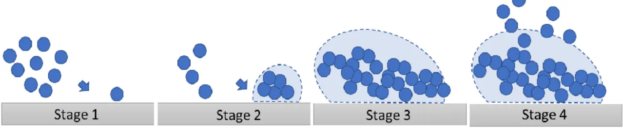
Fig. 1: The four stages of biofilm formation. Stage 1- reversible attachment of bacteria
to the surface; Stage 2- microcolonies formation; Stage 3- biofilm structuring and maturation and Stage 4- biofilm dispersal (risk of infection spreading). Adapted from Salwiczek et al. 2014. 3
Fig. 2: Strategies for coating development. A1- Contact-killing, A2 Antimicrobial -releasing, B- Anti-adhesive. 4
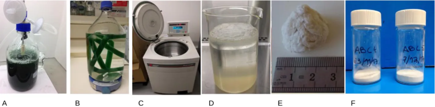
Fig. 3: Representative steps of the isolation of the extracellular polymer from a
Cyanothece sp. CCY 0110 culture. A- Cyanothece sp. culture growing in a 2 L
bioreactor; B- Dialysis; C- Centrifugation; D- RPS precipitation with ethanol; E- Polymer after lyophilization; F- Grounded polymer. 10
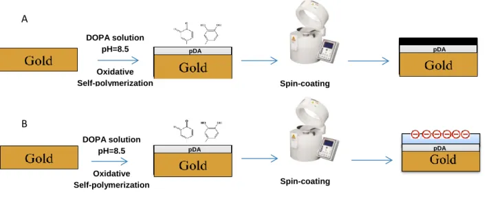
Fig. 4: Squematic representation of the steps involved in the production of PU and RPS
ultrathin coating films. A- PU-coating production; B- RPS-coating production. DOPA- dopamine; pDA- polydopamine. 12
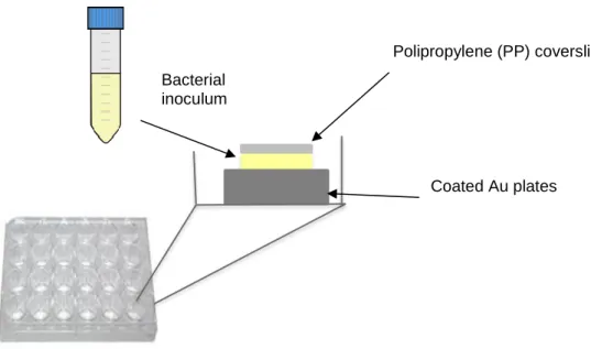
Fig. 5: Squematic representation of the protocol used to perform the anti-adhesive
assays. TSB (tryptic soy broth) was used to prepare bacterial inoculum with or without plasma proteins (1% v/v). 14
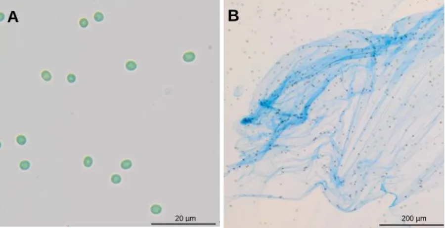
Fig. 6: Light micrographs of Cyanothece sp. CCY 0110. A- without stain; B- stained with
Alcian Blue highlighting the polysaccharides released into culture medium. 19
Fig. 7: Cultures of Cyanothece sp. CCY 0110 in 1 L (left) and 2 L bioreactors (right).
Cultures were grown in ASNIII medium, with a 16 h light (30 µE/m2/s)/8 h dark regimen, at 25 ºC with agitation (150 rpm) and aeration (1.2 L/min). 20
Fig. 8: Growth of Cyanothece sp. CCY 0110 expressed as OD730 nm (blue), mg of
chlorophyll a per L of culture (green) and mg dry weight per L of culture (black). Cultures were grown in 2 L bioreactors with ASNIII medium, a 16 h light (30 µE/ m2/s)/8 h dark regimen, at 25 ºC with aeration (1.2 L/min). The values are means ± standard deviations (n = 3). 20
Fig. 9: Total Carbohydrates (blue) and RPS (red) expressed as mg per L of culture of
FCUP Development of an anti-adhesive coating using an extracellular polymer from a marine cyanobacterium
xv medium, a 16 h light (30 µE/ m2/s)/8 h dark regimen, 25 ºC and aeration (1.2 L/min). The values are means ± standard deviations (n = 3). 22
Fig. 10: A- Total Carbohydrates (blue) and RPS (red) expressed as mg of carbohydrates
per mg of chlorophyll a. B- Total Carbohydrates (blue) and RPS (red) expressed as mg of carbohydrates per mg of DW. Cultures of Cyanothece sp. CCY 0110 were grown in 2 L bioreactors with ASNIII medium, a 16 h light (30 µE/m2/s)/8 h dark regimen, 25 ºC and aeration (1.2 L/min). The values are means ± standard deviations (n = 3). 22
Fig. 11: Surface characterization of the coated samples with pDA layer, PU-coating and
RPS-coating by ellipsometry (n = 9). Statistic analysis were performed by Non-parametric Kruskal Wallis analysis and statistic differences are indicated with *** (p < 0.005). 24
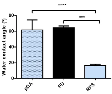
Fig. 12: Surface characterization of the coated samples with pDA layer, PU-coating and
RPS-coating by water contact angle (captive bubble method) (n = 9). Statistic analysis were performed by Non-parametric Kruskal Wallis analysis and statistic differences are indicated with *** (p < 0.005) and **** (p < 0.001). 25
Fig. 13: Adhesion of different bacterial strains after 24 h incubation at 37 °C to
PU-coating (PU applied above pDA-coated Au plates) (control) and RPS-PU-coating (RPS applied above pDA-coated Au plates) (n = 9). Black bars represent bacteria adhered in absence of plasma and blue bars represents adhered bacteria in presence of plasma (1% v/v). A- Staphylococcus epidermidis; B- Staphylococcus
aureus; C- Pseudomonas aeruginosa; D- Escherichia coli (plasma proteins were
not used with this strain). Statistic analysis were performed by Non-parametric Kruskal Wallis analysis and statistic differences are indicated with **** (p < 0.001)
27
Fig. 14: Adhered bacteria to PU-coating (PU applied above pDA-coated Au plates) (left)
and RPS-coating (RPS applied above pDA-coated Au plates) (right) coatings in absence of plasma proteins. PU coated surfaces were used as controls. Magnification: 20x Scale bar: 100 µm. A- Staphylococcus epidermidis; B-
Staphylococcus aureus; C- Pseudomonas aeruginosa; D- Escherichia coli. 29
Fig. 15: Metabolic activity of L929 fibroblasts assessed after 24 h with PU and RPS
FCUP Development of an anti-adhesive coating using an extracellular polymer from a marine cyanobacterium
xvi Essential Media) by the MTT assay as a function of the % of positive control (MEM + L929) (n = 12). 31
Fig. 16: Representative SEM micrographs of platelets adhered to PU-coating
categorized by activation state: A-Non- activated, B- Partial activated and C- Fully activated (magnification: A- 15 000×; B- 10 000x; C- 10 000 x). 32
Fig. 17: Number of adhered platelets to PU-coating (PU applied above pDA-coated Au
plates) and RPS-coating (RPS applied above pDA-coated Au plates) per µm2 after 30 min of incubation at 37 °C in presence or absence of plasma proteins (1% v/v) (n = 9). Statistic analysis were performed by Non-parametric Kruskal Wallis analysis and differences are indicated with **** (p < 0.001). 33
Fig. 18: Number of adhered Escherichia coli per mm2 after 24 h of incubation at 37 °C on PU-coating (PU applied above pDA-coated Au plates) (control), RPS-coating (RPS applied above pDA-coated Au plates) and RPS-coating after 30 days of storage at RT (n = 6). All conditions were performed in the absence of plasma. Statistic analysis were performed by Non-parametric Kruskal Wallis analysis and statistic differences are indicated with **** (p < 0.001). 35
Fig. 19: Surface characterization of RPS-coating and RPS-coating after exposure of
temperature (45 ºC) and two different buffers using ellipsometry (n = 9). A- Surface characterization of coating with pH 5.0; B- Surface characterization of RPS-coating with pH 7.4. Statistic analysis were performed by Non-parametric Kruskal Wallis analysis and statistic differences are indicated with * (p < 0.05). RPS (Control)- RPS-coating at RT without buffer, Day 7 45 ºC- RPS-coating 7 days incubation at 45 ºC without buffer, Day 7 45 ºC pH 5.0- RPS-coating 7 days incubation at 45 ºC in acetate buffer (pH 5.0) and Day 7 45 ºC pH 7.4- RPS-coating 7 days incubation at 45 ºC in PBS (pH 7.4). 36
Fig. 20: Surface characterization of RPS-coating and RPS-coating after exposure of
temperature (45 ºC) and two different buffers using contact angle measurements (n = 9). A- Surface characterization of RPS-coating with pH 5.0; B- Surface characterization of RPS-coating with pH 7.4. Statistic analysis were performed by non-parametric Kruskal Wallis analysis and statistic differences are indicated with ** (p < 0.01). RPS (Control)- coating at RT without buffer, Day 7 45 ºC- coating 7 days incubation at 45 ºC without buffer), Day 7 45 ºC pH 5.0- RPS-coating 7 days incubation at 45 ºC in acetate buffer (pH 5.0) and Day 7 45 ºC pH 7.4 - RPS-coating 7 days incubation at 45 ºC in PBS (pH 7.4). 37
FCUP Development of an anti-adhesive coating using an extracellular polymer from a marine cyanobacterium
xvii
Fig. 21:Elemental chemical composition by XPS of RPS-coating and the various conditions after degradation assay (n = 3). RPS (Control): RPS-coating at RT without buffer, Day 7 45 ºC (RPS-coating 7 days incubation at 45 ºC without buffer), Day 7 45 ºC pH 5.0 (RPS-coating 7 days incubation at 45 ºC in acetate buffer (pH 5.0)) and Day 7 45 ºC pH 7.4 (RPS-coating 7 days incubation at 45 ºC in PBS (pH 7.4)). 38
Fig. 22: Monosaccharides composition (molar %) of samples exposed to pH 5.0 or pH
7.4 and 45 ºC of temperature during 7 days detected by Gas Chromatography. Mannose (Man), glucose (Glc); galactose (Gal); xylose (Xyl); arabinose (Ara); rhamnose (Rha); fucose (Fuc). Day 7 45 ºC pH 5.0 (RPS-coating 7 days incubation at 45 ºC in acetate buffer (pH 5.0)) and Day 7 45 ºC pH 7.4 (RPS-coating 7 days
FCUP Development of an anti-adhesive coating using an extracellular polymer from a marine cyanobacterium
xviii
List of Abbreviations
ASN - Artificial sea water
ATCC - American Type Culture Collection CCY - Culture Collection of Yerseke
CEMUP - Centro de Materiais da Universidade do Porto CFU - Colony forming units
Chl a - Chlorophyll a
DAPI - 4',6-diamidino-2-phenylindole DMSO - DimethylSulfoxide
DOPA - Dopamine DW - Dry weight
EPS - Extracellular polymeric substances
GC FID - Gas Chromatography Flame Ionization Detector GC - Gas chromatography
ISO - International Organization for Standardization MEM - Minimum Essential Media
MTT - 3-(4,5-dimethylthiazol-2-yl)-2,5-diphenyltetrazoliumbromid OCA - Optical contact angle
OD - Optical density
PBS - Phosphate-buffered saline PC - Phosphorylcholine
pDA - Polydopamine PEG - Polyethylene glycol PEO - Poly (ethylene oxide) PP - Polypropylene
PU - Polyurethane
FCUP Development of an anti-adhesive coating using an extracellular polymer from a marine cyanobacterium
xix RPS - Released polysaccharides
RT - Room temperature
SEM - Scanning Electron Microscopy THF - Tetrahydrofuran
TrisHCl - Tris Hydrochloride TSA - Tryptic soy agar TSB - Tryptic soy broth
FCUP Development of an anti-adhesive coating using an extracellular polymer from a marine cyanobacterium
1
1.Introduction
1.1 Medical devices associated-infections
Human life expectancy has increased in last years, and as people live longer, there is an increased demand for medical devices. Medical devices are used to replace missing anatomic structures, improve therapeutic outcomes, optimize appearance or replace the normal physiological functions of the aging human body, prolonging the life and well-being of people around the world (Baveja et al., 2004). Modern devices are used to solve different problems and well-known examples of the devices are heart valves, ventricular assist devices, orthopedic-prostheses, dental implants, drug-eluting stents, central venous and urinary catheters (Cooper, 2015; Neoh et al., 2017). Catheters, in particular, are a ubiquitous and indispensable component inside medical practice (Neoh et al., 2017). However, despite the state-of-the-art technology, medical devices are still greatly susceptible to bacterial contamination (Percival et al., 2015). Medical devices insertion induces a foreign body reaction that impairs normal immunological response and affects blood flow, resulting in local microenvironment modification, enabling bacterial contamination and the establishment of biofilms that can lead to infection (Moraes et al., 2013). The development of medical device associated-infections are influenced by multiple factors, including patient variables (e.g. severity of the illness and overall health status), patient care variables (e.g. antibiotic use, invasive medical device use) and also by the virulence factors of microorganism (Collins, 2008; Moraes et al., 2013). Despite medical devices, such as catheters, being supplied as sterile devices, the moment the packages are opened, and the device is handled and inserted in to a patient, it becomes exposed to the microorganisms from the skin flora of the host, the physician or the hospital environment itself. Moreover, if the patient has an infection in another body area, the bacteria can migrate and develop a new infection around the medical device (Salwiczek et al., 2014). This issue represents a high human and economic burden with an annual mortality of around 100 000 deaths per year in USA alone (Salwiczek et al., 2014). Statistics show that approximately 60% of medical device-associated infections worldwide are attributed to the formation of biofilms (Percival et al., 2015), with about 13 to 39 million of bladder inserted catheters and 150 000 to 400 000 central venous catheters being contaminated each year (Darouiche, 2001).
FCUP Development of an anti-adhesive coating using an extracellular polymer from a marine cyanobacterium
2
1.2 Biofilm formation
Biofilm can be defined as “a microbially derived sessile community characterized by cells that are irreversibly attached to a substratum, interface or to each other; are embedded in a matrix of extracellular polymeric substances that they have produced and exhibit an altered phenotype with respect to growth rate and gene transcription” (Donlan, 2002).
The formation of biofilms on medical device surfaces is a well-characterized process and can be divided into four different stages (Salwiczek et al., 2014), as depicted in Fig.1:
(i) Attachment and monolayer formation: When a medical device is introduced in a body proteins, electrolytes, and other organic molecules deposit on its surface resulting in the formation of a conditioning film that enables bacterial adhesion (Raad and Hanna, 2002). At this point, the adhesion is reversible and the host immune defenses, and/or the use of antimicrobials, can prevent the development of the infection (Moraes et al., 2013; Salwiczek et al., 2014).
(ii) Formation of microcolonies: Irreversible attachment starts, mediated by bacterial surface polymeric substances and/or structures, including capsules, fimbriae, pili and slime (Ribeiro et al., 2012). Within the monolayer, bacteria multiply locally, produce exopolymers and form aggregates, allowing the colonization by other microorganisms. (iii) Maturation and structuring: Development into a mature biofilm consisting of bacterial macrocolonies that eventually converge, encased by extracellular polymeric substances (EPS) (Percival et al., 2010). This complex matrix protects individual bacterial cells from hostile factors such as immunological responses, nutrient limitations and even antibacterial agents (Singha et al., 2017).
(iv) Detachment and return to the planktonic mode of growth: Parts of the biofilm detach from the original spot and colonize new sites (biofilm dispersal), constituting a severe threat to the host (Donlan, 2002). Microbe are translocated through the bloodstream with a risk of spreading the infection, particularly if the host immune response is compromised (Donlan, 2002).
FCUP Development of an anti-adhesive coating using an extracellular polymer from a marine cyanobacterium
3
Fig. 1: The four stages of biofilm formation. Stage 1- reversible attachment of bacteria to the surface; Stage 2- microcolonies formation; Stage 3- biofilm structuring and maturation and Stage 4- biofilm dispersal (risk of infection spreading). Adapted from Salwiczek et al. 2014.
It has been described that biofilm community provide the pathogens with a unique and enclosed environment that is optimal for the exchange of genetic data via plasmids. These plasmids may contain genetic information related to multidrug resistance, with inter-species spread increasing the threat of antimicrobial resistance (Francolini and Donelli, 2010).
The most prevalent microorganisms in medical devices-related biofilms are the positive Staphylococcus aureus, S. epidermidis and Enterococcus spp., the Gram-negative Pseudomonas aeruginosa and Escherichia coli, and the fungi Candida spp. (Kojic and Darouiche, 2004). The development of infections in or around the indwelling devices continues to be a key issue that hinders their utilization, particularly in critically ill patients (Trautner and Darouiche, 2004).
1.3 Preventive care guidelines, treatments and new solutions
Nowadays, standard guidelines for the prevention of indwelling medical device infections includes (i) good hand hygiene, (ii) chlorhexidine skin antisepsis, (iii) prophylactic systemic antibiotic administration, and (iv) removal of the device frequently and immediately when it is no longer needed (Singha et al., 2017). Although these prevention guidelines are being continuously refined, the infection prevalence has not been significantly reduced and is still being fighted by the use of antibiotics. Antibiotic-resistant microorganisms are considered one of the most serious health threats around the world (Brooks and Brooks, 2014). By 2050, it is estimated that antibiotic resistance will kill 10 million people every year (Munita and Arias, 2016). Moreover, current antibiotic therapies are losing their efficacy and not enough new and effective antibiotics are being developed to combat this threat (WHO 2014). The loss of effectiveness of antibiotics will weaken our ability to fight infections and to manage the infections complications in
FCUP Development of an anti-adhesive coating using an extracellular polymer from a marine cyanobacterium
4 vulnerable patients. Therefore, there is an urgent need of antibiotic-free solutions to fight infection establishment (Rather et al., 2017).
The development of new materials or surface coatings that prevent viable bacteria from adhering to medical devices has emerged as a new line of research through different promising strategies (Cloutier et al., 2015; Swartjes et al., 2015). These strategies can be classified as antimicrobial (Fig. 2A1, A2) or anti-adhesive (Fig. 2B) coating strategies. Antimicrobial strategies can be further classified as contact-killing (Fig. 2A1) or antimicrobial-releasing (Fig. 2A2) (Sileika et al., 2011).
Fig. 2: Strategies for coating development. A1- Contact-killing, A2- Antimicrobial-releasing, B- Anti-adhesive.
In the contact-killing strategies (Fig. 2A1), antimicrobials (antibiotics, disinfectants, metals such as silver and copper) are applied to the coating to attribute bactericidal properties to the surface of the device (Rio et al., 2012). In these coatings, antimicrobials are directly immobilized on the surfaces to target bacteria that try to adhere, being expected to provide long-term effects (Neoh et al., 2017). The drawback associated with contact-killing coatings is related to the possible cytotoxicity promoted by the bactericidal agents used (Heidenau et al., 2005) and by the accumulation of death bacteria at the surface, which enables subsequent viable bacteria anchorage (Siedenbiedel and Tiller, 2012).
In the antimicrobial-releasing strategies (Fig. 2A2), the antimicrobials applied in the coating are released over time, killing bacteria or limiting their growth both on the surface of the coating and in its vicinity (Neoh et al., 2017). Over the past decades, a broad range of antibacterial compounds have been developed for release-based
A1 A2 B
: Live bacteria, : Dead bacteria, : Covalent immobilized antimicrobials, : Leachable antimicrobials, : Anti-adhesive polymers
FCUP Development of an anti-adhesive coating using an extracellular polymer from a marine cyanobacterium
5 systems, including antiseptics (e.g. chlorhexidine and triclosan), antibiotics (e.g. rifampicin, nitrofurazone and minocycline), silver (ions and nanoparticles) and nitric oxide (NO) (Cloutier et al., 2015; Neoh et al., 2017). These leachable coatings have inherently limited reservoirs of antibacterial agents, and their action is only temporary (Cloutier et
al., 2015). These coatings have also poor control over the drug release from the medical
device surface. Moreover, as this release may not be sufficient to prevent infection, it may contribute to survival of antimicrobial-resistant strains (Qian et al., 2010).
The anti-adhesive strategies (Fig. 2B) are focused on the prevention of the first step of bacterial colonization, and consequently block biofilm formation. Since the adhesion of bacteria to surfaces is a prerequisite for the formation of biofilm, this strategy represents a promising alternative to inhibit infection establishment without introducing antimicrobials into the equation (Donlan and Costerton, 2002). Anti-adhesive surfaces do not kill bacteria, however, since they block biofilm formation, microorganisms are not protected by the matrix, being more susceptible and exposed to environmental factors and to immune system (Swartjes et al., 2015).
Many strategies have been proposed to develop anti-adhesive surfaces, namely through modification of physico-chemical parameters of the surface (e.g., topography, charge, wettability), consisting namely on the chemically modification of the surface to create negatively charged or hydrophilic surfaces to form anti-adhesive surfaces (Junter
et al., 2016).
In the design of anti-adhesive surfaces, surfactants, neutral polymers, anionic polymers and zwitterionic polymers can be used (Campoccia et al., 2013; Junter et al., 2016). Polymers widely investigated as anti-adhesive coatings are polyethylene glycol
(PEG) and its derivatives, poly(ethylene oxide) (PEO), polyacrylamide,
phosphorylcholine (PC)-based polymers and trimethylsilane (Leckband et al., 1999; Cringus-Fundeanu et al., 2007; Fundeanu et al., 2008; Ma et al., 2012). Besides these synthetic polymers, some natural polymers have also shown promising characteristics that can inhibit bacterial adhesion (Neoh et al., 2017). Development of natural anti-adhesive surfaces include some polysaccharides such as hyaluronic acid and heparin and proteins such as casein and albumin (Morra and Cassineli, 1999; Junter et al., 2016). Other natural polymers produced by bacteria and algae, particularly extracellular polymeric substances (EPS), like alginate, ulvan, agarose, carrageenans and dextran, have also shown anti-adhesive properties against bacterial cells (Elsabee et al., 2008; Jain et al., 2009; Gadenne et al., 2013; Junter et al., 2016).
FCUP Development of an anti-adhesive coating using an extracellular polymer from a marine cyanobacterium
6
1.4 Cyanobacteria and its extracellular polymeric substances
Cyanobacteria are an ancient group of prokaryotes with the ability to perform oxygenic photosynthesis, being important primary producers in many ecosystems (Whitton and Potts, 2000). A large number of strains have also the ability to fix atmospheric nitrogen (N2), therefore cyanobacteria can be considered as the microbial group with the simplest nutritional requirements (Bergman et al., 2008). Owing to their ecological and biochemical diversity, and their ability to grow autotrophically making their cultivation simple and cost-effective, cyanobacteria have attracted an increasing interest of a wide range of industries that use these organisms as source of biomass, polysaccharides, fatty acids, pigments and bioactive secondary metabolites (Karl and Cyril, 2008; Gangl et al., 2015). Moreover, like a wide range of other microorganisms, cyanobacteria are able to synthesize and secrete extracellular polymeric substances (EPS) mainly composed of heteropolysaccharides (but that can also contain proteins, nucleic acids and lipids). These EPS can remain associated with the cell surface as sheaths, capsules and/or slimes, or be released into the surrounding environment as released polysaccharides (RPS). RPS can be derived from sheaths, capsules and slimes or from a divergent biosynthetic process (De Philippis and Vincenzini, 1998; Pereira et
al., 2009). Since, RPS are released into the medium, they can be easily recovered and
purified (De Philippis and Vincenzini, 1998; More et al., 2014).
Cyanobacterial EPS have distinctive characteristics compared to those produced by other microorganisms, as they contain up to 15 different monomers, have a strong anionic nature due to the presence of two different uronic acids and sulfate groups, and have high hydrophobicity conferred by the presence of ester-linked acetyl groups, peptidic moieties and deoxysugars (Pereira et al., 2009; Rossi and De Philippis, 2016). The EPS can have distinct roles, depending on their structure and complexity. They have been reported to be involved in the formation of biofilms, nutrient sequestration and cell protection against desiccation, ultraviolet radiation, elevated salt concentration, predation and external agents such as antibiotics and toxic metal cations (Flemming and Wingender, 2010; Kehr and Dittmann, 2015; Delattre et al., 2016).
Nowadays, cyanobacterial EPS represent interesting alternatives to the plant and macroalgae exopolysaccharides traditionally used in the food, textile, painting, cosmetic, paper, pharmaceutical and oil industries (Li et al., 2001). Recently, cyanobacterial EPS have been extensively studied, and their monosaccharide composition, structure and functional properties reported (Rossi and De Philippis, 2016). Moreover, some activities and/or applications in biomedicine are described in literature, such as anticancer, antiviral, anticoagulant, antimicrobial and antifungal activities (Rechter et al., 2006;
FCUP Development of an anti-adhesive coating using an extracellular polymer from a marine cyanobacterium
7 Majdoub et al., 2009; Challouf et al., 2011; Najdenski et al., 2013; Ou et al., 2014). Furthermore, some studies have also shown that these polysaccharides have anti-adhesive properties (Rendueles et al., 2011) being used for biofilm reduction (Valle et
al., 2006). The polysaccharides produced by the cyanobacterial strains Spirulina, Cyanothece spp. and Cyanospira capsulata have been shown to prevent Helicobacter pylori adhesion to the gastric mucosa (Ascencio et al., 2004; Loke et al., 2007).
Among EPS-producing cyanobacteria, several members of the Cyanothece genus are described as strong EPS producers (De Philippis et al., 1998). Particularly,
Cyanothece sp. CCY 0110 (hereafter Cyanothece) is a marine N2-fixing unicellular cyanobacterium isolated from coastal waters of Zanzibar, and is a highly efficient EPS producer, releasing most of the polymer to the culture medium (Mota et al., 2013). This high EPS-production is probably due to the need of protection from the harsh characteristics of the isolation site, namely from the salinity, high temperature and/or UV light (Mota, 2017).Cyanothece extracellular polymer is composed by nine different
monosaccharides: the hexoses mannose, glucose and galactose; the pentoses xylose and arabinose; the deoxyhexoses rhamnose and fucose; and the acidic hexoses galacturonic and glucuronic acids (Mota et al., 2013). It has a sulphate content around 11% and a peptidic content around 4% (Mota, 2017). This polymer is also remarkably thermostable and mainly of amorphous nature. In addition, RPS produced by
Cyanothece presents a high molecular weight and is soluble in water (Mota, 2017). Cyanothece’s RPS have been previously tested for metal bioremediation and as
agent for controlled drug delivery (Mota et al., 2016; Leite et al., 2017). Regarding the potential as biosorbent, Cyanothece’s RPS was capable of efficiently remove the most common heavy metal pollutants in water bodies - copper, cadmium and lead, especially due to overall negative charge of the polymer that confers a high affinity towards metal cations (Mota et al., 2016). Leite et al., showed that this polymer is a good candidate for the delivery of therapeutic macromolecules, such as BSA (bovine serum albumin), and lysozyme, and that it is not toxic to human dermal neonatal fibroblasts in vitro (Leite
FCUP Development of an anti-adhesive coating using an extracellular polymer from a marine cyanobacterium
8
1.5 Aims of this study
The main goal of this study was the development of an anti-adhesive coating based on a natural extracellular polymer produced by a marine cyanobacterium. This work exploits the unique characteristics of the cyanobacterial biopolymer to decrease bacterial adhesion to surfaces, and in the future be applied to medical devices, such as catheters, envisaging the decreasing of the rate of indwelling medical device-associated infections.
FCUP Development of an anti-adhesive coating using an extracellular polymer from a marine cyanobacterium
9
2. Materials and Methods
2.1 Organism and culture conditions
The unicellular cyanobacterium Cyanothece sp. CCY 0110 (kindly provided by Lucas Stal; Culture Collection of Yerseke, The Netherlands, now available at Culture Collection of Algae and Protozoa (CCAP 1435/2) was grown in 1 L and 2 L bioreactors with ASNIII medium (Rippka et al., 1979), at 25 °C, under a 16 h light (30 μE/m2/s)/8 h dark regimen, with orbital stirring (150 rpm) and with aeration (1.2 L/min) (Mota et al., 2013).
2.2 Light microscopy
Cells were observed using an Olympus X31 light microscope (Olympus) and micrographs were acquired with an Olympus DP25 camera and the Cell B software (Olympus). Cells were also stained with Alcian Blue (0.5% (w/v in 50% etanol) for the visualization of acid carboxylated and sulfated polysaccharides (Thornton et al., 2007).
2.3 Growth measurements
Growth measurements were performed by monitoring optical density, chlorophyll
a and dry weight contents. Optical density (OD) was determined spectrophotometrically
at 730 nm according to Anderson and McIntosh (Anderson and McIntosh, 1991) using a Shimadzu UVmini-1240 (Shimadzu Corp.). Chlorophyll a was extracted by adding 90% (v/v) methanol to the cells and incubating for approximately 24 h in darkness, then the content was determined spectrophotometrically at 630 nm using the equation μg chlorophyll a /mL = 12.7 x Abs 663 nm (Meeks and Castenholz, 1971). Dry weight (DW) content was determined by drying 5 mL of culture at 55 ºC until a constant weight was reached. Results are the average of three replicates of three independent assays.
The specific growth rate (μ) was calculated based on the OD measurements according to (Lopo et al., 2012):
µ=
ln( Xt X0) t−t0where μ = specific growth rate/ per day, Xt = OD730 nm at time t, X0 = OD730 nm at day 0, and t = time (days).
FCUP Development of an anti-adhesive coating using an extracellular polymer from a marine cyanobacterium
10
2.4 Determination of total carbohydrate and RPS contents
Culture samples were placed into dialysis membranes (12 to 14 kDa of molecular mass cutoff; Medicell International Ltd.) and dialyzed against type II water for 24 h in
continuous stirring. Total carbohydrates and RPS were measured
spectrophotometrically at 488 nm by the phenol-sulfuric acid method (Dubois et al., 1956). RPS were obtained by centrifugation of the dialyzed samples at 5000 rpm, during 15 min, at 20 ºC to remove the cells, and then RPS content was measure as described above. Results are the average of three replicates of three independent assays.
2.5 Isolation of Cyanothece’s RPS
The culture was grown until an OD730 nm of approximately 3.0 - 4.0 and dialyzed against a minimum of ten volumes of type II water for 48 h with continuous stirring. Cells were removed by high speed centrifugation (20000 x g, 15 min, 4 ºC) and the supernatant was precipitated with two volumes of 99% ethanol (AGA) at 4 ºC overnight. The RPS were collected with sterile metal forceps, dissolved in type II water, precipitated once more and lyophilized (Mota et al., 2013). All labware used were exclusively dedicated to this work to prevent cross-contamination. The obtained polymer was grounded (mill A10 basic, IKA) and storaged in a desiccator until further steps. Fig. 3 depicts all steps described here from the 2 L culture growth to the grounded polymer.
A B C D E F
Fig. 3: Representative steps of the isolation of the extracellular polymer from a Cyanothece sp. CCY 0110 culture. A- Cyanothece sp. culture growing in a 2 L bioreactor; B- Dialysis; C- Centrifugation; D- RPS precipitation with ethanol; E- Polymer after lyophilization; F- Grounded polymer.
FCUP Development of an anti-adhesive coating using an extracellular polymer from a marine cyanobacterium
11
2.6 Development of RPS-based coating
2.6.1 Substrate preparation
The gold plates (Au) (1x1 cm) were washed twice with acetone (Merck) in a sonication bath (Bandelin Sonorex Digitec Bath 35 kHz) for 3 min to remove the protective photoresist layer, and then rinsed with ethanol (99.9% (v/v); AGA). Then the plates were immersed in “piranha solution” (7 parts of sulfuric acid (95% (v/v); BDH Prolabo; 3 parts of hydrogen peroxide (H2O2)), to remove any organic residues from substrates. (Caution: This solution is highly corrosive due to its composition and reacts violently with many organic materials, so should be handled with suitable protective measures). Afterwards, the plates were sonicated 3 min subsequently with ethanol (99.9% (v/v); Merck), type II water and again with ethanol. Finally, the plates were dried with argon and maintained in an inert atmosphere to prevent samples alteration.
2.6.2 Substrate activation
The Au plates were immersed in freshly prepared dopamine (2-(3,4-Dihydroxyphenyl)ethylamine hydrochloride) (Sigma-Aldrich) solution (2 mg/mL in 10mM TrisHCl pH 8.5) and incubated for 2 h with orbital shaking (70 rpm) in the darkness to allow polymerization of dopamine into a polydopamine (pDA) film on top of the Au plate. Subsequently, samples were sonicated twice with TrisHCl buffer (3 min) and rinsed once with filtered (0.22 m syringe filter) type II water. pDA activated Au plates were dried with argon and used immediately.
2.6.3 Substrate coating
2.6.3.1 Preparation of the polyurethane (PU) solution
Commercially available medical grade polyurethane (PU), Pellethane 2363 80 AE, (Velox) was obtained in pellets form. The pellets were sonicated for 15 min (Bandelin Sonorex Digitec Bath 35 kHz) twice with hexane (Merck) and once with ethanol 99% (Merck). This process was repeated twice to remove any trace of silicone from of the extrusion process of the PU pellets. Afterwards, the pellets were dried in a vacuum oven (Trade Raypa), at room temperature (RT) overnight. A PU solution 0.1% (w/v) was prepared in Tetrahydrofuran (THF) (Merck). This solution was used to prepare coatings on top the Au plates to mimetize PU currently used in medical devices (control).
2.6.3.2 Preparation of the RPS solution
The RPS solution was prepared at 0.5% (w/v) in type II water by stirring for 24 h at RT. The RPS solution was then autoclaved at 110 ºC for 30 min.
FCUP Development of an anti-adhesive coating using an extracellular polymer from a marine cyanobacterium
12
2.6.3.3 Coating production by spin-coating
The PU or the RPS solution were placed at the centre of the pDA activated Au plates and spun at 9000 rpm for 1 min using a spin-coating equipment (Laurell Technologies Corporation), at 4 bar compressed air and 5.5 bar nitrogen. Fig. 4 depicts all the steps involved in the production of the PU (Fig. 4A) and the RPS-based coatings (Fig. 4B).
Fig. 4: Squematic representation of the steps involved in the production of PU and RPS ultrathin coating films. A- PU-coating production; B- RPS-coating production. DOPA- dopamine; pDA- polydopamine.
2.7 Surface characterization
Before surface characterization, samples were dried in a vacuum oven (Trade Raypa), at RT, for at least 1 h and kept in a desiccator with silica until further use.
2.7.1 Ellipsometry
Ellipsometry measurements were performed using an imaging ellipsometer (EP3, Nanofilm Surface Analysis). The ellipsometer operated in a polarizer-compensator-sample-analyzer (PCSA) mode (null ellipsometry). The light source was a solid-state laser with a wavelength of 532 nm. The gold substrate refractive index (n = 0.681) and extinction coefficient (k = 2.478.) were determined by using a delta and psi spectrum with an angle variation between 60º and 81º, and a brewster angle of 70º. These measurements were performed in one zone. The thickness of the RPS-coatings was determined considering n = 0.638 and k = 2. Results are the average of two measurements of three replicates of three independent assays.
A B DOPA solution pH=8.5 Oxidative Self-polymerization Spin-coating pDA pDA DOPA solution pH=8.5 Oxidative Self-polymerization Spin-coating pDA pDA
FCUP Development of an anti-adhesive coating using an extracellular polymer from a marine cyanobacterium
13
2.7.2 Water Contact Angle Measurements
Water contact angle measurements were performed using Captive bubble method with a goniometer model OCA 15, equipped with a video CCD-camera and SCA 20 software (Data Physics). Samples were tape glued to a microscope slide and placed in a quartz chamber filled with type I water. Subsequently, 10 μL bubbles of room air were with a dose rate of 2 µL/s introduced using a J-shaped syringe. Bubble profiles were fitted using tangent formula, to obtain the contact angle. Results are the average of two measurements of three replicates of three independent assays.
2.7.3 X-ray Photoelectron Spectroscopy (XPS)
XPS analysis was performed using a Kratos Axis Ultra HSA spectrometer with Al (Monochomator) X-ray source of 285 eV operating at 15 kV (90W) to estimate the photoemission. Analysis of photoelectrons were performed with a tilt of 70º. Survey scans were acquired with a passing energy of 80 eV. High-resolution carbon (C1s) spectra were collected with an analyzer pass energy of 40 eV. The binding energy (BE) scales were referenced by setting the C1s BE to 285.0 eV. These C1s spectra were resolved into individual Gaussian peaks using CASAXPS software.
2.8 RPS-coating performance evaluation
2.8.1 Anti-adhesive Assays
2.8.1.1 Bacterial strains, media and growth conditions
Staphylococcus epidermidis (ATCC 35981), S. aureus (ATCC 44230), Pseudomonas aeruginosa (PA01 ATCC 27853) and Escherichia coli (ATCC 25922)
were obtained from American Type Culture Collection (ATCC). Bacteria were grown on tryptic soy agar (TSA, Merck) and subsequently on tryptic soy broth (TSB, Merck) overnight at 37 ºC with 150 rpm agitation. OD measurements at 600 nm, allowed to adjust the initial inoculum (see below) and this was subsequently confirmed by count of colony forming units (CFUs).
2.8.1.2 Sample disinfection
Coated Au plates were incubated twice with ethanol 70% (Merck) and twice with (0.22 µm syringe filter) type II water for 15 min. Samples were dried in a flow hood and then transferred to a 24-well plate.
FCUP Development of an anti-adhesive coating using an extracellular polymer from a marine cyanobacterium
14
2.8.1.3 Bacterial adhesion assays
Bacterial adhesion assays were performed accordingly to ISO 22196:2007 (Measurement of antibacterial activity on plastics surfaces). The initial inoculum was adjusted to 1.8x10⁶ CFUs/mL in TSB medium with or without 1% (v/v) human plasma proteins (kindly provided by Hospital de São João). A 5 µL inoculum drop was placed on top of the samples and then covered with a previously sterilized polypropylene (PP) coverslip (Ø 9 mm). Samples were incubated for 24 h at 37 ºC in moisturized condition (Fig. 5). After 24 h, samples were rinsed thrice with Phosphate Buffered Saline (PBS). Adhered bacteria were fixed with paraformaldhehyde 4% (v/v) in PBS, for 30 min, at RT. After rinsing thrice with PBS, samples were stained with 4',6-diamidino-2-phenylindole (DAPI) (0.1 μg/mL) for 30 min, at RT temperature, protected from light and finally, rinsed thrice with PBS. Bacteria adhesion was observed using a high-throughput fluorescence microscope InCell Analyzer 2200 (GE Healthcare Life Sciences) in the DAPI channel (excitation: 365 nm; emission: 420 nm) and images were acquired using 20x objective. The number of adherent bacteria were quantified using the Image J software, and values were converted to bacteria per mm2. Three replicates of each condition were used of three independent assays.
Polipropylene (PP) coverslip
Coated Au plates
TSB or TSB with plasma
Bacterial inoculum
Fig. 5: Squematic representation of the protocol used to perform the anti-adhesive assays. TSB (tryptic soy broth) was used to prepare bacterial inoculum with or without plasma proteins (1% v/v).
FCUP Development of an anti-adhesive coating using an extracellular polymer from a marine cyanobacterium
15
2.8.2 RPS-coating biocompatibility evaluation
RPS-coating cytotoxic potential was evaluated according to ISO 10993-5:2009 (Biological evaluation of medical devices-Tests for in vitro cytotoxicity), using L929 fibroblasts cell line (ATCC) (passage 581). The L929 cells were seeded in Minimum Essential Media (MEM) (supplemented with 10% (v/v) Fetal Bovine Serum and 1% (v/v) penicillin/streptomycin) and maintained at 37 ºC in humified atmosphere of 5% CO2 for 24 h. After this period at pre-confluence, L929 cells were washed twice with PBS, harvested using trypsin solution (0.25% w/v), and the cell concentration was adjusted to 1x10⁵ cells/mL using trypan blue dye and Neubauer chamber. Fibroblast cells at this concentration were seeded on a 96 well plate.
RPS-coating extracts were prepared according to ISO 10993-12:2004 (Biological evaluation of medical devices - Sample preparation and reference materials). Samples were incubated in MEM media (without supplementation) at 37 ºC in humified atmosphere of 5% CO2 for 24 h. Extracts were used without dilution (RPS 100%) or diluted 1:2 in MEM (RPS 50%). DimethylSulfoxide (DMSO) 50% (v/v) was used as control. After 24 h L929 cell growth MEM media was substituted by the extracts/extracts dilutions, and reincubated for another 24 h. Supernatants were removed and 50 µL of MTT [1 mg/mL 3-(4,5-dimethylthiazol-2-yl)-2,5-diphenyltetrazolium bromide freshly prepared in MEM incomplete (non-supplemented with Fetal Bovine Serum and penicillin/streptomycin)] was added to each well and incubated for 2 h at 37 ºC. MTT is metabolically reduced by viable cells to a blue-violet insoluble formazan. Afterwards, MTT was removed and DMSO was added to solubilize formazan. Finally, photometric measurements were performed at 570 nm in a microplate reader using BioTek Synergy Mx (Molecular Devices). Four replicates for each condition were used.
2.8.3 Thrombogenicity assay
The RPS-coating thrombogenicity potential was assessed according to ISO 10993-4:2009 (Biological evaluation of medical devices - Selection of tests for interactions with blood). Hospital de Hospital de São João kindly provided plasma 1% (v/v) and the platelet concentrate used in this assay. RPS-coating samples were pre-immersed in 500 μL of plasma 1% (v/v) in PBS and incubated for 30 min, at 37 ºC and then rinsed with PBS three times. Samples with pre-adsorbed plasma were placed in wells previously blocked with 750 μL of Bovine Serum Albumin 1% (w/v) in PBS. 500 μL of platelet concentrate (3x10⁸ platelets/mL) were added to the samples and incubated for 30 min at 37 ºC, in an orbital shaker at 70 rpm.Three replicates for each condition were used.
FCUP Development of an anti-adhesive coating using an extracellular polymer from a marine cyanobacterium
16
2.8.3.1 Scanning Electron Microscopy (SEM)
For SEM analysis, Au coated plates with adherent platelets were fixed with 1.5% glutaraldehyde (Merck)and dehydrated with a graded series of ethanol (50–99%; v/v) and dried by critical-point. Samples were glued with carbon tape to SEM holders and coated with a gold/palladium thin film, for 60 s and a 15 mA current by sputtering, using the SPI Module Sputter Coater equipment (Structure Probe, Inc). The SEM exam was performed using a high-resolution Scanning Electron Microscope with X-Ray Microanalysis: JSM 6301F (Jeol) at 5 kV and magnification of 1000x, at CEMUP – Centro de Materiais da Universidade do Porto. The number of adherent platelets and, different activation stages were quantified using the Image J software.
2.8.4. RPS-coating stability assay
RPS-coated Au plates were storaged at RT for 30 days. After this time, samples were characterized by ellipsometry and contact angle measurements. Anti-adhesive assays performance was also evaluated as described in section 2.8.1.
2.8.5 RPS-coating degradation assay
RPS-coated samples were submitted to an accelerated degradation assay according to ISO 10993-13:2009 (Biological evaluation of medical devices- Identification and quantification of degradation products from polymeric medical devices). To this purpose, two different pH (5.0 and 7.4) and temperatures (RT and 45 ºC) were applied. Samples were placed on 20 mL propylene flasks and exposed to the following conditions: (i) RPS (Control): RPS-coating at room temperature without buffer;
(ii) Day 7 45 ºC: RPS-coating 7 days incubation at 45 ºC without buffer;
(iii) Day 7 45 ºC pH 5.0: RPS-coating 7 days incubation at 45 ºC in 3 mL acetate buffer (pH 5.0);
(iv) Day 7 45 ºC pH 7.4: RPS-coating 7 days incubation at 45 ºC in 3 mL PBS (pH7.4). Three replicates for each condition were used. Samples from conditions (iii) and (iv) were washed with type II water during 5 min with orbital agitation (70 rpm) three times, to remove the buffer. Subsequently, these samples were dried with argon and maintained in an inert atmosphere to avoid samples alteration. The surface of these samples was characterized by the same techniques mentioned in section 2.7.
At the end of the 7 days of incubation period, supernatants of conditions (iii) and (iv) were collected to analyze possible neutral sugar content by Gas chromatography (GC) at University of Aveiro. For this purpose, supernatants were dialyzed in 1 kDa
FCUP Development of an anti-adhesive coating using an extracellular polymer from a marine cyanobacterium
17 membranes to concentrate, and then lyophilized. The quantification was performed after hydrolysis and derivatization to alditol acetates and analysis by GC-FID (Gas Chromatography – Flame Ionization Detector), using 2-desoxiglucose as pattern (Nunes
et al., 2012).
Anti-adhesive performance of the coatings challenged in the conditions (iii) and (iv), was also evaluated using the methodology described in section 2.8.1.
2.9 Statistical Analysis
Experiments were conducted in triplicate. Numerical data were reported as mean ± standard deviation. Statistical significance was reported by non-parametric Kruskal Wallis analysis, using GraphPad Prism 6.0 software. Significance was defined as having p < 0.05.
FCUP Development of an anti-adhesive coating using an extracellular polymer from a marine cyanobacterium
FCUP Development of an anti-adhesive coating using an extracellular polymer from a marine cyanobacterium
19
3. Results and Discussion
3.1 Growth of Cyanothece sp. CCY 0110
Since the production of the extracellular polymer is a crucial step for the development of this work, Cyanothece sp. CCY 0110 (hereafter Cyanothece) was grown in previously described conditions that promote cell growth/released polysaccharides (RPS) production (for details see Mota et al., 2013 & Fig. 7).
By light microscopy was possible to observe the unicellular morphology of this cyanobacterial strain (Fig. 6A), and in the sample stained with Alcian Blue, a dye specific for acid carboxylated and sulfated polysaccharides (Thornton et al., 2007), it was possible to observe high amount of RPS produced by this cyanobacterial strain (Fig. 6B).
In this work a scale-up from 1 L- to 2 L-cultures was performed (Fig. 7), and different parameters were used to evaluate growth, namely optical density OD730 nm, chlorophyll a (Chl a) and dry weight (DW). OD730 nm is a parameter routinely used to monitoring growth of unicellular cyanobacterial cultures (Anderson and McIntosh, 1991). However, the use of other parameters, such as Chl a and DW, are important to correlate
Cyanothece’s growth with that of other cyanobacterial strains and to allow data
normalization. In fact, as it can be observed in Fig. 8, the three parameters used to evaluate growth followed the same pattern.
Fig. 6: Light micrographs of Cyanothece sp. CCY 0110. A- without stain; B- stained with Alcian Blue highlighting the polysaccharides released into culture medium.
20 µm
A B
FCUP Development of an anti-adhesive coating using an extracellular polymer from a marine cyanobacterium
20
Fig. 7: Cultures of Cyanothece sp. CCY 0110 in 1 L (left) and 2 L bioreactors (right). Cultures were grown in ASNIII medium, with a 16 h light (30 µE/m2/s)/8 h dark regimen, at 25 ºC with agitation (150 rpm) and aeration (1.2 L/min).
Fig. 8: Growth of Cyanothece sp. CCY 0110 expressed as OD730 nm (blue), mg of chlorophyll aper L of culture (green)
and mg dry weight per L of culture (black). Cultures were grown in 2 L bioreactors with ASNIII medium, a 16 h light (30 µE/ m2/s)/8 h dark regimen, at 25 ºC with aeration (1.2 L/min). The values are means ± standard deviations (n = 3).
FCUP Development of an anti-adhesive coating using an extracellular polymer from a marine cyanobacterium
21 From the results obtained for the growth of Cyanothece in 2 L bioreactors was possible to stipulate that:
Moreover, it is possible to observe that this strain does not exhibit a typical bacterial growth curve with a clear exponential phase (Fig. 8). This type of growth is not unusual for cyanobacteria, particularly for strains that produce large amounts of extracellular polysaccharides, and may be related to the substantial amount of energy necessary for the production of EPS, as hypothesized by Yu et al., 2009. Furthermore, the specific growth rate was calculated to compare the culture performance in the 1 L and 2 L bioreactors. For the 1 L bioreactor, a µ=0.018/day was obtained while for 2 L bioreactor the µ=0.012/day. Therefore, the scale-up had a moderate impact in the culture growth rate, but it compensates in terms of the overall amount of polymer produced per unit of time/ space.
3.2 Total carbohydrates and RPS production by Cyanothece sp.
CCY 0110
Total carbohydrates (intracellular carbohydrates plus RPS) and RPS contents were measured, showing that the total carbohydrates reached 582 mg per L of culture and the RPS 464 mg per L of culture after 38 days of culture growth (Fig. 9). The RPS constitute in average 68 ± 10% of the total carbohydrates, showing that the majority of the carbohydrates produced by Cyanothece are released in to the medium, as previously described (Mota et al., 2013). Moreover, these results showed that carbohydrate production in the 2 L culture were in the same range to the previously reported for 1 L culture and maintained a similar pattern of growth (Mota et al., 2013). The amount of carbohydrates produced per chlorophyll a or per DW (and consequently per cell) did not vary significantly during the culture growth (Fig. 10). Therefore, the amount of carbohydrates produced are mainly related to the number of cells (growth), rather than to the amount produced by each cell (Mota et al., 2013).
FCUP Development of an anti-adhesive coating using an extracellular polymer from a marine cyanobacterium
22
Fig. 9: Total Carbohydrates (blue) and RPS (red) expressed as mg per L of culture of Cyanothece sp. CCY 0110. Cultures were grown in 2 L bioreactors with ASNIII medium, a 16 h light (30 µE/ m2/s)/8 h dark regimen, 25 ºC and aeration
(1.2 L/min). The values are means ± standard deviations (n = 3).
Fig. 10: A- Total Carbohydrates (blue) and RPS (red) expressed as mg of carbohydrates per mg of chlorophyll a. B- Total Carbohydrates (blue) and RPS (red) expressed as mg of carbohydrates per mg of DW. Cultures of Cyanothece sp. CCY 0110 were grown in 2 L bioreactors with ASNIII medium, a 16 h light (30 µE/m2/s)/8 h dark regimen, 25 ºC and
aeration (1.2 L/min). The values are means ± standard deviations (n = 3).
The amount of RPS (polymer) produced by Cyanothece CCY0110 grown in 2 L bioreactor was 1.2 ± 0.2 g per L of culture or 1.8 ± 0.2 g per g of DW (n = 3). This amount is lower than that 1.8 g/L of culture reported for 1 L bioreactor by Mota and collaborators (Mota et al., 2013). However, one should bear in mind that the method of purification of the polymer has been also optimized (higher speed centrifugation and two precipitation