THE CHARACTERIZATION OF THE POSSIBLE INTERACTION BETWEEN THE Chlamydia EFFECTOR TARP AND ITS PUTATIVE CHAPERONE CT043
Thesis presented to Escola Superior de Biotecnologia of the Universidade Católica Portuguesa to fulfill the requirements of Master of Science degree in Microbiology
by
António José Santos Tedim Sousa Pedrosa
Place: Imperial College London
Supervision: Dr Rey Carabeo
iii
Resumo
A Chlamydia trachomatis é responsável por uma das infecções bacterianas sexualmente transmissíveis mais comuns na Europa e nos EUA. Esta bactéria Gram negativa é um patogénico intracelular obrigatório, com um ciclo biológico bifásico, no qual se observam duas morfologias bacterianas diferentes, o Corpo Elementar (CE) e o Corpo Reticular (CR). As formas infectantes desta bactéria são os CEs que têm capacidade de se agregar e invadir as células dos mamíferos. Este processo parece ser dependente do Sistema de Secreção Tipo III (T3SS), permitindo a secreção de efectores de Chlamydia para as células do hospedeiro, os quais modulam o esqueleto de actina das células eucariótas promovendo a invasão. Um dos primeiros efectores secretados pela Chlamydia designa-se TarP (Translocated Actin Recruiting Protein). Efectores ortólogos ao TarP estão presentes em todas as espécies do género Chlamydia. Estes ortólogos apresentam polimorfismos associados com alguns subdomínios, como é o caso o domínio rico em tirosina, que apenas se encontra presente no TarP descrito para C. trachomatis. Apesar das diferenças verificadas nos ortólogos das diferentes espécies de Chlamydia, todos têm a capacidade de recrutar e agregar actina permitindo a internalização da bactéria por parte da célula. Uma vez que, numa fase inicial da invasão, a secreção do TarP parece ser indispensável para a mesma, poderia pensar-se que, para que esta secreção seja eficiente, deve ser facilitada por uma chaperona do T3SS. Estas chaperonas são, normalmente, proteínas de baixo peso molecular (13-16 kDa), com um ponto isoeléctrico acídico e uma estrutura secundária com um motivo α-β-β-β-α-β-β. Através do estudo da estrutura secundária previram-se três chaperonas do T3SS, CT043 (Slc1), CT663 (Slc2) CT088 (Scc1), presentes em CEs de Chlamydia. A interacção destas chaperonas com o domínio N-terminal do TarP (aa 1-200) testou-se através de ensaios de co-imunoprecipitação. Observou-se que, das três chaperonas testadas, o TarP unicamente interage de forma específica com a chaperona Slc1. Tendo como base estes resultados, utilizou-se um ensaio de duplo híbrido para avaliar quais os efeitos que mutações a nível do N-ternimal do TarP (1-100) têm na sua interacção com Slc1. Este estudo teve como objectivo identificar os aminoácidos que estão envolvidos na ligação do TarP à chaperona Slc1. Através da utilização de diversos mutantes no N-terminal do TarP, concluiu-se que a interacção entre o TarP e Slc1 parece depender tanto de aminoácidos específicos como de interacções hidrofóbicas entre TarP e Slc1.
v
Abstract
Chlamydia trachomatis is the most common cause of sexually transmitted bacterial infection in Europe and in the United States. It is a Gram negative obligate intracellular pathogen with a unique biphasic development cycle, possessing two distinct morphological forms, the elementary body (EB) and the reticulate body (RB). The EBs are the infectious form and are able to attach and invade mammalian cells. This process is likely dependent on the Type III Secretion System, which allows secretion of Chlamydia effectors that modulate the actin skeleton of the host cell and promote invasion. One of the first effectors to be secreted is TarP (Translocated Actin Recruiting Protein). Orthologs of this effector are present in all Chlamydia with some polymorphisms associated with some subdomains. An example is the tyrosine rich domain, which is only present in the TarP homolog of the C. trachomatis species. Despite this difference, all chlamydial TarP are able to recruit and nucleate actin to promote the internalisation of Chlamydia. Since early secretion of TarP is crucial for invasion it was hypothesized that this secretion, in order to be efficient must be facilitated by a Type III Secretion chaperone. These usually are small proteins (13-16 kDa) with an acidic pI and a secondary structure motif of α-β-β-β-α-β-β. Three putative Chlamydia trachomatis Type III secretion chaperones, CT043 (Slc1), CT663 (Slc2), CT088 (Scc1), which are present in EBs have been predicted by their secondary structures. The interaction of these chaperones with the N-terminal domain of TarP (amino acids 1-200) was tested using pull-down assays. A specific interaction between TarP and Slc1 was observed, but not with Slc2 or Scc1. Using a bacterial two-hybrid assay, we evaluated the effects of mutating TarP(1-100) on its interaction with Slc1. The objective was to identify critical amino acid residues involved in chaperone binding. Using several mutants TarP derivatives, we were able to conclude that the interaction between TarP and Slc1 may depend of both specific amino acids and hydrophobic interactions.
vii
Acknowledgements
I would like to show my gratitude to my mentor Rey Carabeo, who surpassed my expectation in every way, greatly contributing for my development as a scientist and creating optimal work environment. I would also like to thank him for the promptitude in the correction and guidance on my written work, which was invaluable.
Furthermore I also like to thank all the former and Current Lab member: Amanda Brinkworth for the initial guidance in the laboratory and for providing the mutants for my thesis; David Banbury for his friendliness, making me feel more relaxed, for his exchange of ideas about the projects and his patience in showing me how electronic microscopy works; Chris Thompson for being a constant source of knowledge and help whenever I had any doubt and/or problem in a method even after finishing his PhD in Imperial and starting a new job; Tristan Thwaites for his sense of humour shown especially outside the work environment; and Denise Malcolm for her priceless help in the laboratory work, with a special thank, for the help with the pull-down assay.
I specially want to thank my mother and sister for their support and opinion on the thesis.
Finally, I would like to acknowledge Ana for whom a simple thank you is not enough, for her incredible patience, support, for her exchange of ideas on the project, which provided priceless help and for pushing me to grow and finish the thesis.
ix
Contents
Page Resumo iii Abstract v Acknowledgements vii List of figures xiList of tables xiii
List of abbreviations xv
Introduction 1
Materials and Methods 7
Results and Discussion 14
Determination of interaction between TarP and
Type III chaperones from Chlamydia 14
Determination of binding site between effector TarP and
chaperone Slc1 19
General conclusion 33
Future work 36
Annex xvii
xi
List of figures
Page 1. A schematic representation of the Chlamydial developmental cycle. 2 2. Co-imunoprecipitation, NiNTA agarose beads, α-6xHis. 15 3. Co-imunoprecipitation, NiNTA agarose beads, α-FLAG. 16
4. Co-imunoprecipitation, M2 affinity gel, α-6xHis. 17
5. Co-imunoprecipitation, M2 affinity gel, α-FLAG. 18
6. Schematic of Principle of an E. coli two-hybrid system based on
functional complementation of CyaA fragments. 20
7. Analyzes of pUT18C digestion to confirm the functionality of KpnI 22
8. mTarP 1 confirmation in Top10. 23
9. mTarP 2 confirmation in Top10. 23
10. mTarP 3 and 4 confirmation in Top10. 23
11. mTarP 5 confirmation in Top10. 24
12. mTarP 6 confirmation in Top10. 24
13. mTarP 7 confirmation in Top10. 24
14. mTarP 8 confirmation in Top10. 25
15. Slc1 confirmation in DHM1. 26
16. mTarP 1 confirmation in DHM1. 26
17. mTarP 2 confirmation in DHM1. 26
18. mTarP 3 confirmation in DHM1. 27
19. mTarP 4 and 5 confirmation in DHM1. 27
20. mTarP 6 confirmation in DHM1. 27
21. mTarP 7 confirmation in DHM1. 28
22. mTarP 8 confirmation in DHM1. 28
23. Bacterial two-hybrid assay. 29
xiii
List of tables
Page
Strains and plasmids used in this study 12
xv
List of abbreviations
Actin Binding Domain: ABD Chaperone-binding Domain: CBD Dimethyl Sulfoxide: DMSO Elementary Body: EB
Enteropathogenic E. coli: EPEC Kilobase: kb
Luria-Bertani: LB Broth
Phosphate-buffered Saline: PBS Phosphor-inositol 2,3,4,5-P3: PIP3 Polymerase Chain Reaction: PCR Polyvinylidene Difluoride: PVDF Reticulate Body: RB
Sodium Dodecyl Sulphate-polyacrylamide Gel Electrophoresis: SDS-PAGE Specific Chlamydia Chaperone 1: Scc1
Src-homology 2 Domain: SH2 SycE like Chaperone 1: Slc1 SycE like Chaperone 2: Slc2
Translocated Actin Recruiting Phosphoprotein: TarP Type III Secretion System: T3SS
1
Introduction
Pathogenic Chlamydia species can invade human mucosal epithelial cells and cause a diverse number of diseases. C. pneumoniae can infect the majority of humans and cause mild cases of pneumonia in adults and serious respiratory disease can occur in developing countries and in hospitalized individuals. Chronic infection is associated with an enhanced risk of developing atherosclerotic, cerebrovascular and chronic lung diseases. C. psittaci, more recently named Chlamydophila caviae (GPIC), is commonly found among avian species, although it is a highly infectious agent, its infections in the wild only affects a small fraction of birds at a given time. In humans it can cause psittacosis, which is a form of pneumonia associated with high risk groups. Chlamydial infections are also endemic to many mammals, such as koalas, guinea pigs, mice, sheep, and cattle (Darville and Hiltke 2010; Schachter, 1999).
Chlamydia trachomatis is a Gram negative obligated intracellular pathogen it is the most common sexually transmitted bacterial infection in Europe and in the United States, making it an enormous world public health problem. More than 343 thousand cases were reported in EU/EEA in 2009, and of those, approximately two thirds were reported among individuals between 15 and 24 years of age. Still this number is greatly underestimated due to the few countries that report the cases and because two thirds of the infections have few or no symptoms.
Long-term infection of C. trachomatis can lead to severe complications, which depends on the serovar responsible for the infection. C. trachomatis serovars D-K infection can result in cervicitis, urethritis, salpingitis and proctitis. C. trachomatis serovars LGV1-3 are responsible for invasive genital infections, which can cause lymphogranuloma venereum. Infections with serovars A-C result in chronic conjunctivitis, which can progress to blinding trachoma preventable blinding condition (ECDC, 2011; Schachter, 1999; WHO 2011).
A main difference between chlamydial species and other intracellular bacteria is its unique biphasic development cycle in which Chlamydia alternates between two morphological forms, the elementary body (EB) and reticulate body (RB) (Figure 1). EBs are metabolically inert forms, responsible for the dissemination of infection due to
2
their ability to attach and invade susceptible cells. After attachment EBs are internalized in membrane bound vacuoles named inclusions. EBs differentiates into metabolically active forms, termed RBs, and undergoes repeated cycles of binary division (AbdelRahman and Belland, 2005). Cultures are reasonably synchronous until proliferating RBs begin to convert asynchronously to EBs. This conversion occurs at 18–20 hours post infection for C. trachomatis L2 (Moulder, 1991). The host cell then lyses, releasing EBs, which infect neighbouring cells. Under imposed stressful growth conditions, such as the presence of antibiotics and nutrient deprivation the normal Chlamydia development cycle is disrupted, leading to the appearance of large, aberrant RBs (AbdelRahman and Belland, 2005).
The EBs have been linked to a “spore-like” form of the organism. It has a highly compact nucleoid, which is delocalized within the cell body, suggesting an association with bacteria inner membrane or cell wall. Unlike other gram-negative bacteria, Chlamydial EBs have little to no peptidoglycan present in the cell wall. Instead the structural rigidity is thought to be due to a highly cross-linked cysteine rich protein in the outer membrane including OmpA, OmcB, and OmcA (AbdelRahman and Belland, 2005).
Figure 1: A schematic representation of the Chlamydial developmental cycle (AbdelRahman and
3
Electron microscopic examination of EBs using high pressure freezing has showed spike-like hexagonally organized structures on the surface of EBs. Several authors have speculated that this projections correspond to the T3SS (Type III Secretion System) “needle structures”, similar to those found in other gram-negative bacteria like Salmonella enterica serovar typhimurium, EPEC (Enteropathogenic E. coli), Yersinia and Pseudomonas. All this bacteria have several types of bacterial secretion systems already described but only one actively transports proteins directly from the bacteria cytosol to the host cell cytosol (Büttner and Bonas, 2002). This system, Type III Secretion System (T3SS), consists of a needle complex with a cylindrical structure, and a basal structure associated to the bacterial membrane. The needle complex crosses the inner and outer bacterial membrane, and it extends itself to the extracellular space and upon contact with the target cell, effectors/translocators are secreted and form a pore that connects with the needle (Cornelis, 2006).
Conclusive evidence has not yet been found to confirm these hypotheses but Fields et al. (2003) have shown that inclusion membrane protein genes (inc gene) are expressed early in the development cycle, much earlier than the genes encoding components of the T3SS. They have also verified that these proteins appeared in the inclusion membrane before the proteins were produced. These proteins were shown to be T3SS substrates in the heterologous Yersinia system. This strongly supports the conclusion of a pre-formed T3SS in EBs (AbdelRahman and Belland, 2005).
Clifton et al. (2004) have shown that, immediately after the irreversible binding step between the bacteria and the host cell, a T3SS exported effector is translocated to the host cell. This protein is named CT456 or Translocated Actin Recruiting Phosphoprotein (TarP). It contains variable copies of the ENIYE tandem repeat motifs of approximately 50 amino acids in the N-terminal half of the protein. Within this repeats there are tyrosine residues which are phosphorylated upon entry by host cell kinases and is both spatially and temporally associated with the recruitment of actin at the EB site of entry (Clifton et al., 2004; Clifton et al., 2005; Jewett et al., 2008).
TarP orthologs are present in all Chlamydia species examined to date. The alignment analysis of the orthologs from C. trachomatis serovars L2 and D, C. muridarium, C. caviae and C. pneumoniae indicated that only Chlamydia trachomatis ortholog possessed the tyrosine rich domain and further studies revealed that it is also
4
the only one in which phosphorylation occurs. Despite all Chlamydia species demonstrated actin recruitment to the invasion site, suggesting that TarP phosphorylation is not require for actin recruitment (Clifton et al., 2004; Clifton et al., 2005).
Ectopic expression of full-length TarP as well as of the N-terminal, and C-terminal domains confirmed tyrosine phosphorylation of the full-length protein as
well as of the isolated N-terminal region (Clifton et al., 2005).
TarP is able to associate directly with both monomeric actin and filamentous actin, independently of phosphorylation confirming earlier findings and explaining the absence of this domain in the majority of Chlamydia species. Through the comparison of C. trachomatis L2 TarP ortholog and known actin nucleator proteins it was discovered a similar sequence between this effector and the actin binding domain (ABD) of WH2-family proteins (Jewett et al., 2006). The number of ABD varies between TarP orthologs.
The tyrosine residues present in TarP can be phosphorylated by a wide variety of host cell tyrosine kinase containing the Src-homology 2 domain (SH2). Scr, Yes, Fyn, Syk and Ab1 are host cell kinases that have the SH2 domain and all are able to phosphorylate the C. trachomatis TarP residues (Elwell et al., 2006; Jewett et al., 2008; Mehlitz et al., 2008). Furthermore, it was observed that individual knockdowns of each kinase did not completely inhibited tyrosine phosphorylation. This has also been described for other T3SS effectors, from other gram negative bacterial, EPEC effector Tir and Helicobacter pylori CagA, which are all substrates of different kinase of the cell host. Despite of this common promiscuity between effectors and host cell tyrosine kinase the effects of phosphorylation in the function of these proteins vary. In the case of Tir the phosphorilation is necessary in order to activate its function as an actin-nucleator whereas in the case of CagA this phosphorilation is necessary in order for it to induce cytoskeleton changes (Mehlitz et al., 2008).
In Chlamydia trachomatis the phosphorylated tyrosine residues of effector TarP have been shown to be able to recruit the guanine nucleotide exchange factors Sos-1 and Vav-2 (Carabeo et al., 2007; Lane et al., 2008). This factors form a complex with PIP3 (phosphor-inositol 2,3,4,5-P3) and Abi-1, which can activate Rac in a phosphotyrosine dependent manner. Rac can recruit actin by the involvement of Abi-1 and WAVE-2
5
dependent formation of the actin-recruitment Arp 2/3 complex (Carabeo et al., 2007). Every Chlamydial TarPs have proline rich domains that promote TarP aggregation and the ABD that is capable of nucleating actin (Jewett et al., 2006). It has been proposed that the ABD on TarP may further contribute for the polymerization of actin filaments already induced by Arp2/3 complex (Carabeo et al., 2007).
Phosphorylation of CT456 does not appear to be important for this protein activity as an actin nucleator or for the invasion of non-phagocytic cells by Chlamydia. However, it has been shown by Mehlitz et al. (2010) that TarP phosphorylation up regulates the host cell MEK/ERK pathway thereby activating the protein SHC1. This pathway is important for host cell survival, and it’s activation by Chlamydia effector TarP prevents host cell apoptosis.
T3SS secreted effectors and components usually require chaperones to be efficiently translocated. These chaperones that mediate the translocations of T3SS effectors are termed Class I chaperones. They are typically small (13-16 kDa), usually dimers that have a relatively acidic pI (4-5) and have a very distinct secondary structure, α-β-β-β-α-β-β (Brinkworth, 2011; Cornelis, 2006; Kim, 2001; Peters et al., 2007) (Annex A1). These chaperones are known to have multiple roles including stabilization/antiaggregationn, maintenance of secretion-competent state, targeting of the translocation translocation apparatus and conferring temporal hierarchy in secretion and regulation (Page and Parsot, 2002). Also they interact with their cognate effector chaperone-binding domain (CBD), with hydrophobic surface, present in the first 100 amino acids. It should be mentioned that only a few known T3SS chaperones are able to bind to multiple effectors, Spa15 (Shigella spp.), InvB (S. typhimutium) and CesT (EPEC), which suggests that the CBD is not simply the presence of a hydrophobic domain (Brinkworth, 2011). The chaperone CT043-Specific Chlamydia Chaperone 1 (Slc1), previously predicted has a T3SS chaperone, was recently identified and confirmed as being a binding partner of TarP, by bacterial 2-hybrid, pull-down and translocation assays.
In this project it was hypothesized that the binding between the T3SS effector TarP and the chaperone Slc1 is sequence-specific, similar to what is observed in other T3SS-harbouring bacteria. While the binding of Slc1 to TarP may depend on the hydrophobic interactions of the N-terminal domain of TarP with the binding pocket
6
produced by Slc1 dimers, the interaction may also display a greater specificity conferred by a specific sequence of amino acids.
7
Materials and Methods
Culture
Escherichia coli strains were stocked in LB Broth (Luria-Bertani) containing 30% of glycerol at –80 ºC. Initial overnight cultures were performed in LB medium with the appropriate antibiotics (100 μg/ml of Carbenicillin and/or 60 μg/ml of Kanamycin) and incubated at 37 ºC with agitation (200 rpm).
For the bacterial 2-hybrid assays the E. coli DHM1 overnight cultures were inoculated in LB agar with the appropriate antibiotic plus 40 µg/ml of X-gal and 1 M of IPTG, 10 μl of overnight culture using the drop technic, incubated overnight at 37 ºC, them they were left at room temperature to allow the development of colour for a maximum of 72 hours.
For the pull-down assay the E. coli TOP 10 overnight culture was dilute 1:33 into 10 ml of LB with Kanamycin (60 μg/ml) and Carbenicillin (100 μg/ml) and incubated at 37 ºC with agitation (200 rpm) to an optic density at 600 nm (OD600) of 0,4-0,65. At this time, in order to induce the expression of the proteins, 1mM of IPTG was added and the culture was incubated at 37 ºC with agitation (200 rpm).
Polymerase Chain Reaction (PCR)
The primers used for PCR experiments as well as the conditions used for the PCR reaction are listed in Table 2.
For cloning mTarP100aa sequences obtained, the DNA Polymerase with proof reading Accuprime PFX polymerase supermix (Invitrogen Life Technologies) was used. The temperature used for the PCR cycle followed the instructions provided by the manufacturer. The cycle used for polymerase activation was 95 ºC for 5 minutes followed by 31 cycles with the following steps: denaturation at 95 ºC for 15 seconds, annealing at lowest primer temperature minus 5 ºC (calculated using Integrated DNA Technologies) for 30 seconds and elongation phase at 68 ºC, 1 minute per kilobase (kb). After the 31 cycles were completed a final extension set was added, 68 ºC 5 minutes, to prevent incomplete DNA sequences.
8
The PCR fragments were analysed by electrophoresis on 1,5 % agarose gel and SYBR Safe (Invitrogen, Life Technology) staining was performed in order to visualize the DNA fragments obtained.
To confirm the presence of inserts encoding the mTarP100aa sequences in pUT18c and pKT25 and for the confirmation of positive colonies containing the sequences a PCR assays were used according to the instructions provided by the manufacturer for the PCR supermix (Invitrogen Life Technologies). The conditions used were polymerase activation at 95 ºC for 5 minutes followed by 31 cycles with the following steps: denaturation at 95 ºC for 20 seconds, annealing at lowest primer temperature minus 5 ºC (calculated using Integrated DNA Technologies) for 30 seconds and elongation phase at 72 ºC, 1 minute per kb. After the 31 cycles were completed a final extension set was added, 72 ºC for 5 minute, to prevent incomplete DNA sequences. The PCR fragments obtained were analysed as previously described.
PCR Fragments Purification or Extraction Kit
Purification of PCR DNA products obtained was performed using QIAquick Kit (Qiagen) following the manufacturers recommendation in “QIAquick PCR Purification Kit Protocol” or “QIAquick Gel Extraction Kit Protocol” depending on whether the PCR amplicons were purified directly or by gel extraction, respectively.
Plasmids purification
Plasmid DNA was purified from E. coli Top10 genetically modified strains using Qiaprep Spin Miniprep Kit, following manufactures instructions (Qiagen).
Restriction Enzyme Digestion
Restriction enzyme digestion was performed using 2 μl of NEB2 buffer 10x (New England Biolabs), 2 μl of BSA 10x, 1 μl of KpnI restriction enzyme and 10 μl of PCR insert or plasmid in a final volume of 20 μl. Restriction enzyme reaction was performed at 37 ºC for 3 hour, followed by 15 minutes at 65 ºC for deactivation of the
9
enzymes. A small amount of cut plasmid and uncut was analysed using electrophoreses in a 1 % agarose gel to guarantee the functionality of the restriction enzymes.
Determination of Plasmids and Insert Concentration
DNA concentration was measured on NanoDrop UV-Vis spectrophotometer (NanoDrop®) was used, using 1 μl of each sample (PCR insert or plasmid).
Ligation
The ligation between the insert and the plasmid vector was accomplished using T4 ligase (New England Biolabs). The ratio used was 3:1 insert:vector with total amount of DNA ranging between 10 and 100 ng. To achieve ligation, 1 μl of T4 ligase enzyme and 1 μl of the corresponding buffer 10x in a final volume of 10 μl were used. The reaction tube was left at room temperature for 10 minutes, followed by 3 hours at 16 ºC.
Bacterial transformation
Preparation of chemically competent E.coli Top10
Wild type strains of E. coli Top10 were inoculated, using a glycerol stock, in 5ml SOB medium (5 g of NaCl, 20 g of tryptone, 5 g yeast extract, 2,5 ml of 1M KCl solution in 1L of double distilled water (ddH2O)) and incubated overnight at 18 ºC with agitation (200 rpm). The following day OD600 was measured, when it reached a value between 0,4-0,45 the cultures were put on ice. Using pre-cooled 50 ml falcon tubes the cultures were centrifuge at 4000 rpm for 30 min at 4 ºC. Afterwards the supernatant was discard and the pellet was resuspended in 15 ml of ice-cold Inoue solution (10,9 g of MnCl2, 2,2 g of CaCl2, 18,7 g of KCl, 20 ml of 0,5 piperazine-1,2-bis-ethanesulfonic acid, in 1 L of ddH2O). Repeat the centrifugation; discard the supernatant and resuspension of the pellet in 20ml of ice-cold Inoue Solution, twice. Without discarding the supernatant 1,5 ml of dimethyl sulfoxide (DMSO) was added to the 20 ml of cell suspension and incubated on ice for 10 min. The cell suspension was then divided in
10
100 µl aliquots in precooled, sterile 1,5 ml eppendorfs, froze using liquid nitrogen and stored at -80 ºC until used.
Heat shock transformation
DNA (between 1 and 10 μl of insert-plasmid) was mixed with 100 μl of chemically competent bacterial cells and homogenized gently before incubating on ice for 30 minutes. A heat-shock was performed for 45 seconds at 42 ºC followed by 10 minutes on ice. Bacteria were then incubated with 250 μl of SOC medium, at 37 ºC with agitation (200 rpm) for 1 hour. 100 μl of bacterial suspension was plated in LB agar with the appropriate antibiotic and incubated overnight at 37 ºC.
Preparation of electrocompetent cells
E. coli DHM1 overnight cultures in 5 ml of LB medium incubated at 37ºC with agitation (200 rpm) were pelleted by centrifugation (15 minutes, 4000 rpm at 4ºC). Bacterial pellet was resuspended in 1ml of sterile cold 10% glycerol solution. The previously mentioned steps were repeated five times. After the last centrifugation, the bacterial pellet was resuspended in 1 ml of sterile cold 10% glycerol, and divided in 100 µl aliquots.
Transformation by electroporation
After the preparation of electrocompetent cells each aliquot of electrocompetent bacteria were mixed with 1 to 10 µl of insert-plasmid. Electroporation was performed using Gene Pulser II (Biorad) using the following conditions: 25 µFD, 200 Ω, and 1,80 kV; Immediately after electroporation, 300 µl of SOC medium were added and the bacteria were incubated for 1 hour at 37 ºC with agitation (200rpm). The bacterial suspension was plated on LB agar plates containing the appropriate antibiotic and incubated overnight at 37 ºC.
11 Protein pull-down
Overnight culture grown in LB with kanamycin and carbenicillin was diluted by 1:33 into 10 ml of the same medium and grown until OD600 between 0,4-0,65. After protein induction with 1 mM of IPTG for 3,5 hours, bacteria were centrifuged at 4000 rpm for 30 minutes. The supernatant was discarded and the pellets were stored at -20 ºC overnight. The following day the pellets were thawed and for the HIS-tag pull-down it was used the QIAexpressionist protocol 9 “Preparation of cleared E. coli lysate under native conditions” and 11 “Preparation of 6xHis-tagged periplasmic proteins from E. coli” (Qiagen). For the FLAG pull-down anti-FLAG M2 affinity gel was use following manufactures instructions.
Western blotting
Electrophoretic Transfer to PVDF Membranes
Specific secreted proteins were identified by Western blotting analysis. Secreted proteins separated by sodium dodecyl sulphate-polyacrylamide gel electrophoresis (SDS-PAGE) were transferred to a polyvinylidene difluoride (PVDF) membrane. The transfer was perfomed at 180 mA for one hour.
After transfer, the membrane was saturated for 1 hour in phosphate-buffered saline (PBS) containing 5% non-fat milk.
The membrane was then incubated with the primary antibody (1/200 dilution of rabbit polyclonal anti-TarP (kindly provided by Dr. Raphael Valdivia, Duke University Medical Center) for 1 hour at room temperature followed by three washes with PBS 0,1% Tween (5 minutes each at room temperature). The membrane was, then, incubated with the secondary antibody (1/2000 dilution of anti-rabbit peroxidase, Jackson Immunoresearch Laboratoires) for 1 hour at room temperature followed by three washes with PBS 0,1% Tween (5 minutes each at room temperature).
Detection of chemiluminescence was performed using the “ImmobilonTM Western chemilominnescent HRP Substrate” kit from Millipore. The kit is composed by Luminol Reagent and Peroxide solution and was used following the instruction of the manufacturer.
12
Table1: Strains and plasmids used in this study
Strains Characteristics Reference
Top10 E.coli high-level propagation Invitrogen Life
Technologies
BL21star E.coli high-level expression Invitrogen Life
Technologies
DHM1 Adenylate cyclase deficient E.coli Karimova, 1998
Plasmids
pUT18C E.coli 2-hybrid vector - cya fragment 18 fusion
carbenicillin resistance Karimova, 1998 pKT25 E.coli 2-hybrid vector - cya fragment 25 fusion Kanamycin
resistance Karimova, 1998
pUT18C - TarP100 E2H -cya-18 fragment fused to Nterm 100aa TarP L2 This study
pUT18C - ΔTarP1 E2H -cya-18 fragment fused to Nterm 100aa TarP L2
(Ser17Thr, Ser23Thr) carbenicillin resistance This study
pUT18C - ΔTarP2
E2H -cya-18 fragment fused to Nterm 100aa TarP L2 (Gln9His, Ser26Tyr, Asn39Ile, Ser56Pro, Ile57Asn,
Asn66Leu) carbenicillin resistance
This study
pUT18C - ΔTarP3 E2H -cya-18 fragment fused to Nterm 100aa TarP L2
(Gln44Leu, Thr68Ala) carbenicillin resistance This study pUT18C - ΔTarP4 E2H -cya-18 fragment fused to Nterm 100aa TarP L2
(Gln44Leu) carbenicillin resistance This study
pUT18C – ΔTarP5
E2H -cya-18 fragment fused to Nterm 100aa TarP L2 (Thr13Ser, Ser21Thr, Thr41Ala, Glu62Val, Thr80Ile,
Thr85Ser) carbenicillin resistance
This study
pUT18C – ΔTarP6 E2H -cya-18 fragment fused to Nterm 100aa TarP L2
(Ser17Thr, Asn92Asp) carbenicillin resistance This study pUT18C – ΔTarP7 E2H -cya-18 fragment fused to Nterm 100aa TarP L2
(Ser23Thr, Asn69Ile) carbenicillin resistance This study
pUT18C – ΔTarP8
E2H -cya-18 fragment fused to Nterm 100aa TarP L2 Δ (Gln9Leu, Ser32Pro, Thr43Ala, Gln44Leu, Ala71Thr)
carbenicillin resistance
This study
pKT25 - CT043 E2H -cya-25 fragment fused to CT043 Kanamycin
resistance Brinkworth, 2011
pET200-TarP200 Directional TOPO expression vector, kanamycin
resistance, expressing N-terminus 200aa TARP-HIS Brinkworth, 2011 pET101-Slc2 Directional TOPO expression vector, ampicillin resistance,
13
pET101-CT043 Directional TOPO expression vector, ampicillin resistance,
expressing CT043-FLAG Brinkworth, 2011
pET56-DEST-Scc1 Bacterial expression destination vector expressing
Scc1-FLAG Brinkworth, 2011
Table 2: Primers used in this study
Primer Sequence 5’→3’ (restriction enzyme)
TarP100aa-REV (KpnI) GAACTAACGTATGGAGAAAGGTACCCAAGCTTCCAGCAACGGCTT TarP100aa-FWD (KpnI) ATCACCGGTATACGGGTACCGATGATGACGAATTCTATA CT043-REV (SalI) AATGTTAATGATATGTTGTCGACTTATTAGTCCTTATCGTCATCGTC CTTGTAATCAGCACGGATTCCTGCTGG CT043-FWD (KpnI) CTATTTCTAATACTAATGGTACCGAAGGAAGATTCCCTCATGTCCA GGCAGAATGCT
14
Results and Discussion
Determination of interaction between TarP and Type III
chaperones from Chlamydia
To evaluate the interaction between TarP and 3 hypothetical T3SS chaperone, Slc1, CT088 (Specific Chlamydia Chaperone 1, “Scc1”) and CT663 (Specific Chlamydia Chaperone 2, “Slc2”), each target sequence was marked with one of two tags. TarP was tagged with 6xHIS-tag, which consists in the addition of 6 histidine amino acids before the stop codon. On the other hand, the chaperones were tagged with a FLAG-tag, which the sequence is N-DYKDDDDK-C. Reciprocal tagging of TarP and chaperones was also performed.
The TarP insert with the 6xHIS-tag was cloned into Champion™ pET200 Directional TOPO® from Invitrogen Life Technologies, which is an expression System that is designed for high-yield protein production in E. coli. This plasmid encodes for resistance to kanamycin antibiotic. Slc1 and Slc2 with the FLAG-tag were inserted into Champion™ pET101 Directional TOPO® from Invitrogen Life Technologies, which has the same backbone as pET200 except for the antibiotic resistance cassette, which is for ampicillin. Scc1 was cloned into Gateway Nova pET-56-DEST Vector (Millipore). This vector is designed to create expression clones that are fused to a C-terminal His•Tag This plasmid has an ampicillin resistance cassette.
The plasmids were transformed into E. coli Top10 and 3 different recombinants were made. The first with TarP200 and pET101-Slc1, the second with pET200-TarP200 and pET101-Slc2 and the last one with pET200-pET200-TarP200 and pET36-DEST-Scc1 (Brinkworth, 2011).
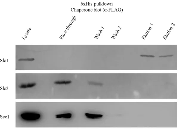
To determine if TarP interacted with the chaperones, pull-down assays were performed. Initially, a nickel column (Ni-NTA-agarose, Qiagen) was used to pull-down the TarP bait. In the elution fraction, the presence of FLAG-tagged chaperones were monitored by Western blotting against the FLAG-tag. Different fractions from the pull-down experiments were run on SDS-PAGE, transferred to PVDF membranes for Western blotting. The chaperone that interacted with 6xHis-TarP should be present in
15
the elution fraction. In the Figure 2 we can see that the pull-down assay was successful because 6xHis-TarP appeared in the lysate and in the elution fractions. The presence of TarP in the flow through and washes in two of the membranes could be due to a saturation of α-6xHis antibody. It is also clear that, from the three chaperones under investigation, only Slc1 interacted with TarP. This chaperone only appeared in the elution fraction while Slc2 and Scc1 appeared in the flow through and in the washes fractions and are completely absent in the fraction corresponding to the elutions (Figure 3).
Figure 2: Co-imunoprecipitation, NiNTA agarose beads, α-6xHis.
NiNTA pull-down of His tag marked protein TarP analysed by Western blot using α-6xHis antibody. The bands represented TarP: The top membrane represents the E. coli coexpressing TarP and Slc1; the middle coexpresses TarP and Slc2; the botttom membrane is from the clone coexpressing TarP and Scc1.
16
Figure 3: Co-imunoprecipitation, NiNTA agarose beads, α-FLAG.
NiNTA pull-down of His tag marked proteins TarP analysed by Western blot using α-FLAG antibody. The bands represent the different chaperones: The top membrane represents the E. coli coexpressing TarP and Slc1; the middle coexpresses TarP and Slc2; the bottom membrane is from the clone coexpressing TarP and Scc1.
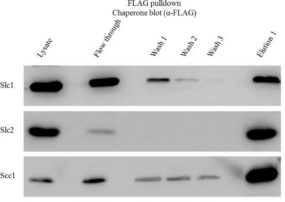
To confirm this results the inversed pull-down was also made, using the M2-affinity gel (Sigma). The chaperones marked with FLAG-tag interact with the antibodies in the M2-affinity gel. The polyacrylamide gels were analyze again by Western blotting with the same antibodies used previously (Figures 4 and 5). We can see that these results are concordant with the first pull-down assay. The Figure 4, which shows the membranes of the M2 affinity pull-down using α-6xHis antibody, reveals that TarP only appears in the elution fraction when it is coexpressed with Slc1. When it is coexpressed with Slc2 and Scc1 the Western blot reveal that TarP only appears in the Lysate and flow through fractions, and thus not specifically retained in the column. The only chaperone that shows an interaction with TarP is Slc1.
17
Figure 4: Co-imunoprecipitation, M2 affinity gel, α-6xHis.
M2 affinity gel pull-down of FLAG tag marked proteins chaperones analysed by Western blot using α-6xHis antibody. The bands represent TarP: The top membrane represents the E. coli coexpressing TarP and Slc1; the middle coexpresses TarP and Slc2; the bottom membrane is from the clone coexpressing TarP and Scc1.
In Figure 5, we can confirm that the M2 affinity pull-down assays were done correctly showing the majority of the chaperones in the elution fraction with some appearing in the flow through and washes but this is probably due to over saturation of the affinity gel. These results were further confirmed using the bacterial two-hybrid assay and the β-galactosidase (Brinkworth, 2011).
18
Figure 5: Co-imunoprecipitation, M2 affinity gel, α-FLAG.
M2 affinity pull-down of FLAG tag marked proteins chaperones analysed by Western blot using α-FLAG antibody. The bands represent the different chaperones: The top membrane represents the E. coli coexpressing TarP and Slc1; the middle coexpresses TarP and Slc2; the bottom membrane is from the clone coexpressing TarP and Scc1.
19
Determination of binding site between effector TarP and
chaperone Slc1
Bacteria two-hybrid assay
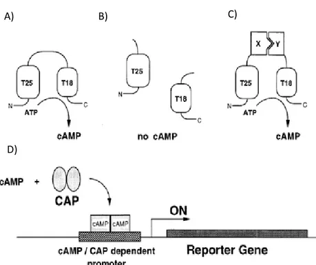
This assay is based on a protein encoded by a cyaA gene in Bordetella pertussis, which converts ATP to cAMP. Through biochemical studies it was discovered that the catalytic domain could be proteolytically cleaved into two complementary fragments T25, corresponding to amino acids 1-224, and T18, corresponding to amino acids 245-399. It was also demonstrated that the two fragments alone were not able to reassociate. The rationale behind this method is if the two fragments fused to putative interacting proteins were expressed in a adenylate cyclase-deficient E. coli, they would reassociate and lead to cAMP synthesis. The function and activity of adenylate cyclase can be monitored in E. coli because cAMP binds to a transcriptional activator, CAP, which regulates the expression of several genes, including genes involve in the catabolism of lactose and maltose (Figure 6) (Karimova, et al. 1998).
For experimental purposes Karimova, et al. (1998) constructed plasmids that expressed either the T25 fragment or the T18. For this study the pUT18C (Annex A2) was fused with several sequences of mutant TarP, and pKT25 (Annex A3) fused with Slc1. The mutant sequences of TarP (mTarP) were obtained using random mutagenesis by error prone PCR (McCullum, et al. 2010; Pritchard, et al. 2005). Afterwards for the isolation of the different sequences they were treated with restriction enzymes, clone into pCX340 plasmid, transformed into chemically competent E. coli Top10 and finally sent out for sequencing to identify and exclude mutants with frame shift mutations. Error prone PCR was optimized by adjusting the Mg2+ concentrations a decrease in MnCl2, a higher concentration than usual of one of the dNTPs to obtain at least one mutation, but no more than six simultaneous mutations within the insert, without obtaining frame shift mutations. Random PCR mutagenesis was used instead of site-directed mutagenesis methods for the following reasons – a) there were no distinguishing motifs that could be considered as chaperone binding sites, unlike in other pathogens (e.g. Salmonella) (Stebbins and Galân, 2001) and b) single mutations rarely results in detectable loss of binding in other effector-chaperone interactions.
20
Figure 6: Schematic of principle of an E. coli two-hybrid system based on functional complementation of CyaA fragments.
In A, the full-length catalytic domain when expressed in E. coli, exhibits a basal activity that results in cAMP synthesis. In B, the two fragments, when coexpressed as independent polypeptides, are unable to interact and no cAMP synthesis occurs. In C the two frangements are fused to two interacting proteins, X and Y, are brought into close proximity, resulting in functional complementation followed by cAMP production. D represents a schematic of promoter activation of the complementation. cAMP, synthesized in an E. coli cya strain by the complementing T25 and T18 pairs, binds to the catabolite gene activator protein, CAP. The cAMPyCAP complex then can recognize specific promoters such as lacZ and mal (Karimova, et al. 1998).
Slc1 protein is highly conserved among chlamydial species. However, there is a lack of amino acid similarity amongst the N-termini region of TarPs belonging to different Chlamydia species. When analyzing the alignment obtained for the last 200 amino acids using ClustalW2 Multiple Sequence Alignment of TarP homologs belonging to C. trachomatis L2, C. muridarum, GPIC, and C. pneumonia some conserved or semi-conserved regions were found. Among these conserved regions several spatially conserved hydrophobic sites flanked by conserved threonines and serines were found. Furthermore, the previous analysis revealed that the conserved regions were located in the N-terminal 100 amino acids of the different TarP homologs, which may indicate a possible chaperone binding site. There may be some
A) B) C)
21
interchangeability involved in the binding of TarP from one species by the Slc1 chaperone from another, as evidenced by the ability of C. trachomatis Slc1 to specifically bind C. caviae TarP (Brinkworth 2011) (Annex A4).
So in order to determine which amino acid residues were indeed important for TarP interaction with the chaperone, we proceeded to the construction of several mutants in this region. These mutants were sequence-verified to identify the mutations, catalogued, and functionally evaluated using the bacterial two-hybrid assay.
Transformation of E. coli Top10
The mTarP sequences were kindly provided by Doctor Amanda Brinkworth and they were amplified by PCR using an Accuprime PFX polymerase, a DNA polymerase with proof reading to avoid further mutations. Afterwards the PCR products were purified using a PCR purifying kit. The plasmids were propagated in E. coli Top10, and stored at -80ºC. They were extracted using a Miniprep Kit (Qiagen).
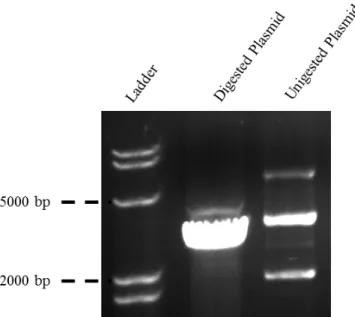
Both the plasmids and the purified inserts were digested using KpnI restriction enzyme, which cuts the DNA sequence forming sticky ends. This allows for the extremities of the insert and the plasmid to interact and ligate. The digestion was followed by PCR purification because KpnI is not deactivated by temperature. The mTarP sequences were separately ligated to pUT18C. The agarose gel of cut and uncut plasmid analyses is shown in Figure 7 and from it we can assure that the restriction enzyme is working properly. Any uncut plasmid could potentially yield false-positive colonies on selective media.
22
Figure 7: Analyzes of pUT18C digestion to confirm the functionality of KpnI.
Since the plasmid circular DNA in the undigested lane we observe the 3 bands, which is common. The top band corresponds to the relaxed circular form, the middle band is when the plasmid is linearized, and the bottom band represents the supercoiled circular DNA. In the Digested lane one band clearly appears demonstrating a good cleavage by KpnI.
The initial transformation was done in chemically competent cells of E. coli Top10. This was due to its high transformation efficiency (1 x 10⁹ cfu/µg supercoiled DNA) and is ideal for high-efficiency cloning and plasmid propagation, allowing stable replication of high-copy number plasmids.
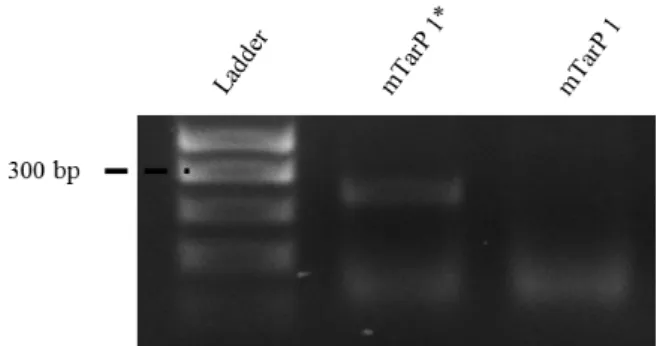
The pUT18C plasmid has an AmpR cassette so the transformed E. coli Top10 bacteria were plated in LB media with carbenicillin. This antibiotic was used instead of ampicillin, because it is more stable in solution letting us keep stocks ready for much longer periods of time, without having to worry about loss of function. Between 5 to 6 colonies obtained after the overnight incubation were tested for the presence of the insert by colony PCR and analysed by agarose gel electrophoresis. Primers for colony PCR immediately flanked the insert. The results obtained are summarized in Figures 8 to 14. The DNA band of the positive colonies is around the 300 bp mark, which is expected for a protein with 100 amino acids.
23
Figure 8: mTarP 1 confirmation in E. coli Top10.
Analysis of E. coli Top10 for the presence of pUT18C plasmid with mTarP, using primers TarP100aa-FW and TarP100aa-REV (Table 2). Amplification products were resolved in 1,5% agarose gel and visualized by staining with SYBR SAFE Legend: mTarP 1* - positive result for the presence of the insert; mTarP 1 - negative result for the presence of the insert. The lower band are background bands.
Figure 9: mTarP 2 confirmation in E. coli Top10.
Analysis of E. coli Top10 for the presence of pUT18C plasmid with mTarP 2, using primers TarP100aa-FW and TarP100aa-REV (Table 2). Amplification products were resolved in 1,5% agarose gel and visualized by staining with SYBR SAFE. Legend: mTarP 2* - positive result for the presence of the insert; mTarP 2 - negative result for the presence of the insert.
Figure 10: mTarP 3 and 4 confirmation in E. coli Top10.
Analysis of E. coli Top10 for the presence of pUT18C plasmid with mTarP 3 and plasmid with mTarP 4, in the case of mTarP 4, using primers TarP100aa-FW and TarP100aa-REV (Table 2).
Amplification products were resolved in 1,5% agarose gel and visualized by staining with SYBR SAFE. Legend: mTarP 3* - positive result for the presence of the insert; mTarP 3 - negative result for the presence of the insert; mTarP 4*- positive result for the presence of the insert; mTarP 4- negative result for the presence of the insert.
24
Figure 11: mTarP 5 confirmation in E. coli Top10.
Analysis of E. coli Top10 for the presence of pUT18C plasmid with plasmid with mTarP 5, using primers TarP100aa-FW and TarP100aa-REV (Table 2). Amplification products were resolved in 1,5% agarose gel and visualized by staining with SYBR SAFE Legend: mTarP 5* - positive result for the presence of the insert; mTarP 5 - negative result for the presence of the insert.
Figure 12: mTarP 6 confirmation in E. coli Top10.
Analysis of E. coli Top10 for the presence of pUT18C plasmid with mTarP 6, using primers TarP100aa-FW and TarP100aa-REV (Table 2). Amplification products were resolved in 1,5% agarose gel and visualized by staining with SYBR SAFE Legend: mTarP 6*- positive result for the presence of the insert; mTarP 6 - negative result for the presence of the insert.
Figure 13: mTarP 7 confirmation in E. coli Top10.
Analysis of E. coli Top10 for the presence of pUT18C plasmid with mTarP 7, using primers TarP100aa-FW and TarP100aa-REV (Table 2). Amplification products were resolved in 1,5% agarose gel and visualized by staining with SYBR SAFE Legend: mTarP 7* - positive result for the presence of the insert; mTarP 7 - negative result for the presence of the insert.
25
Figure 14: mTarP 8 confirmation in E. coli Top10.
Analysis of E. coli Top10 for the presence of pUT18C plasmid with mTarP 8, using primers TarP100aa-FW and TarP100aa-REV (Table 2). Amplification products were resolved in 1,5% agarose gel and visualized by staining with SYBR SAFE Legend: mTarP 8* - positive result for the presence of the insert.
The positive colonies were grown overnight, and part of this culture was stored at -80 ºC with 30% glycerol as stocks. The rest was used to extract the plasmid using the Qiagen miniprep kit and sent for sequencing, using the TarP100aa-REV primer, to ascertain if no further mutations were added, because of the PCR amplification reaction, which could lead to frame shift mutation. Another reason to do this procedure was to assure that the insert was fused in the right direction within the plasmid. This was verified using the BLAST nucleotide alignment tool to assure that prior to the TarP gene there was the cya gene from Bordetella (data not shown).
In the Figures 8 to 14, only the mTarP sequences, which were proven to have the insert in the right location and orientation, are shown.
Transformation of E. coli DHM1
The pKT25 plasmid was fused with hypothetical type III chaperone CT043 and electroporated into E. coli DHM1. The non-mutated N-terminous 100 amino acids of TarP was also fused to pUT18C, and electroporeted into E. coli DHM1 pKT25-Slc1 to serve as a positive control for the bacterial two-hybrid assay done by Amanda Brinkworth.
The mTarP 1 to 8 that were previously transformed into E. coli Top 10 were extracted using the miniprep kit from Qiagen and electroporated into electrocompetent E. coli DHM1 with pKT25-Slc1 plated in LB agar with carbenicillin and kanamycin.
26
Like the previous transformation 5 to 6 colonies obtained were analysed by colony PCR to confirm the presence of the Slc1 (Figure 15) as well as mTarP inserts (Figures 16 to 22).
Figure 15: Slc1 confirmation in E. coli DHM1.
Analysis of E. coli DHM1 for the presence of pKT25 plasmid with Slc1, using primers CT043-FW and CT043-REV (Table 2). Amplification products were resolved in 1,5% agarose gel and visualized by staining with SYBR SAFE Legend: Slc1 – example of two positive result for the presence of the Slc1 insert.
Figure 16: mTarP 1 confirmation in E. coli DHM1.
Analysis of E. coli DHM1 for the presence of pUT18C plasmid with mTarP 1, using primers TarP100aa-FW and TarP100aa-REV (Table 2). Amplification products were resolved in 1,5% agarose gel and visualized by staining with SYBR SAFE Legend: mTarP 1 - positive result for the presence of the insert.
Figure 17: mTarP 2 confirmation in E. coli DHM1.
Analysis of E. coli DHM1 for the presence of pUT18C plasmid with mTarP 2, using primers TarP100aa-FW and TarP100aa-REV (Table 2). Amplification products were resolved in 1,5% agarose gel and visualized by staining with SYBR SAFE Legend: mTarP 2 - positive result for the presence of the insert.
27
Figure 18: mTarP 3 confirmation in E. coli DHM1.
Analysis of E. coli DHM1 for the presence of pUT18C plasmid with mTarP 3, using primers TarP100aa-FW and TarP100aa-REV (Table 2). Amplification products were resolved in 1,5% agarose gel and visualized by staining with SYBR SAFE Legend: mTarP 3 - positive result for the presence of the insert.
Figure 19: mTarP 4 and 5 confirmation in E. coli DHM1.
Analysis of E. coli DHM1 for the presence of pUT18C plasmid with mTarP 4 and plasmid with mTarP 5, using primers TarP100aa-FW and TarP100aa-REV (Table 2). Amplification products were resolved in 1,5% agarose gel and visualized by staining with SYBR SAFE Legend: mTarP 4 - positive result for the presence of the insert; mTarP 5 - positive result for the presence of the insert.
Figure 20: mTarP 6 confirmation in E. coli DHM1.
Analysis of E. coli DHM1 for the presence of pUT18C plasmid with mTarP 6, using primers TarP100aa-FW and TarP100aa-REV (Table 2). Amplification products were resolved in 1,5% agarose gel and visualized by staining with SYBR SAFE Legend: mTarP 6 - positive result for the presence of the insert.
28
Figure 21: mTarP 7 confirmation in E. coli DHM1.
Analysis of E. coli DHM1 for the presence of pUT18C plasmid with mTarP 7, using primers TarP100aa-FW and TarP100aa-REV (Table 2). Amplification products were resolved in 1,5% agarose gel and visualized by staining with SYBR SAFE Legend: mTarP 7 - positive result for the presence of the insert.
Figure 22: mTarP 8 confirmation in E. coli DHM1.
Analysis of E. coli DHM1 for the presence of pUT18C plasmid with mTarP 8, using primers TarP100aa-FW and TarP100aa-REV (Table 2). Amplification products were resolved in 1,5% agarose gel and visualized by staining with SYBR SAFE Legend: mTarP 8 - positive result for the presence of the insert.
Analysis of mTarP sequences.
The analyses of mTarP sequences was done using the software Geneious Basic 5.4.4 program, which allows the alignment and comparison between the different sequences with a reference sequence in this case it was the first 100 amino acids of C. trachomatis serovar L2 TarP. The software also provides a graphic figure showing the hydrophobicity of each amino acid allowing for an easy comparison, of the hydrophobicity changes, between the mutants and the reference sequence (Figure 24). Sequence analysis was followed by bacterial two-hybrid assay to functionally evaluate the effects of the mutations on Slc1 interaction with TarP.
29
Bacteria two-hybrid assay was performed using the eight colonies mentioned above. LB plates supplemented with antibiotics carbenicillin and kanamycin were used to prevent the growth of contaminants and the loss of both plasmids. IPTG and X-gal were also added to the medium. IPTG is used to induce the expression from the ptac promoter that regulates the expression in both plasmids of the cya fragment fused with
the proteins of interest (T18-mTarP and T25-Slc1). X-gal is an indicator of
-galactosidase activity, which should increase if there was induction of lacZ expression. lacZ expression should only occur if the two subunits of cya were reconstituted by virtue of interaction between the two fused proteins. The interaction is detected by the development of blue colonies. In Figure 23 it is possible to see the result for this assay.
Figure 23: Bacterial two-hybrid assay.
Colonies mark 1-8 represent the E. coli DHM1 transformed with pUT18C.mTarP 1 to 8 respectably and Slc1; the colony marked as TarP has the plasmids pUT18C-TarP, and pKT25-Slc1; The colony marked as empty has both the pUT18C and pKT25 with no inserts.
As we can see mTarP 1 only has two different amino acid substitutions from a serine to a threonine (Ser17Thr, Ser23Thr). Analysing the hydrophobicity chart we can
30
see that the difference in hydrophobicity is not significant, which means that if the binding between TarP and Slc1 was due to hydrophobic interactions the colonies colour should resemble the positive control.
In the mTarP 2 sequence there are six amino acid substitution mutations at residues 9, 26, 39, 56, 57, and 66. On one hand, we have the mutation from a glutamine to a histidine (Gln9His), which as can be seen in the figure, does not alter significantly the hydrophobicity. One mutation decreased the hydrophobicity in position 57 by substituting the amino acid isoleucine to asparagine (Ile57Asn). On the other hand, there are the mutations that changed serine to tyrosine (Ser26Tyr), asparagine to isoleucine (Asn39Ile, Asn66Leu), and serine to proline (Ser56Pro), which together greatly increase the overall hydrophobicity of the protein, which in theory should strengthen the binding between mTarP and Slc1. From such a more stable binding, one would expect colonies on X-gal plates with similar colouration as that of wild-type. However, this is not the case. The colony had less blue colour than the wild-type colony, but had a slightly higher blue colouration when compared to the negative control. This result indicates that together, the mutations slightly affected binding of Slc1 to mTarP 2. A similar colony phenotype was observed for mTarP 3. Two amino acid substitution mutations are present in mTarP 3. The first is a change from glutamine to leucine (Gln44Leu) and the other one from threonine to alanine (Thr68Ala). The Gln44Leu change greatly increased the hydrophobicity and Thr68Ala slightly increases it as well. So the expected result would be a colony colour similar to the positive control or a darker colour due to a stronger binding. As with mTarp 2, the binding was less efficient that wild-type or mTarP 1. The mTarP 4 mutant has the same mutation in the amino acid 44 that mTarP 3 and the expected results should be the same. The results for mTarP 2, 3, and 4 could mean that increasing the overall hydrophobicity of the first 100 amino acid residues may have a negative effect on Slc1 binding. On the other hand, some of the six mutations may have hit residues essential for Slc1 binding, thus giving an intermediate phenotype.
The mTarP 5 and 8 mutants had multiple mutations. Sequencing of the inserts revealed six amino acid substitution mutations (amino acids 13, 21, 41, 62, 80, 85) for mTarP 5 and five for mTarP 8 at positions 9, 32, 43, 44, and 71. Four of those mutations in mTarP 5 do not change significantly the hydrophobicity of the overall protein, those
31
mutations are: from a threonine to a serine (Thr13Ser; Thr85Ser), from a serine to threonine (Ser21Thr) and from a threonine to alanine (Thr41Ala). The other mutations significantly increased the hydrophobicity of the protein, namely a glutamine-to-valine change (Glu62Val) and a threonine to isoleucine (Thr80Ile) substitution. A similar predicted increase in net hydrophobicity could be inferred from the amino acid sequence of mTarP 8. Two mutations Thr43Ala and an Ala71Thr substitution did not result in an increase in hydrophobicity, but when combined with other substitutions (i.e. from a glutamine to a leucine (Gln9Leu) at position 9, an alanine to a proline (Ser32Pro) and a glutamine to a leucine (Gln44Leu)), a predicted net increase in hydrophobicity is apparent. Therefore, similar to mTarP 2, 3, and 4, it was not possible to distinguish if the decreased interaction of Slc1 with these mutants could be attributed to an increase in net hydrophobicity or the changes to essential amino acid residues.
Two mutants that were informative were mTarP 6 and mTarP 7, whose phenotypes did not seem to be associated with increased hydrophobicity, unlike mTarP 2, 3, 4, 5 and 8. Two amino acid substitutions were observed in the m TarP 6. One is a serine to a threonine (Ser17Thr) substitution and the other was an asparagine to an aspartic acid (Asn92Asp) substitution. Both mutations did not significantly change the protein hydrophobicity. The same could be said for mTarP 7 mutations, which consisted of a serine-to-threonine (Ser23Thr) and an asparagine-to-tyrosine (Asn69Ile) substitution. Their respective colonies displayed intermediate phenotypes, indicating that Ser17, Asn92, Ser 23, and Asn69 may be essential for Slc1 binding.
Taken together, it is possible that TarP may rely on both specific sequences, and hydrophobicity in its interaction with Slc1.
32
Figure 24: Sequences of mTarP.
mTarP sequences were compared with the reference sequence from C. trachomatis serovar L2 TarP, with the mutations highlighted. Underneath each sequence there is a graphic, which represents the hydrophobicity of each amino acid.
33
General conclusion
A characteristic of many bacterial T3S effectors is their need of a specific chaperone for efficient secretion into the host cell cytosol (Knodler, et al., 2006). Most chaperones bind only to one, or a maximum of two effector with some sequence similarities, the exception being Spa15 from S. flexneri, which was found to be able to bind with several effectors with no sequence similarity (Page, et al., 2001; Page and Parsot, 2002).
Is this project, it was found using pull-down assays that chaperone Slc1 interacts with chlamydial effector TarP, one of the first effectors to be secreted and essential for the invasion of Chlamydia into the host cell (Clifton et al., 2004; Clifton et al., 2005). Interestingly, the other two putative chaperones present in EBs did not bind TarP, indicating that amino acids 1-100 has the necessary sequence information to confer specific interaction with Slc1 and not with the structurally homologous Slc2 and Scc1 proteins. To investigate this specificity, we undertook a random mutagenesis experiment to introduce mutations within TarP (1-100). Using the bacteria two-hybrid assay, the mutated derivatives were evaluated for their abilities to interact with Slc1 in a prokaryotic cytosolic milieu. Data indicated that binding of Slc1 to TarP may be determined by both general hydrophobic interactions and specific amino acids. This work provided the first report of a systematic investigation of the interaction between an EB-associated effector (TarP), with its recently identified chaperone Slc1. This avenue of research is important as an optimal interaction is likely required for efficient translocation of the effector from the bacterial cytosol into the host cell cytoplasm, as recently demonstrated using a heterologous T3SS (Brinkworth 2011).
The co-imunoprecipitation experiments, which were successful in demonstrating TarP interaction with Slc1 involved the co-expression of both chaperone and effector. This is particularly important as it mimics the potential environment within the reticulate body, where TarP and Slc1 are co-expressed prior to RB-to-EB differentiation. Therefore, we can confidentially say that the specificity of TarP for Slc1 displayed in this experimental system accurately reflects the true situation in Chlamydia. Also, the interaction was more efficient when compared with pre-purified components mixed in vitro (Brinkworth, 2011). In addition, co-expression allows for
34
the co-translational interaction between newly made Slc1 proteins with the growing polypeptide chain of TarP as it emerges from the ribosome. It is possible that under this condition, like with other Type III effectors, the N-terminal end could be maintained in a relatively disorganised conformation, which in turn may promote chaperone binding by exposing the hydrophogic residues, as well as critical amino acid side chains.
The characterization of this chaperone-effector binding was investigated using a bacterial two-hybrid system to evaluate eight mutant sequences of TarP. Five of these sequences (mTarP 2, 3, 4, 5 and 8) had mutations, which greatly increased the net hydrophobicity of the polypeptide. These revealed an intermediate phenotype (i.e. the blue coloration was significantly less than the wild-type, but noticeably greater than the negative control) suggesting that either one or more mutations in these mutants were in amino acids essential for the interaction between TarP and Slc1 or that the net increase in hydrophobicity had a negative effect in this interaction. The other three mutants (mTarP 1, 6 and 7) showed no significant differences in the overall hydrophobicity of the protein relative to the wild-type sequence, yet they presented an intermediate phenotype suggesting that the partial loss of binding could be attributed not to effects on hydrophobicity, but to mutations that possibly targeted essential amino acids for the interaction. One particular mutation in mTarP 6 (Asn92Glu) involved a change from a polar and uncharged side chain to a polar acidic one. This change could affect local hydrogen bonding of neighbouring residues with this new side chain. Interestingly, both mTarP 6 and 7 displayed Ser-to-Thr substitution. Other mutants also displayed such changes, but it was difficult to ascribe altered functions because of the other mutations that significantly changed hydrophobicity. However, in the context of the other mutations in mTarP 6 and 7, without the confounding effects of changes in net hydrophobicity, these Ser-to-Thr substitutions may indicate that Ser residues may be necessary for Slc1 binding to TarP. Indeed, one of the common features within the otherwise relatively variable N-terminal region amongst the different TarP homologs is the presence of a number of serine residues. Thus it is possible that serine in some or all of these positions are necessary, and even a conservative substitution of Ser to Thr could not be tolerated. Thus, it may be worth investigating, using the same bacterial two-hybrid assay, in conjunction with site-directed mutagenesis, the role of these serine residues.
35
It is known that SptP, a Yersinia class I T3S chaperones interact with its effector YopE using two types of interactions, hydrophobic, which provide the binding strength, and hydrogen bonds, which is believed to provide specificity to between the chaperone-effector (Ghosh, 2004). Thus, it is possible that some of the residues targeted in the mutagenesis experiments form crucial hydrogen bonds with Slc1 amino acid residues, and by substituting these residues for different ones, as in mTarP 6, altered hydrogen bonds that are not compatible with Slc1/TarP interaction are formed and thereby diminishing the interaction, which manifests as intermediate phenotypes in the bacterial two-hybrid assay. One of the potential pitfalls of mutagenesis experiments is the effect on protein conformation, which in turn would be expected to dramatically affect the quality of protein-protein interactions. However, this may not be applicable in this particular case because a number of studies have shown that the N-terminus region of other effectors tended to remain in a disorganised conformation. This disorganisation may allow the docking of a relatively linear polypeptide into the hydrophobic groove formed by the dimerisation of two chaperone molecules (Yip and Strynadka, 2006).
To our knowledge, this is the first attempt to identify the nature of chaperone binding site on TarP. With this knowledge, we advance the efforts to define the consensus recognition sequence for Slc1.