https://doi.org/10.1590/0004-282X20180080
VIEWS AND REVIEWS
Twenty-five years since the identification of
the first SCA gene: history, clinical features
and perspectives for SCA1
Vinte e cinco anos desde a identificação do primeiro gene das SCAs: história, aspectos
clínicos e perspectivas para a SCA1
Carlos Roberto Martins Junior1, Fabrício Castro de Borba1, Alberto Rolim Muro Martinez1, Thiago Junqueira
Ribeiro de Rezende1, Iscia Lopes Cendes2, José Luiz Pedroso3, Orlando Graziani Povoas Barsottini3,
Marcondes Cavalcante França Júnior1
1Universidade de Campinas, Faculdade de Ciências Médicas, Departamento de Neurologia, Campinas SP, Brasil; 2Universidade de Campinas, Faculdade de Ciências Médicas, Departamento de Genética Médica, Campinas SP, Brasil; 3Universidade Federal de São Paulo, Unidade de Ataxia, Departamento de Neurologia, São Paulo SP, Brasil.
Correspondence: Marcondes Cavalcante França Jr.; Departamento de Neurologia da UNICAMP; Rua Tessália Vieira de Camargo, 126; 13083-887 Campinas SP, Brasil; E-mail: [email protected]
Conflict of interest: There is no conflict of interest to declare.
Support: Fundação de Amparo à Pesquisa do Estado de São Paulo (FAPESP) (Grant #13/01766-7 and #2014/19786-7) and Coordenação de Aperfeiçoamento de Pessoal de Nível Superior (CAPES).
Received 18 February 2018; Received in final form 27 May 2018; Accepted 04 June 2018.
ABSTRACT
Spinocerebellar ataxias (SCA) are a clinically and genetically heterogeneous group of monogenic diseases that share ataxia and autosomal dominant inheritance as the core features. An important proportion of SCAs are caused by CAG trinucleotide repeat expansions in the coding region of different genes. In addition to genetic heterogeneity, clinical features transcend motor symptoms, including cognitive, electrophysiological and imaging aspects. Despite all the progress in the past 25 years, the mechanisms that determine how neuronal death is mediated by these unstable expansions are still unclear. The aim of this article is to review, from an historical point of view, the first CAG-related ataxia to be genetically described: SCA 1.
Keywords: Spinocerebellar ataxias; ataxin 1; spinocerebellar degenerations.
RESUMO
As ataxias espinocerebelares (SCA) são um grupo clínico e geneticamente heterogêneo de doenças monogênicas que compartilham ataxia e herança autossômica dominante como características principais. Uma proporção importante de SCAs é causada por expansões de repetição de trinucleotídeos CAG na região de codificação de diferentes genes. Além da heterogeneidade genética, os aspectos clínicos transcendem os sintomas motores, incluindo aspectos cognitivos, eletrofisiológicos e de imagem. Apesar de todo o progresso feito nos últimos 25 anos, os mecanismos que determinam como se dá a morte neuronal mediada por essas expansões instáveis ainda não estão claros. O objetivo deste artigo é revisar, de um ponto de vista histórico, a primeira ataxia geneticamente relacionada com o CAG descrita: SCA 1.
Palavras-chave: ataxias espinocerebelares; ataxina 1; degenerações espinocerebelares.
EARLY STUDIES
In 1863, the German pathologist Nikolaus Friedreich described a new spinal disease, which turned out to be the first description of a hereditary ataxia1. The disease was character
-ized by cerebellar ataxia, dysarthria and deep areflexia, generally with an early age of onset and presenting an autosomal recessive pattern of inheritance1. Over 30 years later, Pierre Marie reported
on a group of patients presenting with hereditary ataxia, but with late disease onset, mostly presenting with increased tendon
reflexes and occasionally with an autosomal dominant pattern of inheritance2. It was later pointed out that Marie’s cohort was
both clinically and pathologically heterogeneous and several classifications throughout the 20th century failed to outline
prop-erly the full clinical and pathological spectrum of the autosomal dominant cerebellar ataxias (ADCA)3,4. The term spinocerebellar
HISTORY OF SCA1 GENETICS
In the first half of the 20th century, many researchers
pub-lished papers on small families segregating ADCA, raising the most diverse hypotheses on the genetic basis of the dis-ease, usually with conflicting results5. Nonetheless, in the late
1940s, a North American group started following a large fam-ily of patients (n = 342) in which 45 individuals presented with the disease. At that time, the authors attempted to determine the genetic basis of the disease using linkage analyses, but they failed to uncover the responsible gene/mutation8.
Despite the initial disappointing results, the investigators kept following these kindred. From the 1970s to the 1980s, strong evidence came out supporting that the causative gene of at least that form of ADCA was related to the HLA human complex. Further studies mapped the disease locus to the short arm of chromosome 69. Finally, 100 years after
Marie’s original publication, in 1993, a collaborative study led by Zoghbi et al. was published, describing an unstable CAG trinucleotide repeat expansion at the coding region of the implicated gene as the first causative mutation of ADCA10.
The normal SCA1 allele contained 6-38 CAG repeats inter -rupted by the CAT trinucleotide, in contrast with mutation carriers that had 39-44 continuous CAG repeats (Table 1). These authors also pointed out that the size of the repeat inversely correlated with the age of onset of the symptoms (anticipation phenomenon)10,11,12.
Other researchers such as Harry T. Orr and Sandro Banfi played key roles in the description and characterization of the ATXN1 gene10,13. The disease locus was named ATXN1
and those presenting with the mutation would thereafter be definitively classified as having SCA type 14. Curiously, the
gene related to Friedreich’s ataxia was only described three years later. Hence, SCA1 was indeed the first hereditary ataxia to have its genetic basis elucidated3,10,13.
The ATXN1 gene encodes a protein called ataxin 1, which has nuclear location inside neurons, but cytoplasmic loca-tion in other peripheral tissues. Mutaloca-tion carriers produce an abnormal form of ataxin 1 with an excessive number of consecutive glutamine residues. Less than five years after the gene description, experimental studies with transgenic mice showed that the pathogenesis by which the mutation causes disease is a toxic gain of function4. Zoghbi’s group
then revealed the specific role of protein misfolding in the pathogenesis of SCA1, a finding that has proved relevant to
other polyglutamine disorders14. After this, the group started
looking at potential pharmacological interventions in animal models15.
DEMOGRAPHIC DATA AND PHENOTYPIC CHARACTERIZATION
The SCAs are rare diseases with prevalence rates rang -ing from 1-4 per 100,000 andSCA1 accounts for between 3% and 16% of all autosomal dominant SCAs16. The search for
relative frequencies of SCAs in Brazilian cohorts have shown that SCA1 represents 4.2% of all SCAs; generally, it is the 4th
or 5th most frequent SCA, after SCA3, SCA2 and SCA717,18,19,20.
The first descriptions of Brazilian patients with SCA1 are relatively recent. All patients attending our reference center in southeastern Brazil have descended from Italian immi-grants21. The relative frequencies of SCA1 patients in differ
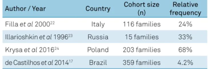
-ent populations are shown in Table 222,23,24.
Clinical aspects
The up-to-date clinical description of SCA1 is a late-onset cerebellar syndrome, with ocular movement disturbances, in particular, hypermetric saccades, pyramidal or extrapyrami-dal signs and peripheral neuropathy3,4. A phenomenon of
pro-gressive worsening of the phenotype across generations has been observed previously4. The first symptoms often include
gait disturbances, later followed by diplopia, dysarthric speech, uncoordinated handwriting and episodic vertigo. Patients may less commonly present with vocal cord abduc-tor paralysis or psychosis3. The average age of onset of SCA1
is between the third and fourth decades, but there is clear variability even within families. Survival from disease onset is around 15 years, but again there is remarkable variability (range 10 to 28 years)3,5.
In a recent series of Brazilian patients17, the most frequent
clinical findings were, in descending order: ataxia (100%), pyra -midal findings (89%), dysarthria and/or dysphagia (89%), alter -ations in ocular movements (44%), nystagmus (33%), rigidity (33%), palpebral retraction (22%), sensory loss (11%), dystonic movements (11%), seizures (11%). Absent reflexes, amyotro -phy, visual loss, tremor and cognitive decline were not found.
Disease progression has recently been assessed in a large multicentric European study25. Authors found that SCA1
pro-gresses faster than other types of SCAs, with a mean annual
Table 1.ATXN1 allele ranges.
Allele types (CAG) repeat length Penetrance
Normal 6–38; 39–44 CAT interrupted None
Intermediate 44 CAT interrupted* Partial
Expanded 39–44 CAGs uninterrupted; 45–91 Full
*A woman with 44 CAG repeats with CAT repeat interruptions had an affected father but was herself asymptomatic at age 66 years9.
Table 2. Relative frequency of SCA1 in different populations.
Author / Year Country Cohort size (n)
Relative frequency
Filla et al 200022 Italy 116 families 24%
Illarioshkin et al 199623 Russia 15 families 33%
Krysa et al 201624 Poland 203 families 68%
progression in ataxia score of 2.11 (Scale for the Assessment and Rating of Ataxia). Within the SCA1 group, patients who had a shorter duration of follow up, older age at initial evalu-ation and longer CAG repeats progressed at faster rates. It remains to be investigated whether such rates of progression also take place in SCA1 patients coming from other regions. Although ataxia is the main and most disabling symptom in most patients with SCA1, many have additional non-ataxia symptoms, which have a great impact on the quality of life of these patients.
Cognitive impairment
It is known that the cerebellum contributes to cognitive tasks, including executive and language functions26. In
par-ticular, patients with isolated acute or chronic focal cere-bellar lesions show frontal-like and parietal-like symptoms; this clinical picture is referred to as the cerebellar cognitive affective syndrome26. The neuroanatomical basis for this syn
-drome is not fully clear, but at least two hypotheses have been raised: it may stem either from the interruption, as in a disconnection syndrome, or from a dysfunctional contri-bution of the cerebellum in the neocerebellar–neocortical reverberation network. This syndrome is characterized by impairments in executive functions (deficiency in planning, set-shifting, abstract reasoning, working memory, verbal flu -ency), language disorders, disturbances in spatial cognition, and personality changes26.
Several studies have reported the neuropsychological features of patients with SCA1, and most show impaired executive function, attention, visuospatial perception, ver-bal fluency, immediate and delayed memory27. Fancellu et
al. reported that patients had significant deficits compared to controls, mainly in executive functions (phonemic and semantic fluencies, attentional matrices) and the disso -ciation in the progression of motor disability and cognitive impairments, suggesting that motor and cognitive functions might be related to different progression rates in SCA128.
Ma et al. reported that SCA1 causes mild impairment in executive function, temporal orientation and logical thinking. The cognitive deficits were correlated with clinical severity of ataxia symptoms, but not age, age of onset, years of educa-tion and disease duraeduca-tion29. Klinke et al. showed that
asymp-tomatic SCA1 carriers have normal cognitive test scores, thus suggesting that cognitive dysfunction does not seem to precede cerebellar ataxia30. While some phenotypic traits,
such as age of onset, progression rate and disease sever-ity are directly related to genotype (CAG expansion length), it is unclear whether the occurrence and severity of cogni-tive impairment also depend on it. Moriarty et al. reported impairments in executive functions, speed, attention, visual memory and theory of mind in SCA1 patients. These authors also found SCA1 patients to have the fastest cognitive decline when compared with the other SCA patients, a finding in line with the motor deterioration pattern in SCAs30.
The pattern of alterations in executive and visuospatial functions described in SCA127 suggests a frontoparietal
dys-function that could be caused by an alteration in the fronto-ponto-cerebello-thalamo-cortical circuits26. A direct
dysfunc-tion of frontoparietal areas could be hypothesized; however, impairments in these cortical areas have not yet been proven in SCA1. In addition, the involvement of subcortical struc-tures, such as the basal ganglia, could play a role in the devel-opment of cognitive dysfunction31.
Depression
Patients with cerebellar degenerative diseases have greater apathy, depression, anxiety and personality changes than normal individuals, especially when the cerebel-lar degeneration is associated with basal ganglia involve-ment26,27.It is hypothesized that these symptoms could be
caused by a dysfunction in the connections between the prefrontal cortex (dorsolateral, orbitomedial, dorsomedial) and the basal ganglia32. Fancellu et al. reported that SCA1
patients had significantly higher depression scores and apa -thy than controls28. Klinke et al. showed a mildly depressed
mood (Beck Depressive Inventory score 11–18) in 50% of SCA1 patients, though no patient showed scores indicating a severely depressed mood (score ≥18)27. McMurtray et al.
iden-tified depressive and memory symptoms as heralding fea -tures of SCA1 in 25% and 42% of the patients, respectively33.
Sleep disorders and fatigue
Hypersomnolence in SCA1 patients can interfere with daytime functioning and periodic limb movements can be a cause of sleep fragmentation in these patients34. Obstructive
sleep apnea can coexist with SCA1, possibly as a manifesta-tion of pharyngeal dilator muscle incoordinamanifesta-tion in response to impending airway collapse34. According to Abele et al.,
rest-less legs syndrome is significantly more frequent in patients with SCA1 than in the general population and is present in 28% of patients35. Fatigue is a major manifestation in SCA1
patients and related to disease duration, depression and probably disease severity36.
Electrophysiological features
Multisystem involvement in SCA1 is common and elec-trophysiology is a potent tool to uncover impairment of multiple neuronal systems and even to decipher subclini-cal affective functions. Electrooculography reveals quanti -tatively abnormal eye movements, such as gaze-evoked nys-tagmus and slowing of saccades37. Visual-evoked potentials
reveal decreased amplitude and/or prolonged latency of P100 wave38. Abnormal acoustic-evoked potential findings include
loss of waves as well as increased interpeak intervals38.
to show subclinical affective functions of the corticospinal tracts, which is very frequent in SCA138. Almost all patients
presented with prolonged central motor conduction times and peripheral motor conduction times in all extremities38,39.
Nerve conduction studies reveal mildly reduced nerve conduction velocities in sensory and motor nerves, but amplitude reduction is much more prominent in sensory nerves than motor nerves39. Electromyography often reveals
signs of chronic denervation affecting proximal and distal muscles. The pattern may resemble a distal neuropathy in some patients, but in others, it is much like a diffuse motor neuronopathy39. In a recent study, Linnemann et al. reported
on the features of peripheral neuropathy in SCA1, in which mixed neuropathy was the most frequent pattern (83%), fol-lowed by axonal (11%) and demyelinating (6%) disease40.
Neuropathology
Spinocerebellar ataxia type 1 is characterized macroscopi-cally by olivoponto-cerebellar atrophy41. On microscopy, there
is dramatic loss of cerebellar Purkinje cells as well as of neu-rons of the dentate nuclei, basal pontine and other brainstem areas, including the red nuclei, vestibular and motor cranial nerve nuclei41,42. In contrast, the pars compacta of the
sub-stantia nigra, the basal ganglia, thalamus, cerebral cortex and hippocampus are usually spared41. Hence, SCA1
neuropathol-ogy can involve components of the cerebello-thalamocortical loop, the basal ganglia-thalamocortical loop, the visual system and the somatosensory system43. The impairment of the spi
-nal cord is conspicuous. It is characterized by gross atrophy, caused by loss of anterior horn cells, fibers in the posterior col -umns and neurons from the Clarke’s column44.
Neuroimaging
Schulz et al. evaluated volumetric changes in SCA1 using voxel-based morphometry in a multicenter study with 48 patients. The authors reported gray matter volume loss in the cerebellar hemispheres, vermis, brainstem and white matter loss in the midbrain, pons, middle cerebellar peduncles, and cerebellar hemispheres. The brainstem and pons volumes correlated inversely with ataxia severity45.
Analyses of supratentorial structures revealed gray mat-ter volume loss in the caudate nuclei, putamina and temporal lobes, but no correlations with clinical or molecular data were found45. In a two-year prospective study, Reetz et al. evaluated
37 patients with SCA1. Cross-sectional analyses found a sig-nificant decline in gray matter volume for the whole brain -stem, left anterior and posterior cerebellum and the right putamen and pallidum compared to healthy controls. The length of the expanded CAG repeat allele correlated with cer-ebellum and pons volume loss in these patients. Longitudinal analyses identified progressive atrophy in the brainstem, pons, putamen, left caudate and left cerebellar hemisphere after two years. The length of the expanded CAG repeat allele correlated with the rate of cerebellar volume loss. In the same
study, authors attempted to compare the rates of atrophy within different SCA subtypes. The SCA1 group indeed had an increased rate of volume loss in the whole brainstem, left cerebellar hemisphere, and putamen, when compared with the SCA3 group46.
Other studies using voxel-based morphometry also reported volume loss in the cerebellum and brainstem involving both gray and white matter47. Regional damage to
cerebral white matter in individuals with SCA1 has repeat-edly been demonstrated by diffusion tensor imaging in sev -eral studies48.
A multicenter observational European study, with 26 pre-symtomatic carriers of SCA1 mutations reported gray mat-ter loss in the medulla oblongata extending to the pons and to the lobule IX of the cerebellum in these individuals com-pared with matched healthy controls. These results indi -cate that anatomical damage takes place before symptom onset in SCA149. Patients with SCA1 also have cervical
spi-nal cord atrophycombined with anteroposterior flatten -ing50. Interestingly, the spinal cord area correlates with ataxia
severity, disease duration, and CAG expansion50.
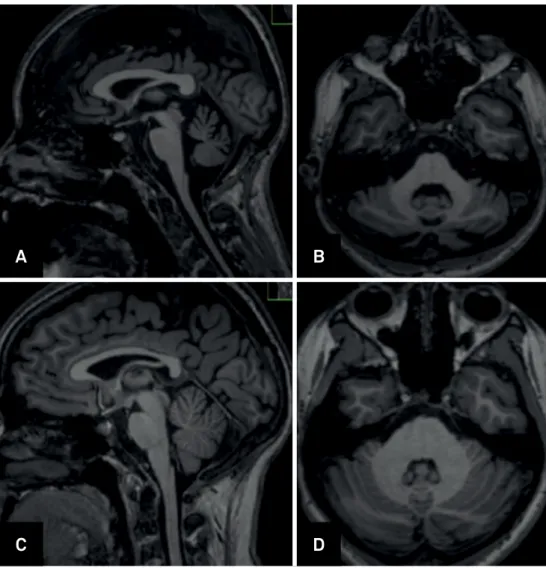
Regarding neurochemical alterations detected by proton magnetic resonance spectroscopy, measurements of metab-olites such as N-acetylaspartate and myoinositol indicate neuronal loss in the cerebellum and pons in SCA1 patients. These parameters correlated with motor disability51. Figure 1
shows some aspects of imaging in SCA1.
DISEASE MECHANISMS AND FUTURE THERAPEUTIC PERSPECTIVES
Wild-type ATXN1 includes a polyQ tract that normally con-tains 6-34 glutamines. A monopartite nuclear-localization signal motif near the carboxyl terminus directs the localization of the protein to the nucleus52. ATXN1 containing a wild-type polyQ
tract can move between the cytoplasm and nucleus inside neu-rons. On the other hand, ATXN1 containing an expanded polyQ tract is transported to the nucleus but cannot be exported to the cytoplasm53. Such abnormal and obligatory localization inside
the nucleus seems to be important for SCA1 pathogenesis, since the substitution of a single amino acid within the nuclear-local-ization signal prevents the expanded ATXN1 from entering the cell nucleus and this mitigates its toxicity54.
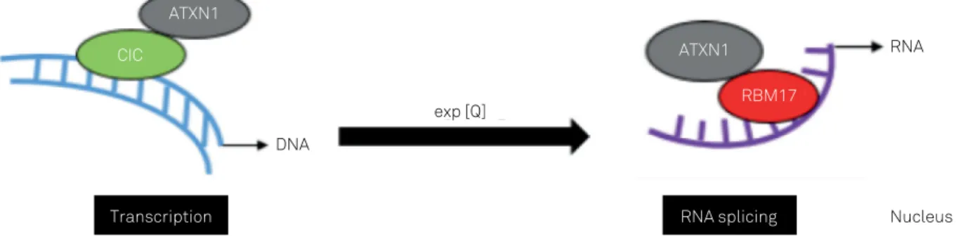
Wild-type and expanded ATXN1 interact with vari-ous nuclear elements, such as RNA53,54, several regulators
of transcription, capicua (CIC)54, and others. An important
On entering the nuclei of Purkinje cells, ataxin-1 binds to CIC. Although ataxin-1 alone does not bind to DNA54,55,
the ataxin-1-CIC complex is targeted to chromatin and tran-scription sites, binding DNA. Noticeably, as trantran-scription pro-ceeds, ataxin-1 is hypothesized to shuttle between complexes with CIC and the splicing factor RNA-binding motif protein (RBM)17. Ataxin-1 can be found in two high-molecular-weight soluble complexes: one that includes CIC and another that includes RBM1755. These two ataxin-1 complexes could be in
a dynamic equilibrium and the dynamics of the complexes affect gene expression. An altered balance in the interaction of expanded ataxin-1 with CIC and RBM17 could drive patho-genesis in SCA1. In this way, expansion of the polyQ tract in ataxin-1 favors ataxin-1-RBM17 complex formation56,
impli-cating this complex in SCA1 pathogenesis (Figure 2).
Like other neurodegenerative disorders, such as Huntington disease57, mechanistic target of rapamycin
(mTOR) signaling is altered in SCA1. In the cerebellar pro-teomes of transgenic mice with ataxin-1[82Q]-expressing Purkinje cells, mTOR levels were decreased before the onset
of symptoms58. The DNA-damage and DNA-repair pathways
have also been implicated in the pathogenesis of SCA159. In
Sca1154Q/2Q mice, enhanced expression of proteins that func-tion in several DNA-repair pathways and restorafunc-tion of mito-chondrial DNA repair by expressing the DNA-repair protein60,
could improve motor performance, Purkinje cell morphology, molecular functions and attenuate SCA1-like phenotypes.
Furthermore, early changes in the expression of specific receptors and ion channels that are important for regulat-ing membrane excitability, such as the dysregulation of cal-cium-activated potassium channel physiology, contribute not only to motor dysfunction but also to structural changes in neurons that consistently precede cell death, changing the intrinsic excitability of Purkinje neurons61.
Several aspects of SCA1 pathogenesis still remain unclear 25 years after the gene/mutation description. Moreover, the studies have shown that long polyglutamine tracts expressed by the mutated gene have an increased tendency to aggre-gate, creating nuclear inclusion bodies in affected neurons that are ubiquitinated3,4. Screening is in progress to find polyQ
A
B
C
D
aggregate inhibitors and also to check whether silencing gene expression can be an interesting therapeutic option3,4.
However, as described above, several cellular pathways are implicated in SCA1 pathogenesis and this multipathway pic-ture makes the development of new therapies difficult. To date, there is no curative treatment for SCA1. Nevertheless, symptomatic therapy may ameliorate symptoms, especially the nonmotor aspects, like depression and sleep disorders.
There are several lines of research on therapeutic agents for SCA1, usually based on animal and cell-based mod-els. Given that mutant polyQ disease proteins can disturb gene expression, RNA homeostasis and protein homeo-stasis through diverse pathways with largely downstream consequences, acting proximally in the disease cascade to lower the levels of the toxic protein can be a useful strat-egy towards developing a disease-modifying therapy. In this sense, endeavors to target the expression of polyQ SCA pro-teins have used antisense oligonucleotides or virus-mediated delivery of short hairpin RNAs or artificial miRNAs to inter -fere with translation. Preclinical tests of antisense oligonu-cleotides or miRNA-based drugs for SCA156 show
consider-able promise, making clinical trials increasingly likely. Experimental data suggested that lithium treatment improves motor coordination and multiple behavioral mea-sures, and might lead to neuroprotective properties in the cerebellum of SCA1 mouse models63. Based on this, lithium
could be a potential treatment for human SCA1. A Phase I trial of oral lithium has been completed as an intramural National Institutes of Health study, but results have not yet been pub-lished (NCT00683943). Riluzole has been shown to provide some symptomatic relief of ataxia in a mixed group of indi-viduals including people with SCA164; however, further
inves-tigation is needed, particularly long-term disease-specific tri -als. A double-blind randomized placebo controlled trial of a riluzole prodrug BHV4157 is ongoing (NCT02960893).
In line with other therapies, Matsuura et al. reported that intrathecal injection of mesenchymal stem cells into the men-ingeal covering of the cerebellum improved Purkinje cell orga-nization, reduced cerebellar dendrite atrophy and normalized behavior and motor deficits in B05 mice injected at five weeks of age65. Thus, further investigation is needed to confirm the
therapeutic potential of stem cells in SCA1. Gene therapy is based on reducing expression of the mutant ATXN1 through gene silencing or overexpressing a paralog of ATXN1, ataxin-1-like (ATXN1L), to competitively inhibit the formation of toxic complexes by polyQ-ataxin-166. This approach appears to be
promising, but further clinical studies are still needed.
In conclusion, in over 100 years of research, there is still a long way to fully understanding and treating patients diag-nosed with SCA1. Nevertheless, recent progress and the genetic outbreaks of the 1990s have given neurologists and neuroscientists valuable hints on where to search for answers. exp [Q]
DNA
CIC
ATXN1
ATXN1
RBM17
Transcription RNA splicing Nucleus
RNA
Figure 2. Illustration of a model of SCA1 pathogenesis that incorporates the crucial nuclear interactions and functions of ATXN1. On entering the nuclei of Purkinje cells, ATXN1 readily binds to CIC (transcriptional regulator capicua). Although ATXN1 alone does not bind to DNA, CIC can bind DNA. ATXN1 can be found in two high-molecular-weight soluble complexes: one that includes CIC and another, distinct complex that includes RBM17 (RNA splicing factor). Importantly, expansion of the polyQ tract in ATXN1 favors ATXN1–RBM17 complex formation, implicating this complex in SCA1 pathogenesis. PolyQ expansion is depicted by ‘exp[Q]’.
References
1. Friedreich, N. Ueber degenerative Atrophie der spinalen
Hinterstränge. Arch Pathol Anat Physiol KlinMed. 1863;26:391-419. 2. Marie P. La semaine médicale: sur l’hérédo-ataxie cérébelleuse.
Semaine Médicale. 1893.
3. Finsterer J. Ataxias with autosomal, X-chromosomal or maternal inheritance. Can J Neurol Sci. 2009 Jul;36(4):409-28. https://doi.org/10.1017/S0317167100007733
4. Cummings CJ, Orr HT, Zoghbi HY. Progress in pathogenesis studies of spinocerebellar ataxia type 1. Philos Trans R Soc Lond B Biol Sci. 1999 Jun;354(1386):1079-81. https://doi.org/10.1098/rstb.1999.0462
5. Schut JW. Hereditary ataxia: clinical study through six generations. Arch NeurPsych. 1950;63(4):535-68. https://doi.org/10.1001/archneurpsyc.1950.02310220002001 6. Konigsmark BW, Weiner LP. The olivopontocerebellar atrophies:
a review. Medicine (Baltimore). 1970 May;49(3):227-41. https://doi.org/10.1097/00005792-197005000-00003
8. Matson GA, Schut JW, Swanson J. Hereditary ataxia: linkage studies in hereditary ataxia. Ann Hum Genet. 1961 May;25(1):7-23. https://doi.org/10.1111/j.1469-1809.1961.tb01492.x
9. Rich SS, Wilkie P, Schut L, Vance G, Orr HT. Spinocerebellar ataxia: localization of an autosomal dominant locus between two markers on human chromosome 6. Am J Hum Genet. 1987 Oct;41(4):524-31. 10. Orr HT, Chung MY, Banfi S, Kwiatkowski TJ Jr, Servadio A, Beaudet
AL et al. Expansion of an unstable trinucleotide CAG repeat in spinocerebellar ataxia type 1. Nat Genet. 1993 Jul;4(3):221-6. https://doi.org/10.1038/ng0793-221
11. Goldfarb LG, Vasconcelos O, Platonov FA, Lunkes A, Kipnis V, Kononova S et al. Unstable triplet repeat and phenotypic variability of spinocerebellar ataxia type 1. Ann Neurol. 1996 Apr;39(4):500-6. https://doi.org/10.1002/ana.410390412
12. Zühlke C, Dalski A, Hellenbroich Y, Bubel S, Schwinger E, Bürk K. Spinocerebellar ataxia type 1 (SCA1): phenotype-genotype correlation studies in intermediate alleles. Eur J Hum Genet. 2002;10(3):204-9. https://doi.org/10.1038/sj.ejhg.5200788 13. Banfi S, Servadio A, Chung MY, Kwiatkowski TJ Jr, McCall AE, Duvick
LA et al. Identification and characterization of the gene causing type 1 spinocerebellar ataxia. Nat Genet. 1994 Aug;7(4):513-20. https://doi.org/10.1038/ng0894-513
14. Cummings CJ, Mancini MA, Antalffy B, DeFranco DB, Orr HT, Zoghbi HY. Chaperone suppression of aggregation and altered subcellular proteasome localization imply protein misfolding in SCA1. Nat Genet. 1998 Jun;19(2):148-54. https://doi.org/10.1038/502
15. Emamian ES, Kaytor MD, Duvick LA, Zu T, Tousey SK, Zoghbi HY et al. Serine 776 of ataxin-1 is critical for polyglutamine-induced disease in SCA1 transgenic mice. Neuron. 2003 May;38(3):375-87. https://doi.org/10.1016/S0896-6273(03)00258-7
16. Manto M, Marmolino D. Cerebellar ataxias. Curr Opin Neurol. 2009 Aug;22(4):419-29. https://doi.org/10.1097/WCO.0b013e32832b9897 17. Castilhos RM, Furtado GV, Gheno TC, Schaeffer P, Russo A, Barsottini
O et al. Rede Neurogenetica. Spinocerebellar ataxias in Brazil: frequencies and modulating effects of related genes. Cerebellum. 2014 Feb;13(1):17-28. https://doi.org/10.1007/s12311-013-0510-y 18. Silveira I, Lopes-Cendes I, Kish S, Maciel P, Gaspar C, Coutinho P et al. Frequency of spinocerebellar ataxia type 1, dentatorubropallidoluysian atrophy, and Machado-Joseph disease mutations in a large group of spinocerebellar ataxia patients. Neurology. 1996 Jan;46(1):214-8. https://doi.org/10.1212/WNL.46.1.214
19. Trott A, Jardim LB, Ludwig HT, Saute JA, Artigalás O, Kieling C et al. Spinocerebellar ataxias in 114 Brazilian families: clinical and molecular findings. Clin Genet. 2006 Aug;70(2):173-6. https://doi.org/10.1111/j.1399-0004.2006.00656.x
20. Teive HA, Munhoz RP, Arruda WO, Lopes-Cendes I, Raskin S, Werneck LC et al. Spinocerebellar ataxias: genotype-phenotype correlations in 104 Brazilian families. Clinics (São Paulo). 2012;67(5):443-9. https://doi.org/10.6061/clinics/2012(05)07
21. Lopes-Cendes I, Steiner CE, Silveira I, Pinto Júnior W, Maciel JA, Rouleau GA. Clinical and molecular characteristics of a Brazilian family with spinocerebellar ataxia type 1. Arq Neuropsiquiatr. 1996 Sep;54(3):412-8. https://doi.org/10.1590/S0004-282X1996000300009 22. Filla A, Mariotti C, Caruso G, Coppola G, Cocozza S, Castaldo I et al.
Relative frequencies of CAG expansions in spinocerebellar ataxia and dentatorubropallidoluysian atrophy in 116 Italian families. Eur Neurol. 2000;44(1):31-6. https://doi.org/10.1159/000008189 23. Illarioshkin SN, Slominsky PA, Ovchinnikov IV, Markova ED, Miklina
NI, Klyushnikov SA et al. Spinocerebellar ataxia type 1 in Russia. J Neurol. 1996 Jul;243(7):506-10. https://doi.org/10.1007/BF00886871 24. Krysa W, Sulek A, Rakowicz M, Szirkowiec W, Zaremba J. High relative
frequency of SCA1 in Poland reflecting a potential founder effect. Neurol Sci. 2016 Aug;37(8):1319-25. https://doi.org/10.1007/s10072-016-2594-x 25. Jacobi H, Montcel ST, Bauer P, Giunti P, Cook A, Labrum R et al.
Long-term disease progression in spinocerebellar ataxia types 1, 2, 3, and
6: a longitudinal cohort study. Lancet Neurol. 2015 Nov;14(11):1101-8. https://doi.org/10.1016/S1474-4422(15)00202-1
26. Schmahmann JD, Sherman JC. The cerebellar cognitive affective syndrome. Brain. 1998 Apr;121(Pt 4):561-79. https://doi.org/10.1093/brain/121.4.561
27. Klinke I, Minnerop M, Schmitz-Hübsch T, Hendriks M, Klockgether T, Wüllner U et al. Neuropsychological features of patients with spinocerebellar ataxia (SCA) types 1, 2, 3, and 6. Cerebellum. 2010 Sep;9(3):433-42. https://doi.org/10.1007/s12311-010-0183-8 28. Fancellu R, Paridi D, Tomasello C, Panzeri M, Castaldo A,
Genitrini S et al. Longitudinal study of cognitive and psychiatric functions in spinocerebellar ataxia types 1 and 2. J Neurol. 2013 Dec;260(12):3134-43. https://doi.org/10.1007/s00415-013-7138-1 29. Ma J, Wu C, Lei J, Zhang X. Cognitive impairments in patients with
spinocerebellar ataxia types 1, 2 and 3 are positively correlated to the clinical severity of ataxia symptoms. Int J Clin Exp Med. 2014 Dec;7(12):5765-71.
30. Moriarty A, Cook A, Hunt H, Adams ME, Cipolotti L, Giunti P. A longitudinal investigation into cognition and disease progression in spinocerebellar ataxia types 1, 2, 3, 6, and 7. Orphanet J Rare Dis. 2016 Jun;11(1):82. https://doi.org/10.1186/s13023-016-0447-6 31. Jacobi H, Hauser TK, Giunti P, Globas C, Bauer P, Schmitz-Hübsch T et al.
Spinocerebellar ataxia types 1, 2, 3 and 6: the clinical spectrum of ataxia and morphometric brainstem and cerebellar findings. Cerebellum. 2012 Mar;11(1):155-66. https://doi.org/10.1007/s12311-011-0292-z 32. Leroi I, O’Hearn E, Marsh L, Lyketsos CG, Rosenblatt A, Ross CA et al.
Psychopathology in patients with degenerative cerebellar diseases: a comparison to Huntington’s disease. Am J Psychiatry. 2002 Aug;159(8):1306-14. https://doi.org/10.1176/appi.ajp.159.8.1306 33. McMurtray AM, Clark DG, Flood MK, Perlman S, Mendez MF.
Depressive and memory symptoms as presenting features of spinocerebellar ataxia. J Neuropsychiatry Clin Neurosci. 2006;18(3):420-2. https://doi.org/10.1176/jnp.2006.18.3.420 34. Dang D, Cunnington D. Excessive daytime somnolence in
spinocerebellar ataxia type 1. J Neurol Sci. 2010 Mar;290(1-2):146-7. https://doi.org/10.1016/j.jns.2009.12.007
35. Abele M, Bürk K, Laccone F, Dichgans J, Klockgether T. Restless legs syndrome in spinocerebellar ataxia types 1, 2, and 3. J Neurol. 2001 Apr;248(4):311-4. https://doi.org/10.1007/s004150170206 36. Martins CR Jr, Martinez AR, D’Abreu A, Lopes-Cendes I, França
Junior MC. Fatigue is frequent and severe in spinocerebellar ataxia type 1. Parkinsonism Relat Disord. 2015 Jul;21(7):821-2. https://doi.org/10.1016/j.parkreldis.2015.04.015
37. Buttner N, Geschwind D, Jen JC, Perlman S, Pulst SM, Baloh RW. Oculomotor phenotypes in autosomal dominant ataxias. Arch Neurol. 1998 Oct;55(10):1353-7. https://doi.org/10.1001/archneur.55.10.1353 38. Abele M, Bürk K, Andres F, Topka H, Laccone F, Bösch S
et al. Autosomal dominant cerebellar ataxia type I. Nerve conduction and evoked potential studies in families with SCA1, SCA2 and SCA3. Brain. 1997 Dec;120(Pt 12):2141-8. https://doi.org/10.1093/brain/120.12.2141
39. Warrenburg BP, Notermans NC, Schelhaas HJ, Alfen N, Sinke RJ, Knoers NV et al. Peripheral nerve involvement in spinocerebellar ataxias. Arch Neurol. 2004 Feb;61(2):257-61. https://doi.org/10.1001/archneur.61.2.257
40. Linnemann C, Montcel ST, Rakowicz M, Schmitz-Hübsch T, Szymanski S, Berciano J et al. Peripheral Neuropathy in Spinocerebellar Ataxia Type 1, 2, 3, and 6. Cerebellum. 2016 Apr;15(2):165-73. https://doi.org/10.1007/s12311-015-0684-6 41. Globas C, Montcel ST, Baliko L, Boesch S, Depondt C, DiDonato S et
al. Early symptoms in spinocerebellar ataxia type 1, 2, 3, and 6. Mov Disord. 2008 Nov;23(15):2232-8. https://doi.org/10.1002/mds.22288 42. Schut JW, Haymaker W. Hereditary ataxia: a pathologic study of
43. Rüb U, Schöls L, Paulson H, Auburger G, Kermer P, Jen JC et al. Clinical features, neurogenetics and neuropathology of the polyglutamine spinocerebellar ataxias type 1, 2, 3, 6 and 7. Prog Neurobiol. 2013;104:38-66. https://doi.org/10.1016/j.pneurobio.2013.01.001 44. Iwabuchi K, Tsuchiya K, Uchihara T, Yagishita S. Autosomal dominant
spinocerebellar degenerations. Clinical, pathological, and genetic correlations. Rev Neurol (Paris). 1999 Apr;155(4):255-70.
45. Schulz JB, Borkert J, Wolf S, Schmitz-Hübsch T, Rakowicz M, Mariotti C et al. Visualization, quantification and correlation of brain atrophy with clinical symptoms in spinocerebellar ataxia types 1, 3 and 6. Neuroimage. 2010 Jan;49(1):158-68. https://doi.org/10.1016/j.neuroimage.2009.07.027 46. Reetz K, Costa AS, Mirzazade S, Lehmann A, Juzek A, Rakowicz M
et al. Genotype-specific patterns of atrophy progression are more sensitive than clinical decline in SCA1, SCA3 and SCA6. Brain. 2013 Mar;136(Pt 3):905-17. https://doi.org/10.1093/brain/aws369 47. Guerrini L, Lolli F, Ginestroni A, Belli G, Della Nave R, Tessa C et al.
Brainstem neurodegeneration correlates with clinical dysfunction in SCA1 but not in SCA2. A quantitative volumetric, diffusion and proton spectroscopy MR study. Brain. 2004 Aug;127(Pt 8):1785-95. https://doi.org/10.1093/brain/awh201
48. Della Nave R, Ginestroni A, Tessa C, Salvatore E, De Grandis D, Plasmati R et al. Brain white matter damage in SCA1 and SCA2. An in vivo study using voxel-based morphometry, histogram analysis of mean diffusivity and tract-based spatial statistics. Neuroimage. 2008 Oct;43(1):10-9. https://doi.org/10.1016/j.neuroimage.2008.06.036 49. Jacobi H, Reetz K, Montcel ST, Bauer P, Mariotti C, Nanetti L et al.
Biological and clinical characteristics of individuals at risk for spinocerebellar ataxia types 1, 2, 3, and 6 in the longitudinal RISCA study: analysis of baseline data. Lancet Neurol. 2013 Jul;12(7):650-8. https://doi.org/10.1016/S1474-4422(13)70104-2
50. Martins Junior CR, Martinez AR, Rezende TJ, Branco LM, Pedroso JL, Barsottini OG et al. Spinal cord damage in
spinocerebellar ataxia type 1. Cerebellum. 2017 Aug;16(4):792-6. https://doi.org/10.1007/s12311-017-0854-9
51. Oz G, Hutter D, Tkác I, Clark HB, Gross MD, Jiang H et al.
Neurochemical alterations in spinocerebellar ataxia type 1 and their correlations with clinical status. Mov Disord. 2010 Jul;25(9):1253-61. https://doi.org/10.1002/mds.23067
52. Klement IA, Skinner PJ, Kaytor MD, Yi H, Hersch SM, Clark HB et al. Ataxin-1 nuclear localization and aggregation: role in polyglutamine-induced disease in SCA1 transgenic mice. Cell. 1998 Oct;95(1):41-53. https://doi.org/10.1016/S0092-8674(00)81781-X
53. Irwin S, Vandelft M, Pinchev D, Howell JL, Graczyk J, Orr HT et al. RNA association and nucleocytoplasmic shuttling by ataxin-1. J Cell Sci. 2005 Jan;118(Pt 1):233-42. https://doi.org/10.1242/jcs.01611 54. Yue S, Serra HG, Zoghbi HY, Orr HT. The spinocerebellar ataxia type 1
protein, ataxin-1, has RNA-binding activity that is inversely affected by the length of its polyglutamine tract. Hum Mol Genet. 2001 Jan;10(1):25-30. https://doi.org/10.1093/hmg/10.1.25
55. Fryer JD, Yu P, Kang H, Mandel-Brehm C, Carter AN, Crespo-Barreto J et al. Exercise and genetic rescue of SCA1 via the transcriptional repressor Capicua. Science. 2011 Nov;334(6056):690-3. https://doi.org/10.1126/science.1212673
56. Lim J, Crespo-Barreto J, Jafar-Nejad P, Bowman AB, Richman R, Hill DE et al. Opposing effects of polyglutamine expansion on native protein complexes contribute to SCA1. Nature. 2008 Apr;452(7188):713-8. https://doi.org/10.1038/nature06731 57. Lee JH, Tecedor L, Chen YH, Monteys AM, Sowada MJ, Thompson LM
et al. Reinstating aberrant mTORC1 activity in Huntington’s disease mice improves disease phenotypes. Neuron. 2015 Jan;85(2):303-15. https://doi.org/10.1016/j.neuron.2014.12.019
58. Sánchez I, Balagué E, Matilla-Dueñas A. Ataxin-1 regulates the cerebellar bioenergetics proteome through the GSK3β-mTOR pathway which is altered in Spinocerebellar ataxia type 1 (SCA1). Hum Mol Genet. 2016 Sep;25(18):4021-40. https://doi.org/10.1093/hmg/ddw242 59. Bettencourt C, Hensman-Moss D, Flower M, Wiethoff S, Brice A, Goizet
C et al. DNA repair pathways underlie a common genetic mechanism modulating onset in polyglutamine diseases. Ann Neurol. 2016 Jun;79(6):983-90. https://doi.org/10.1002/ana.24656 PMID:27044000 60. Taniguchi JB, Kondo K, Fujita K, Chen X, Homma H, Sudo T et
al. RpA1 ameliorates symptoms of mutant ataxin-1 knock-in mice and enhances DNA damage repair. Hum Mol Genet. 2016 Oct;25(20):4432-47. https://doi.org/10.1093/hmg/ddw272 61. Hourez R, Servais L, Orduz D, Gall D, Millard I, Exaerde AK et al.
Aminopyridines correct early dysfunction and delay neurodegeneration in a mouse model of spinocerebellar ataxia type 1. J Neurosci. 2011 Aug;31(33):11795-807. https://doi.org/10.1523/JNEUROSCI.0905-11.2011 62. Miyazaki Y, Du X, Muramatsu S, Gomez CM. An miRNA-mediated
therapy for SCA6 blocks IRES-driven translation of the CACNA1A second cistron. Sci Transl Med. 2016 Jul;8(347):347ra94. https://doi. org/10.1126/scitranslmed.aaf5660
63. Watase K, Gatchel JR, Sun Y, Emamian E, Atkinson R, Richman R et al. Lithium therapy improves neurological function and hippocampal dendritic arborization in a spinocerebellar ataxia type 1 mouse model. PLoS Med. 2007 May;4(5):e182. https://doi.org/10.1371/journal.pmed.0040182
64. Romano S, Coarelli G, Marcotulli C, Leonardi L, Piccolo F, Spadaro M et al. Riluzole in patients with hereditary cerebellar ataxia: a randomised, double-blind, placebo-controlled trial. Lancet Neurol. 2015
Oct;14(10):985-91. https://doi.org/10.1016/S1474-4422(15)00201-X 65. Matsuura S, Shuvaev AN, Iizuka A, Nakamura K, Hirai H.
Mesenchymal stem cells ameliorate cerebellar pathology in a mouse model of spinocerebellar ataxia type 1. Cerebellum. 2014 Jun;13(3):323-30. https://doi.org/10.1007/s12311-013-0536-1 66. Keiser MS, Boudreau RL, Davidson BL. Broad therapeutic benefit