Diana Cláudia Martins da Costa Barros
Study of the dimorphic fungus
Paracoccidioides brasiliensis:
development of molecular tools and
morphology evaluation
Mestrado em Genética Molecular
Diana Cláudia Martins da Costa Barros
Outubro de 2010
Universidade do Minho
Escola de Ciências
Study of the dimorphic fungus
Paracoccidioides brasiliensis:
development of molecular tools and
morphology evaluation
Trabalho efectuado sob a orientação de:
Orientador: Professor Doutor Fernando Rodrigues
Co-orientador: Professor Doutor Agostinho Almeida
Número do Bilhete de Identidade: 12838302
Título da Dissertação: Study of the dimorphic fungus Paracoccidioides brasiliensis: development of molecular tools and morphology evaluation
Orientador: Professor Doutor Fernando Rodrigues Co-orientador: Professor Doutor Agostinho Almeida Ano de conclusão: 2010
Designação do Mestrado: Mestrado em Genética Molecular
É AUTORIZADA A REPRODUÇÃO INTEGRAL DESTA TESE APENAS PARA EFEITOS DE INVESTIGAÇÃO, MEDIANTE DECLARAÇÃO ESCRITA DO INTERESSADO, QUE A TAL SE COMPROMETE.
Universidade do Minho, Outubro de 2010
_________________________________________
iii
A
CKNOWLEDGEMENTSO curso de Mestrado em Genética Molecular teve um significado muito importante para mim, não apenas pela realização pessoal e conquista de mais um grau académico, mas também porque foi um período deveras marcante na minha vida.
Antes de mais, e porque uma tese de mestrado não seria possível de ser iniciada antes de um ano curricular, quero agradecer a todos os que fizeram parte desse mesmo ano. Agradeço por isso:
A todos os docentes do Mestrado em Genética Molecular, pela qualidade de ensino e conhecimentos transmitidos.
A todos os meus colegas de Mestrado, pelos momentos agradáveis que passamos juntos e, em especial à Virgínia pela sua amizade e companheirismo.
Ao Departamento de Biologia da Escola de Ciências, Universidade do Minho, em particular à Professora Doutora Maria João Sousa, na qualidade de directora do curso de Mestrado em Genética Molecular.
Porém, este trabalho não teria sido concretizado sem a orientação, participação, ajuda e sugestões de várias pessoas. Gostaria, neste sentido, de expressar a minha gratidão e reconhecimento a todos que de uma forma directa ou indirecta ajudaram a tornar real este projecto.
Ao meu orientador Professor Doutor Fernando Rodrigues, em primeiro lugar por me conceder a oportunidade de desenvolver este trabalho. Por outro lado, estou-lhe também muito agradecida por toda a compreensão e apoio disponibilizados, não esquecendo os conhecimentos científicos transmitidos assim como toda a sua contribuição científica neste trabalho.
Ao meu co-orientador Professor Doutor Agostinho Almeida, por todo o apoio, amizade e conselhos dados. Estou-lhe também muitíssimo agradecida pela disponibilidade que sempre demonstrou, por todos os ensinamentos bem como todas as sugestões e recomendações transmitidas durante a elaboração deste trabalho.
Ao Instituto de Ciências da Vida e da Saúde (ICVS), Escola de Ciências da Universidade do Minho, nomeadamente a Professora Doutora Cecília Leão, na qualidade de Directora e ao Professor Doutor Jorge Pedrosa, na qualidade de Coordenador do Domínio Microbiologia e Infecção.
Á minha família, em especial aos meus queridos pais e irmã. Sem vocês este percurso não teria sido possível. Agradeço-vos do fundo do coração por todo o apoio, incentivo e acima de tudo por nunca terem deixado de acreditar em mim. Esta vitória também é vossa!
Para o Hugo, que foi a luz nos meus dias mais sombrios. Sem ti não teria chegado ao fim deste percurso. Obrigada por acreditares em mim. Esta vitória também é tua.
O trabalho apresentado foi desenvolvido no âmbito do projecto PTDC/BIA-MIC/108309/2008 - “Unraveling the specific involvement of the small Rho-like GTPase Cdc42 in the highly polymorphic nature of Paracoccidioides
brasiliensis yeast cells”, pelo qual se agradece o suporte
v
A
BSTRACTParacoccidioides brasiliensis, a thermal dimorphic fungus, is the etiological agent of
paracoccidioidomycosis, the most common systemic mycosis affecting the rural population of Latin America. During the last decade, molecular approaches have allowed a broader insight into P. brasiliensis. However, knowledge regarding fundamental biology of this fungus has been greatly hampered by the absence of appropriate genetic tools. We present results concerning the development of a strategy for gene targeted mutagenesis that could increase the probability of homologous recombination (HR) in P. brasiliensis, using antisense RNA (aRNA) technology to down-regulate the KU80 gene, a key component of the nonhomologous end joining (NHEJ) pathway. However, the aRNAs developed within this work were not able to downregulate PbKU80’s expression, thus other targeted sequences should be employed. The present work also focuses on the analysis of different morphological features (mother cell size and form; bud number, size and form) and possible correlations that might exist among them, and between the expression of the Rho-like GTPase Cdc42 (Pbcdc42p), a pivotal molecule in cellular division and shape. We used real-time PCR (RT-PCR) to determine PbCDC42 transcript levels and stereomicroscopy (by contour measurements of 150 mother cells and buds) for morphological analysis of exponentially growing batch culture yeast cells from the 3 described cryptic species (S1, PS2, and PS3), using both clinical and environmental isolates. We show that cell size and cell form of both the mother and bud cells are very heterogeneous both within the same strain and among the strains. Regarding bud number per mother cell, most strains showed no predominance or budding pattern. Moreover, no associations were detected between mother cell and bud cell size and form, suggesting that P. brasiliensis yeast cells do not obey the critical mass rule between mother and daughter cells conversely to other yeast cells like
Saccharomyces cerevisiae. Interestingly, expression of PbCDC42, although heterogeneous
among the groups of the 3 cryptic species, seems to show a negative correlation with bud number in the tested strains, but not with any of the other analyzed morphological features. Altogether, this study provides a quantitative assessment of morphological traits of P.
brasiliensis yeast cells, supporting the hypothesis that P. brasiliensis does not follow standard
Paracoccidioides brasiliensis, um fungo termo-dimórfico, é o agente etiológico da
paracoccidioidomicose, a micose sistémica mais comum que afecta a população rural da América Latina. Durante a última década, abordagens moleculares têm permitido uma visão mais ampla sobre P. brasiliensis. No entanto, o conhecimento acerca da biologia fundamental deste fungo tem sido dificultado pela ausência de ferramentas moleculares adequadas. Neste trabalho apresentamos resultados relativos ao desenvolvimento de uma estratégia para mutagénese dirigida que poderia aumentar a probabilidade de recombinação homóloga (RH) em P. brasiliensis, aplicando a tecnologia de RNA antisense (aRNA) para diminuir a expressão do gene KU80, um interveniente chave na via nonhomologous end joining (NHEJ). No entanto, as moléculas antisense desenvolvidas no âmbito deste trabalho não foram capazes de reduzir a expressão de PbKU80, assim, outras sequências antisense deverão ser desenvolvidas. O presente trabalho também se debruçou na análise de diferentes características morfológicas (tamanho e forma da célula mãe; número, tamanho e forma das gémulas) e possível correlação que possa existir entre elas, e entre a expressão da Rho-like GTPase CDC42 (Pbcdc42p), uma molécula crucial na forma e divisão celulares. Usámos a técnica de PCR em tempo real para determinar os níveis de expressão de PbCDC42 e a estereomicroscopia (por medição do contorno de 150 células mãe e suas gémulas) para a análise morfológica de células de levedura em crescimento exponencial pertencentes a 3 espécies crípticas descritas (S1, PS2 e PS3), usando tanto isolados clínicos como ambientais. Mostrámos que tanto o tamanho como a forma de ambas as células mãe e respectivas gémulas são muito heterogéneos, tanto para a mesma estirpe, como entre estirpes diferentes. Quanto ao número de gémulas por célula mãe, na maioria das estirpes não se verificou qualquer predominância ou padrão de gemulação. Além disso, não foram encontradas associações entre o tamanho e a forma das células mãe e respectivas gémulas, sugerindo que as células leveduriformes de P. brasiliensis não obedecem ao modelo ideal de gemulação exibido entre célula mãe e gémula, ao contrário de outras leveduras como Saccharomyces cerevisiae. Curiosamente, a expressão de PbCDC42, embora heterogénea entre as 3 espécies crípticas, parece apresentar uma correlação negativa com o número de gémulas nas estirpes testadas, mas não com qualquer das outras características morfológicas analisadas. Em suma, este estudo faculta uma avaliação quantitativa das características morfológicas das células leveduriformes de P. brasiliensis,
apoiando a hipótese de que este fungo dimórfico não segue as regras standard do crescimento celular.
vii
Í
NDICECHAPTER 1: GENERAL INTRODUCTION 1
1.1 Paracoccidioides brasiliensis:ABRIEF OVERVIEW 3
1.1.1 PARACOCCIDIOIDOMYCOSIS:BIOLOGICAL AGENT AND DISEASE 3
1.1.2 PATHOGENESIS AND CLINICAL MANIFESTATIONS 6
1.1.3 STRIKING CHARACTERISTICS OF THERMODIMORPHISM 7
1.1.4 TRENDS IN GENOME AND GENETIC TOOLS 10
1.1.5 LABORATORY DIAGNOSIS AND MOLECULAR IDENTIFICATION 13
1.1.6 AIM AND OUTLINE OF THE THESIS 14
CHAPTER 2: DEVELOPMENT OF TARGETED GENE DISRUPTION IN Paracoccidioides
brasiliensis: DOWNREGULATION OF THE NON-HOMOLOGOUS END JOINING PATHWAY 15
2.1 INTRODUCTION 17
2.2 MATERIALS AND METHODS 18
2.2.1 MICROORGANISMS AND CULTURE MEDIA 18
2.2.2 CLONING VECTORS 19
2.2.3 NUCLEOTIDE AND PROTEIN SEQUENCE ANALYSIS 19
2.2.4 CLONING OF P. brasiliensisaRNAPbKU80 OLIGONUCLEOTIDES AND BINARY VECTOR CONSTRUCTION 19
2.2.5 Paracoccidioides brasiliensisATMT PROCEDURES 22
2.2.6 ANALYSIS OF TRANSFORMANTS BY PCR 22
2.2.7 MITOTIC STABILITY 23
2.2.8 REAL-TIME POLYMERASE CHAIN REACTION (RT-PCR) 23
2.3 RESULTS AND DISCUSSION 24
2.3.1 ISOLATION OF PbKU80 AND CONSTRUCTION OF AKU80 24
2.3.2 CLONING STRATEGY 28
2.3.3 A. tumefaciens-MEDIATED TRANSFORMATION OF P. brasiliensis 30
2.3.4 DOWNREGULATION OF PbKU80 USING ANTISENSE TECHNOLOGY 31
CHAPTER 3: YEAST CELLULAR MORPHOLOGY OF P. brasiliensis AND CDC42 EXPRESSION 35
3.1 INTRODUCTION 37
3.2 MATERIALS AND METHODS 38
3.2.1 MICROORGANISMS AND CULTURE MEDIA 38
3.2.2 REAL-TIME POLYMERASE CHAIN REACTION (RT-PCR) 39
3.2.3 MICROSCOPIC COUNT METHODS 39
3.2.4 REPRODUCIBILITY OF THE RESULTS AND STATISTICAL ANALYSIS 40
3.3 RESULTS AND DISCUSSION 40
3.3.1 EVALUATION OF MORPHOLOGICAL PARAMETERS REVEALS HIGH HETEROGENEITY AMONG P. brasiliensis STRAINS 40
3.3.2 EFFECT OF PbCDC42 EXPRESSION ON THE CELLULAR MORPHOLOGY OF P. brasiliensis 47
CHAPTER 4: CONCLUDING REMARKS AND FUTURE PROSPECTS 53
Chapter 1
Chapter 1
3
1.1
Paracoccidioides brasiliensis
: A Brief Overview
1.1.1 Paracoccidioidomycosis: Biological Agent and Disease
Fungi are ubiquitous organisms occurring in almost all habitats. While more than one hundred thousand fungal species are known to this date, only a minority is responsible for causing disease in mammals and, particularly, in humans. When inside the host, morphological transitions are frequently required to express a certain degree of virulence in particular fungi. Notable among these important dimorphic fungal pathogens are Candida
albicans, Histoplasma capsulatum, Cryptococcus neoformans and Paracoccidioides brasiliensis whose
morphogenetic processes are the subjects of intense studies (Momany 2005, Berman 2006).
P. brasiliensis grows as a saprophytic mycelium at environmental temperatures while
at the temperature of the infected host it switches to the pathogenic yeast form. Adolfo Lutz was the first to isolate this fungus in 1908, officially recognized in 1971 as the causative agent of paracoccidioidomycosis (PCM), one of the most frequent systemic mycoses affecting the rural population of Latin America (Lutz 1908, Restrepo and Tobón 2005).
To understand the pathophysiology of any infectious agent, one key piece of knowledge is the determination of its habitat. In the case of P. brasiliensis, its precise ecological micro-niche remains undefined, mainly due to the difficulty to isolate P.
brasiliensis mycelia form (Montenegro and Franco 1994). It is important to emphasize that
the PCM "reservarea", i.e., the location where the pathogen habitat and infection coincide, is influenced by several abiotic factors such as water availability (Simões et al. 2004). Epidemiologic studies suggest that PCM‟s endemic area is located in regions with a certain ecological characteristics which the following factors favor the presence of P. brasiliensis: temperatures between 17 to 24 °C, high humidity with annual pluviosities ranging from 500 to 2,500 mm3, abundant forests and watercourses, short winters and rainy summers
(Brummer et al. 1993, Restrepo-Moreno 2003). Therefore, P. brasiliensis’ microenvironment is normally defined as exogenous to humans with several pieces of evidence pointing out the soil of humid tropical and subtropical forests of Central and South America as its most probable habitat (Hogan et al. 1996). Consistently, P. brasiliensis has frequently been isolated from the nine-banded armadillo Dasypus novemcinctus in Brazil (Bagagli et al. 1998, Bagagli et
al. 2003; Silva-Vergara and Martinez 1999, Silva-Vergara et al. 2000) and from the
naked-tailed armadillo Cabassous centralis in Colombia (Corredor et al. 1999, Restrepo et al. 2000). It was found in 75-100 % of armadillos from hyperendemic PCM areas (Bagagli et al. 1998,
Bagagli et al. 2003, Restrepo et al. 2000), and was recovered from young and older adult animals with no sign of disease (Bagagli et al. 1998, Bagagli et al. 2003). By having a low body temperature, ranging from 32.7 to 35.3ºC (Boily 2002), a weak immunological response system (Purtilo at al. 1975) and constant putative contact with P. brasiliensis in the soil, armadillos may acquire the pathogen repeatedly. The demonstration of a high incidence of P. brasiliensis in these animals has opened new perspectives for comprehending this pathogen‟s ecology and evolution. It is becoming clear that this group of fungal pathogens has evolved to a lifestyle associated with animal hosts, with the maintenance of the saprobic phase in the environment. This reinforces the hypothesis that parasitism could not be considered a blind alley, but rather a two-way street for dimorphic pathogenic fungi, with the preservation of the saprobic form-coupled with a parasitic form in animals (Bagagli et al. 2006).
Although for many years classic systematic has classified P. brasiliensis as an imperfect fungus within the phylum Deuteromycota in the class of Hyphomycetes mainly due to lack of identification of sexual structures (San-Blas et al. 2002) recent advance of molecular methodologies (including caryotyping, multilocus enzyme electrophoresis, RAPD, RFLP among others; Bowman et al. 1996) based on epidemiological typing and population genetics have proved helpful in the resolution of taxonomic ambiguity. Phylogenetic comparisons based on the rDNA 28S ribosomal subunit, has more precisely placed P. brasiliensis as belonging in the order Onygenales, family Onygenaceae (phylum Ascomycota), together with other human pathogenic fungi such as Blastomyces dermatitidis,
Histoplasma capsulatum, and Coccidioides immitis (Leclerc et al. 1994, Bagagli et al. 2006).
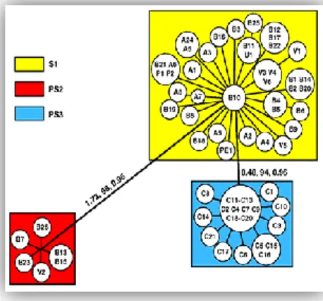
Recently, based on phylogenetic analysis of 65 isolates, three different phylogenetic species (S1, PS2, and PS3) of P. brasiliensis were recognized using a combined data set of eight regions in five nuclear loci (Matute et al. 2006) (Figure 1).
Figure 1. Unrooted tree showing the partitions found in P.
brasiliensis, based on combined data of five nuclear loci obtained with weighted maximum parsimony. S1 consists of a major group of isolates from Brazil, Argentina, Venezuela, Peru and Paraguay, PS2 represents a cryptic phylogenetic species of P. brasiliensis (so far, six isolates from Brazil and Venezuela) and PS3 forms a separate group of Colombian isolates. The values above the branches represent their individual support: the first is the tree length, the second is the weighted high bootstrap, and the third is the posterior probabilities (adapted from Matute et al. 2006).
Chapter 1
5 In addition, a clade of 17 genotypically similar isolates, including Pb01, a P.
brasiliensis isolate that has been the subject of intense molecular studies for many years, was
shown to exhibit great genomic and morphological divergence from the S1/PS2/PS3 species clade. This “Pb01-like” group was even recommended to be considered a new phylogenetic species, since it is strongly supported by all independent and concatenated genealogies, increasing enormously the genomic variation known in the Paracoccidioides genus (Carrero et al. 2008, Teixeira et al. 2009).
PCM has been found in 14 Latin American countries, with the highest incidence in Brazil (80%) followed by Venezuela and Colombia (Figure 2). Over 10 million people are estimated to be infected by this fungus but only up to 2% of them develop the disease (McEwen et al. 1995). Notheless, mainly due to world globalization and intense migratory connections, PCM has also been reported in other areas such as North America, Asia and Europe (Gushulak and MacPherson 2000).
Figure 2. Geographic distribution of PCM in Central and South America.
Incidence (low, medium and high) is labeled by different colours (adapted from Shikanai-Yasuda et al. 2006).
P. brasiliensis belongs to the Group 3 Biological Agents characterized by those
organisms that: i) can cause severe human disease and present a serious hazard to workers, ii) may present a risk of spreading to the community, but iii) there is usually efective prophylaxis or treatment available (DIRECTIVE 2000/54/EC. L 262-21-L 262/45. 18-9-2000). For these reasons, research developed on P. brasiliensis has to be carried out at a Biosafety Level 3 Facility characterized by special safety practices, equipment and facilities.
1.1.2 Pathogenesis and Clinical Manifestations
The long latency period of the disease and the lack of epidemic outbreaks also create difficulties in determining under which circumstances the primary infection occurs (Franco et al. 2000). However, with the accumulating epidemiological, clinical and experimental evidence, the pathogenic process is thought to occur via inhalation of airborne fungal propagules (e.g., mycelial fragments or conidia). Once inside the host, the propagules produced reach the pulmonary alveolar epithelium and transform into the parasitic yeast form (Franco 1987, McEwan et al. 1987, Brummer et al. 1993) that may lead to diverse clinical manifestations (Montenegro and Franco 1994).
Depending on host factors, strain-level virulence differences, and environmental conditions, a wide spectrum of clinical and pathological manifestations can be observed in these patients (Montenegro and Franco 1994), where the vast majority (up to 60 % in areas of endemicity) develop only asymptomatic or subclinical PCM (Souza et al. 2000), which sometimes progresses into a disease with a diversity of clinical forms. The disease presents two main clinical forms: the acute or sub-acute form (juvenile type), and the chronic form (adult type) (Franco et al. 1987, Lacaz 1994) (Figure 3). The acute or sub-acute clinical form affects mainly children and young adults of both sexes, representing only 3 to 5% of all cases. It is characterized by a rapid and severe evolution (weeks to months) with dissemination to the reticulo-endothelial system leading to a marked involvement and hypertrophy of spleen, liver, lymph nodes, and bone marrow. On the other hand, the chronic clinical form occurs mainly in adult males (approximately 80 to 90%) and can be restricted to only one organ or disseminated to several organs and tissues, occurring more frequently in the lungs, oral and laryngeal mucous membranes, skin, lymph nodes, and adrenal glands with a slower course of infection (months to weeks). Furthermore, whereas the acute or sub-acute clinical form of PCM results in a significant rate of mortality, the chronic clinical form leads to a considerable patient morbidity (Brummer et al. 1993). Nevertheless, other clinical settings have been recently more frequently detected. For example, the central nervous system has also been identified as a target for PCM (neuroparacoccidioidomycosis, NPCM) (Tristano et al. 2004, Fagundes-Pereyra et al. 2006, Pedroso et al. 2009).
Chapter 1
7
Figure 3. Clinical forms of Paracoccidioidomycosis. (A) Acute or juvenile form of PCM (patient with
mucocutaneous lesions). (B) Chronic or adult form of PCM (patient with extensive skin involvement and ulcerated lesions) (Corporación para Investigaciones Biológicas‟, CIB, archives).
One important feature of this systemic mycosis is its marked incidence in males, with an estimated ratio of male:female of approximately 48:1 (Restrepo and Greer 1983). Several studies have addressed this sex bias, suggesting that the mechanism underlying this process involves hormonal regulation. Exposure of P. brasiliensis to female hormones, such as estrogen, blocks the conidia- or mycelium-to-yeast transition (Restrepo et al. 1984, Salazar et al. 1988, Clemons et al. 1989), probably via a cytosolic steroid-binding protein (Loose et al. 1983), and thus contributing to a lower percentage of infected female (Aristizabal et al. 1998). In spite of these evidences, the molecular events that rule this particular phenomenon are still unknown.
1.1.3 Striking Characteristics of Thermodimorphism
An important feature of several fungal pathogens is their inherent ability to assume either a filamentous or a unicellular morphology in response to changes in environmental conditions when they infect host tissues (San-Blas at al. 2002). This process, broadly referred to as dimorphism, is an intrinsic genetic property of certain fungi, and appears to be linked to pathogenicity, since strains unable to undergo the morphologic transition are mostly avirulent (Maresca and Kobayashi 2000).
Temperature, nutritional factors, or both, are usually the agents that activate this change in morphology (San-Blas et al. 2002). However, in P. brasiliensis the ability to conduct a transition between the yeast and mycelial forms is only dependent on the temperature, making it amenable to study the molecular and biochemical events that regulate this phenomenon (Szaniszlo et al. 1983).
P. brasiliensis’ morphological shift is accompanied by extensive modifications in the
cell membrane and cell wall composition (Blas and Blas 1977, Blas and San-Blas 1994). Regarding cell wall composition, the mycelia-to-yeast transition is accompanied
by the switch in glucan polymer linkage in the cell wall from -1,3-glucan to -1,3-glucan, not only the quantity but also the spatial arrangement of these polysaccharides (San-Blas and San-Blas 1977, San-Blas and San-Blas 1994). Moreover the cell membrane lipid composition changes, particularly glycosphingolipids (Toledo et al. 1995). Although several efforts have been made to better understand the morphologic alterations, particularly those depending on the temperature, the lack of molecular tools to study this fungus has drastically hampered this line of research.
P. brasiliensis grows as a yeast form at 36-37 ºC in host tissues and culture media
such as Brain Heart Infusion agar or Sabouraud agar. Yeast colonies become visible within 3 to 7 days of incubation at 36 ºC (depending on the isolate) appearing soft, wrinkled and tan to cream (Figure 4A). Microscopically, yeast cells are multinucleated and of varying sizes (4-35 m), usually oval to elongated, and have a thick refractile cell wall and a cytoplasm that contains prominent lipid droplets (Restrepo and Jimenez 1980) (Figure 4C).
P. brasiliensis mycelia is usually cultured in modified synthetic McVeigh Morton agar
at temperatures ranging from 19 ºC to 28 ºC, producing within 15 to 30 days small, irregular, and white to tan colonies with short hairy looking mycelia (Figure 4B). Microscopically, hyphae are thin (1 to 3 m), multinucleated and septated structures (Figure 4D) from which arthroconidia and pedunculated and single-celled conidia are produced when specific isolates are cultured under conditions of nutritional deprivation. These conidia are uninucleated and round shaped structures measuring less than 5 m in diameter (Figure 4E-F), and respond to temperature changes, transforming either into yeast cells at 36 ºC or producing hyphae at lower temperatures.
Figure 4. P. brasiliensis yeast (36 ºC), mycelial (18 ºC) and conidial forms. Macroscopic features of colonies
from yeast (A) and mycelial form (B). Microscopic features of cells grown in batch culture from yeast (C) and mycelial form (D). Scanning electron microscopy analysis of conidia cells (E) and (F)(white arrows indicate intercalary conidia) (adapted from Wanke and Londero 1994 and Almeida, A.J et al. unpublished data).
Chapter 1
9
P. brasiliensis yeast form is generally characterized by a multiple budding phenotype
and a polymorphic cell growth, leading to the formation of cells with extreme variations in shape and size, as well as in the number of buds that are produced depending on the environmental nutritional media (Figure 4C). In compliance with these cellular features, conclusive histological diagnosis of PCM has traditionally relied on the identification of the most characteristic feature of the yeast form of P. brasiliensis, i.e., the pilot‟s wheel appearance of the mother cell surrounded by multiple peripheral daughter cells (Restrepo-Moreno 2003). More recently, it has been suggested that P. brasiliensis may follow an alternative control mechanism during cell growth and possess an unusual scenario for polarity establishment and maintenance. Consequently, there is the question of whether or not an accurate regulation of the cellular division exists.
According with P. brasiliensis features and the role of Ccd42p, a pivotal molecule in numerous cellular events, such as polarity signaling during growth and morphogenesis (Cotteret and Chernoff 2002), Almeida et al. (2009) evaluated the role of this Rho-like GTPase in the polymorphic morphology and virulence of this pathogenic fungus. In fact, they showed that a lower expression of PbCDC42 results in a decrease in the size of buds and mother cells, characterized by being more spherical, less elongated and polymorphic than wild-type cells, suggesting a differential control of the apical-isotropic switch during growth. This study also showed that silencing PbCDC42 facilitates P. brasiliensis phagocytosis and decreases virulence. Although Cdc42p has been implicated in a wide variety of cellular processes, there is still little insight into the mechanisms of action or the conservation of function for Cdc42p within these processes. While interacting with multiple regulators and effectors, Cdc42p seem to be conserved in most cell types, however it is dangerous to extrapolate precise Cdc42p functions or mutational phenotypes from one organism to another, given, for instance, the differences in phenotypes seen between analogous cdc42 mutants in Saccharomyces cerevisiae and Schizosaccharomyces pombe (Adams et al. 1990, Ziman et al. 1991, Miller et al. 1994). It is unfortunate that despite all the detailed information about Cdc42 functions and interacting proteins, there little experimental data addressing the specific mechanism(s) of action for Cdc42p in these different cellular processes (Johnson 1999).
1.1.4 Trends in Genome and Genetic Tools
Research on PCM and its etiological agent, P. brasiliensis, has promoted a fast growth with the introduction of molecular and chemical tools, opening new roads for a better understanding of this health problem. Research fields have been mostly directed towards the study of host-pathogen interactions and morphogenesis (San-Blas and Nino-Vega 2008), although the knowledge in other areas such as molecular diagnosis, epidemiology, immunology, taxonomy and genetics has also been enhanced. As in other pathogenic fungi, advances in pulsed-field gel electrophoresis (PFGE) methodologies and confocal fluorescence microscopy have allowed the genomic characterization and chromosomal mapping of P. brasiliensis (Montoya et al. 1997, Cano et al. 1998, Feitosa et al. 2003). The nuclear genome size was estimated by PFGE at around 30 Mb, and confocal fluorescence microscopy provided strong evidence to the haploid/diploid or even aneuploid nature of P. brasiliensis (Feitosa et al. 2003). DNA sequencing of ~50 Kb showed a density of one gene per 3.0-3.7 Kb, suggesting that 7500-9000 genes were present in its genome (Reinoso et al. 2005). More recently, new insights regarding P. brasiliensis genome size and ploidy were obtained by Almeida et al. (2007a). They used a flow cytometry (FCM) protocol to characterize cellular morphology and nuclei content of the various subpopulations discriminated during cell cycle profile analysis, a technique not only further informative concerning genome size and ploidy but also complementary to other DNA content quantification methods. Cell cycle analysis revealed a genome size ranging from 26.3 to 35.5 Mb per uninucleated yeast cell. The ploidy, assessed by comparing genome size by FCM with the average haploid size obtained from electrophoretic karryotyping, showed that P. brasiliensis present a haploid or at least an aneuploid DNA content. Cell cycle progression of P. brasiliensis yeast cells analyzed by flow cytometry, under different environmental conditions, seem to indicate that P. brasiliensis yeast cells may possess alternative control mechanisms during cell growth to manage multiple budding and its multinucleate nature (Almeida et al. 2006). Moreover, it is also imperative to mention the recent advances made by the Broad Institute, specifically the Fungal Genome Initiative (FGI) Project. This project, has already sequenced the genome of 3 P. brasiliensis‟s species, namely Pb01 (a clinical isolate from an acute form of PCM in an adult male), Pb03 (one of the better-studied isolates of PS2) and Pb18 (the representative of the major phylogenetic group S1).
Regarding genetic manipulation, crucial for establishing a link between in vitro analysis of DNA and its in vivo function (Magee et al. 2003), classical genetic tools as
Chapter 1
11 electroporation and protoplasting among others have proved inefficient in P. brasiliensis. In the light of this, Agrobacterium tumefaciens-mediated transformation system (ATMT) with transformation efficiency of 78 ± 9 transformants/co-cultivation (5 ± 1 transformants/106
target cells), was developed and used as molecular toolbox for this dimorphic fungus (Almeida et al. 2007b).
One specific approach to study gene function and molecular genetics in eukaryotic organisms has been direct gene targeting. For some time now, genetic manipulation of prokaryotes and eukaryotes and the technologies involved have been the focus of many research labs, promoting ongoing development of new methodologies ranging from gene knock-out/in, point mutations, and RNA silencing (Bertling 1995). Gene targeting is a method for modifying the structure of a specific gene without removing it from its genomic environment. This process involves the construction of a piece of DNA which is then introduced into the cell where it replaces or modifies the normal locus, and for several model organisms, such as S. cerevisiae, Drosophila melanogaster, Caenorhabditis elegans, Arabidopsis
thaliana and mice, the generation of knock-out/in mutants is already standardized (Bertling
1995). The method of disrupting, modifying or replacing a target gene is supported by the cellular machinery that requires the action of a branch pathway of DNA double-strand break (DSB) repair mechanism (Bertling 1995, Ninomiya et al. 2004). Eukaryotes have developed two main pathways to deal with this type of DNA damage: nonhomologous end-joining (NHEJ) and homologous recombination (HR), often described as “error-prone” and “error-free” respectively. The first, NHEJ, involves direct ligation of the strand ends independent of DNA homology, whereas the second, HR, involves interaction between homologous sequences (Shrivastav et al. 2008, Christmann et al. 2003).
The usage of NHEJ and HR depends on the phase of the cell cycle. NHEJ occurs mainly in G0/G1, whereas HR occurs during the late S and G2 phases (Takata et al. 1998, Johnson and Jasin, 2000). The NHEJ system involves the recognition of and binding to damaged DNA occurs by the Ku70–Ku80 complex. Thereafter, the Ku heterodimer binds to DNA–PKcs, forming the DNA–PK holoenzyme. DNA–PK activates XRCC4–ligase IV, which links the broken DNA ends together. Before re-ligation by XRCC4–ligase IV, the DNA ends are processed by the MRE11–Rad50–NBS1 complex, presumably involving FEN1 and Artemis. On the other hand, HR starts with nucleolytic resection of the DSB in the 5‟ → 3‟ direction by the MRE11–Rad50–NBS1 complex, forming a 3‟ single-stranded DNA fragment to which Rad52 binds. Rad52 interacts with Rad51, provoking a DNA strand exchange with the undamaged, homologous DNA molecule. Assembly of the Rad51
nucleoprotein filament is facilitated by different Rad51 paralogues (such as Rad51B, Rad51C and Rad51D, XRCC2 and XRCC3). After DNA synthesis, ligation and branch migration, the resulting structure is resolved (Christmann et al. 2003).
These mechanisms are conserved in evolution, but have different contribution to overall DSBs repair within different eukaryotic cells (Pastwa and Błasiak 2003). For example, the budding yeast S. cerevisiae repairs DNA DSB mainly by the HR system. However, it seems that most fungi and higher eukaryotes, including plants and mammals, have an “inefficient homologous integration system” using the NHEJ pathway to repair DNA DSB (Kooistra et al. 2004). Due to either high activity of NHEJ or poor efficiency of HR, targeting of genes at a desired locus in the dimorphic fungus P. brasiliensis is difficult being a rate-limiting step for further progress in this area.
The NHEJ has been blocked/downregulated in order to increase the rate of homologous integration of exogenous DNA in several fungal models including Neurospora
crassa, different Aspergillus spp., Cryptococcus neoformans, Claviceps purpurea, Magnaporthe grisea
and Sordaria macrospora among many others (Ninomiya et al. 2004, Goins et al. 2006, Pöggeler and Kück 2006, da Silva Ferreira et al. 2006, Haarmann et al. 2008, Villalba et al. 2008, Guangtao et al. 2009).
Very recently, another technique has been used in P. brasiliensis, namely antisense aRNA technology (Almeida et al. 2009, Hernández et al. 2010). Antisense refers to short DNA or RNA sequences, termed oligonucleotides, which are designed to be complementary to a specific gene sequence. The goal is to alter specific gene expression resulting from the binding of the antisense oligonucleotide to a unique gene sequence. Antisense technology was first effectively used in plants to alter the levels of various degradative enzymes or plant pigments, but was rapidly applied to mammalian cells. The exact mechanism by which antisense technology prevent protein production from a targeted gene remains uncertain. Proposed mechanisms include triplex formation, blocking RNA splicing, preventing transport of the mRNA antisense complex into the cytoplasm, increasing RNA degradation, or blocking the initiation of translation. Delivery of antisense oligonucleotides into target cells or the cell nucleus has been problematic. Currently, the most problematic aspect associated with antisense technology revolves around the specificity of their action. In some cases, non-specific antisense sequences, in other words, sequences which do not bind to the targeted gene or RNA, have prevented gene expression to the same degree as their sequence-specific antisense counterparts. This has led to considerable complication in data interpretation and requires detailed and careful data
Chapter 1
13 analysis. Since antisense technology focuses on preventing gene expression, it has been most widely applied to cancer gene therapy (Lichtenstein and Nellen 1997, Morcos 2007).
1.1.5 Laboratory Diagnosis and Molecular Identification
Histopathologic diagnosis is typically made through morphological criteria, mainly upon the identification of the characteristic multiple-budding feature of the yeast form (San-Blas et al. 2002). The isolation of the causative agent from clinical samples is essential to confirm accurate identification but is time-consuming since microbiological procedures may take over 20 days (Brummer et al. 1993). Alternatively, the diagnosis of PCM by indirect serological methods (e.g., complement fixation, agar-gel immunodiffusion and immunoenzymatic assays) that rely on antibody detection and is of considerable value due to the wide range of clinical presentations and the time-consuming procedures for the isolation of P. brasiliensis from clinical specimens (de Camargo et al. 1984, Cano and Restrepo 1987, Taborda and Camargo 1994, Ortiz et al. 1998). However, antibody levels may be absent in immunocompromised patients, or may remain present months after successful therapy (San-Blas et al. 2002). Nevertheless, serological diagnosis has sometimes shown altered sensitivity and specificity (may vary from 65 to 100 %) due to cross-reactivity with other fungi (e.g., H. capsulatum), or due to the use of undefined antigenic proteins derived from diverse cellular components of both yeast and mycelia forms of more than one P. brasiliensis strain (Lacaz et al. 1991, Freitas-Silva and Roque-Barreira 1992). However, advances in molecular biology have allowed the production of more reproducible and defined antigenic proteins through gene cloning and sequencing.
Molecular diagnosis of PCM has also been under development, with several primers being proposed as specific probes for clinical and field uses. For example, Goldani
et al. (1995) reported the cloning and sequencing of a species-specific 110 bp DNA
fragment generated by PCR from P. brasiliensis. Additionally, the specific DNA fragment from three different isolates of P. brasiliensis was amplified by PCR with primers mostly complementary to non-actin sequences of the 110 bp DNA fragments (Goldani et al. 1995). The literature shows that P. brasiliensis DNA samples have not only been applied in standard polymerase chain reactions (sPCR), but also in other amplification methods such as PCR-enzyme immunoassay (PCR-EIA), real time PCR (RT-PCR) and nested PCR (nPCR) in an attempt to improve test specificity/sensitivity (Goldani et al. 1995, Sandhu et
al. 1995, Sandhu et al. 1997, Diez et al. 1999, Bialek et al. 2000, Gomes et al. 2000,
1.1.6 Aim and outline of the thesis
The work presented during this thesis, was developed within the research line of the Microbiology and Infection Research Domain of the Life and Health Sciences Research Institute (ICVS), School of Health Sciences, University of Minho.
Despite recent advances, knowledge regarding the fundamental biology of P. brasiliensis has been hampered by the absence of appropriate genetic tools. Therefore to further contribute to elucidate biological phenomena of this pathogen, we aimed to develop a strategy for gene targeted mutagenesis. Moreover, to better understand the relationship between the mechanisms that regulate cellular division of P. brasiliensis and its distinctive multiple budding phenotype and particular morphological trait, we analyzed different biological parameters of yeast cell morphology of several isolates and expression of the Rho-like GTPase Cdc42.
Chapter 1 provides an overview on the current knowledge concerning the etiological agent P. brasiliensis and the systemic mycosis it causes, paracoccidioidomycosis. The methodologies used to study this dimorphic pathogenic fungus as well as molecular developments on P. brasiliensis are also addressed, leading the reader to Chapters 2 and 3.
In Chapter 2, the development of an efficient methodology for gene disruption in
P. brasiliensis was attempted. In summary, work developed throughout this chapter proposes
to down-regulate the non-homologous end joining pathway, by decreasing the expression levels of the Ku80 gene, a component of the DNA-dependent protein kinase catalytic subunit (the Ku70-Ku80 heterodimer), would increase homologous recombination, an essential tool to produce strains suitable to HR allowing future functional genomics studies. Chapter 3 focuses on the quantification of different morphologic aspects that the yeast form of this dimorphic fungus presents, both inter- and intra-strain. Moreover, the hypothetical role of PbCDC42 in the heterogeneous cell size and form of several P.
brasiliensis strains from the described cryptic species was addressed.
Finally, Chapter 4 presents the main conclusions bringing together Chapter 2 and 3 in the context of the initially proposed objectives, in order to provide a global perspective of the work and possible lines for future research.
Chapter 2
Development of Targeted Gene Disruption in
Paracoccidioides brasiliensis: Downregulation
Chapter 2
17
2.1 Introduction
The incidence of fungal infections has been increasing, particularly in patients who are immunocompromised by human immunodeficiency virus infection. Systemic fungal infections in such patients are often life threatening. Paracoccidioides brasiliensis, the causative agent of PCM is among this group of pathogenic fungi that are of clinical significance and scientific interest (Guarro et al. 1999, Restrepo-Moreno 2003, Chakrabarti and Shivaprakash 2005). Even though during the last decade molecular approaches have allowed a broader insight into P. brasiliensis, knowledge regarding the fundamental biology of this pathogenic fungus has been hampered by the absence of appropriate genetic tools (San-Blas et al. 2002, Felipe et al. 2005, Almeida et al. 2007). In particular, the development of an efficient gene targeted mutagenesis for gene disruption, vital to understand the role of specific genes in several biological processes such as dimorphism, pathogenesis, among others. In order to accomplish disruption, modification or replacement of a target gene investigators usually take advantage of the cellular machinery that requires the action of a DNA double-strand break (DSB) repair mechanism (Haarmann et al. 2008 Villalba et al. 2008). DSBs are the most severe form of DNA damage, mostly induced by ionizing radiation, some chemicals, like anticancer drugs, or arise spontaneously during DNA replication, and can result in cell death or a wide variety of genetic alterations including large- or small-scale deletions, loss of heterozygosity, translocations, and chromosome loss (Pastwa and Błasiak 2003, Shrivastav et al. 2008).
Eukaryotes have two main pathways to deal with this type of DNA damage: nonhomologous end-joining (NHEJ) and homologous recombination (HR) (Christmann et
al. 2003, da Silva Ferreira et al. 2006). In Saccharomyces cerevisiae the most important DSB
repair pathway is HR, while other organisms, such as humans, preferentially use NHEJ (Ninomiya et al. 2004). The NHEJ machinery for DSBs repair is mediated by the essential DNA binding Ku70-Ku80 heterodimer, which directs the DNA-PK to the end of a DSB, thus stabilizing its DNA binding, that along with other components execute crucial steps in NHEJ repair (Pöggeler and Kück 2006). Recently, da Silva Ferreira et al. (2006) disrupted
Aspergillus fumigatus gene homologous to Neurospora crassa KU80. Transformation of the A. fumigatus KU80 disruption yielded 80% transformants exhibiting integration at the
homologous site, compared to 3 to 5% for a wild-type recipient. To our knowledge, the rate-limiting step for further progress in the dimorphic fungus P. brasiliensis is the low percentage of homologous integration.
Thus, we decided to downregulate the P. brasiliensis KU80 gene in order to increase the integration of introduced exogenous DNA fragments by homologous recombination. Specifically, we amplified a DNA fragment from the cDNA library using degenerated primers designed based on Ku80p sequence alignment from Ajellomyces capsulatus,
Coccidioides immitis, Penicillium marneffei and Microsporum canis to isolate PbKU80 gene and
applied antisense RNA (aRNA) technology using oligonucleotides targeting the 5‟ untranslated region of PbKU80 to knockdown its expression in P. brasiliensis yeast cells.
2.2 Materials and Methods
2.2.1 Microorganisms and culture media
Escherichia coli XL-1-Blue
E. coli XL-1-Blue strain (Bullock et al. 1987) was used as host for plasmid amplification and
cloning. E.coli was grown on Luria Bertani (LB) medium (Sambrook et al. 1998) at 37 ºC. For bacterial selection, kanamycin, 50 mg/L, or ampicillin, 100 mg/ml were used as supplements.
Agrobacterium tumefaciens LBA1100
A. tumefaciens LBA1100 (C58C1 with a disarmed octopine-type pTiB6 plasmid) (Beijersbergen et al. 1992) was used as recipient for binary vectors. A. tumefaciens was maintained at 28 °C on LB medium containing spectinomycin, 250 mg/L and rifampicin, 20 mg/L. For selective purposes, kanamycin, 100 mg/L was employed.
During Agrobacterium tumefaciens-mediated transformation (ATMT) procedures, A.
tumefaciens LBA1100 containing the binary vector pUR5750 (conferring kanamycin
resistance in A. tumefaciens and E. coli) was applied (de Groot et al. 1998).
Paracoccidioides brasiliensis
P. brasiliensis yeast cells, strain ATCC 60855, Pb18 and Garcia, were maintained at 36 ºC by
periodic subculturing in brain heart infusion (BHI) solid medium (1.5% wt/vol agar). For the assays carried out in this study, yeast cells were grown in both BHI and modified synthetic McVeigh Morton (MMcM) liquid media (Restrepo and Jimenez 1980), at 36 ºC with aeration on a mechanical shaker (200 rpm).
Chapter 2
19
2.2.2 Cloning vectors
pCR35
The plasmid pCR35, kindly provided by C. A. Rappleye (Department of Microbiology, Ohio State University, Columbus, Ohio, USA) (Rappleye et al. 2004) is a 6823 bp vector that contains the GFP gene downstream from Histoplasma capsulatum promoter region calcium-binding protein (CBP1). Additionally it contains the neomycin phosphotransferase II gene (nptII), the E. coli selectable kanamycin resistance marker, the Podospora anserina URA5 gene (PaURA5) which encodes orotidine-5‟-monophosphate pyrophosphorylase that leads to uracil auxotrophy in Histoplasma capsulatum and one Ori.
pUR5750
The plasmid pUR5750 (de Groot et al. 1998) is a 15731 bp binary vector. This vector contains a transferred DNA (T-DNA) harbouring, between left and right border sequences, an E. coli hygromycin B phosphotransferase (HPH) gene driven by the
Aspergillus nidulans glyceraldehydes 3-phosphate (GPD) promoter and transcriptional
terminator (TRPC) from pAN7-1 and the A. tumefaciens selectable kanamycin resistance marker, neomycin phosphotransferase II gene (nptII), flanked by the nopaline synthase promoter (Pnos) and terminator (Tnos). Additionally it contains the neomycin phosphotransferase III gene (nptIII), the E. coli selectable kanamycin resistance marker.
2.2.3 Nucleotide and protein sequence analysis
Sequence similarity searches were performed using BLAST
(http://blast.ncbi.nlm.nih.gov/Blast.cgi). Both nucleotide and protein sequence alignments were carried out with the DNAMAN Version 4.0 program (Lynnon BioSoft).
2.2.4 Cloning of
P. brasiliensis
aRNA PbKU80 oligonucleotides and
binary vector construction
DNA and total RNA from exponentially growing P. brasiliensis yeast cultures were extracted using Trizol standard procedures (van Burik et al. 1998).
Recombinant DNA manipulations, agarose gel electrophoresis and polymerase chain reaction (PCR) were conducted according to Sambrook et al. (1998). Plasmid purification was carried out using the Quiagen® plasmid purification procedure.
E. coli transformation by heat shock was performed as described elsewhere
Binary vector was mobilized to A. tumefaciens LBA1100 by electroporation as described by Dulk-Ras and Hooykaas (Dulk-Ras and Hooykaas 1995). Briefly, after preparation of A. tumefaciens cells for electroporation, bacterium cells were thawed on ice. 2 to 3 L of plasmid DNA were added to 40 L of cell suspension and transferred to an electroporation cuvet. Electroporation was done applying an electric pulse at 2.5 kV, 25 F and 200 Ω with a time constant of approximately 4.7 ms. After A. tumefaciens cells recovery in 1 mL of SOC medium at 29 ºC for 2 h with shaking (180 rpm), aliquots of cell mixtures were inoculated onto LB selection plates and incubated at 28 ºC for 3 days.
The parent plasmids used in this study are summarized in Table 2. The parent plasmid for pCR35::A1ku80 and pCR35::A2ku80 was pCR35, and the parent plasmids for pUR5750::A1 and pUR5750::A2ku80 were pCR35::A1, pCR35::A2ku80 and pUR5750.
PCR amplification was performed using degenerate primers (Deg1 and Deg2, Table 1) designed based on protein sequence alignment of the Ku80p from Ajellomyces capsulatus (GenBank accession number GG663365), Coccidioides immitis (ac. no. XP_001248033),
Penicillium marneffei (ac. no. XP_002151653) and Microsporum canis (ac. no. EEQ38520) using
the cDNA library of P. brasiliensis produced in our laboratory. The sequence of the 1315-bp PCR product (STAB VIDA, Oeiras, Portugal) was used to design 4 primers to obtain 2 antisense molecules (A1ku80 and A2ku80) from the 5‟ UTR region using the primers in Table 1 (F1/R1; F2/R2).
The obtained antisense fragments were digested with AscI and XhoI and cloned into the AscI/XhoI site of pCR35. The ligated DNA was introduced in E. coli by heat shock and positive clones selected on LB kanamycin plates. To identify pCR35::A1ku80 and pCR35::A2ku80 colony PCR was performed using the external primers (P3/P4) for A1/A2ku80 cassette. 200 ng of DNA were added to 18 µL of reaction mixture defined by reaction buffer 1×, 2 mM MgCl2, 200 µM dNTP, 200 µM of each primer and 1 unit of Taq
polymerase. The PCR reaction cycling was as follows: 1 cycle at 94 ºC for 10 min, 35 cycles at 94 ºC for 15 s, 58 ºC for 30 s, 72 ºC for 2 min, and 1 final cycle at 72 ºC for 10 min. Some pCR35::A1/A2ku80 positive clones were then selected for a second screening of colony minipreps by digesting the DNA with KpnI, which originated one fragment with 1823 bp for pCR35::A1ku80 and 1809 bp for pCR35::A2ku80, confirming the insertion of the A1/A2ku80 fragments on pCR35.
Binary vectors were constructed by cloning the KpnI fragments from pCR35::A1/A2ku80 into the KpnI site of the binary vector pUR5750. The ligated DNA was introduced in E. coli. Selection was performed on LB kanamycin plates. In order
Chapter 2
21 identify positive clones, colony PCR was performed using the external primers (P5/P6) for A1/A2ku80 cassette as described above. Positive pUR5750::A1/A2ku80 clones were selected for a colony miniprep sceening digesting the DNA with KpnI, which provided a digestion pattern of 2 fragments of 15731 and ≈1800 bp, respectively. The obtained binary vector was mobilized to A. tumefaciens LBA1100 ultracompetent cells by electroporation as described above and transformants were isolated by kanamycin selection at 50 mg/L.
Table 1. Primers used in this study for molecular cloning, analysis of transformants,
and real-time polymerase chain reaction (RT-PCR). Primer sequences (5’-3’) a Deg1-AARGTNCCNCCNAARGCNAAR Deg2-GGNTTYAARGARGARGAYAAR F1-ccgctcgagTTATGATCAATTTTTCCTCC R1-ggcgcgccTACCATGGTGACAATTGGCC F2-ccgctcgagAGGAAAACCGCGAATCTCGG R2-ggcgcgccATTAATCTGGCTTATGTCTT P3-ggggtaccCCGCGGATCACGGTATCGATGA P4-ggggtaccCCGGTACCTAGGTGGATCCAAT P5-ggggtaccGATCGGTGCGGGCCTCTTCG P6-ggggtaccCATGACGGCCATCATGCCAA Hyg1-ATGCCTGAACTCACCGCGAC Hyg2-TTCTACACAGCCATCGGTCC RT-PCR P1 Pbku80-GGAGCTACAAGCTCAAAGCAA P2 Pbku80-GCCTGAAAGTGGCTTTTCTG P1 Pbtub2-AGCCTTGCGTCGGAACATAG P2 Pbtub2-ACCTCCATCCAGGAACTCTTCA
a Low caps indicate restriction enzyme sequence recognition sites.
bAntisense primers.
Table 2. Features of the parent plasmids used in this study. Plasmid Features between RB and LB Resistance gene for selection in
E. coli
Resistance gene for selection in A.
tumefaciens
Resistance gene for selection in P.
brasiliensis
pCR35 n.a. Kanamycin n.a. n.a.
pUR5750 Pnos::ntpII::Tnos Ttef::hph::Ptef Kanamycin Kanamycin Hygromycin B
Hph, hygromycin B phosphotransferase gene; LB, left border; n.a., not applicable; nptII, neomycin phosphotransferase II gene; Pnos, nopaline synthase promoter; Ptef, translation elongation factor-1 α promoter; RB, right border; Tnos, nopaline synthase terminator, Ttef, translation elongation factor-1 α terminator.
2.2.5
Paracoccidioides brasiliensis
ATMT procedures
A. tumefaciens LBA1100 carrying the desired binary vector was grown overnight on
LB liquid medium with antibiotics in a water bath, at 28 °C with shaker (180 rpm). 1 ml of the cell culture was spun down and washed with induction medium (IM) (Bundock et al., 1995) with acetosyringone (AS), 200 M, and antibiotics. Bacterial cells were diluted in IM to an OD660nm of 0.30, and re-incubated at 28 °C until an OD660nm of approximately 0.80.
P. brasiliensis yeast cells were grown in BHI or MMcM batch cultures to the exponential
growth phase (48 to 60 h).
P. brasiliensis yeast cell samples were centrifuged (4500 g for 15 min), washed with
IM and adjusted to a final concentration of 1 × 10⁸ cells/ml using direct microscopic
counts (Neubauer counting chamber procedures). Different ratios of A. tumefaciens and P.
brasiliensis cells were mixed (to a final volume of 120 l) and inoculated onto sterile Hybond
N membrane (Amersham Biosciences, Piscataway, NJ, USA) on solid IM for co-cultivation at 25 ºC for 3 days. Prior to incubation, co-cultivation plates with cellular mixtures were air dried in a safety cabinet for 30 min. Following co-cultivation, membranes were transferred to tubes with 2 mL non-selective BHI liquid medium containing cefotaxime, 200 mg/L, for growth inhibition of A. tumefaciens, and cells were dislodged by aid of a spatula and vortexing for 1 min. The cell suspension was recovered for 48 h at 36 ºC, 200 rpm, before plating in selective media (HygB 50 g/ml). Selection plates were incubated at 36 ºC for 15 to 20 days and monitored for colony forming ability.
Transformation efficiency was determined by taking into account P. brasiliensis yeast cell concentration at the moment of co-cultivation and the number of hygromycin resistant (HygR) colonies
.
2.2.6 Analysis of transformants by PCR
Randomly selected HygR putative transformants were tested for the presence of the
T-DNA by PCR amplification of the HPH gene using total DNA as template. Fungal DNA extractions were performed on 72 h cultures using mechanical disruption with glass beads as described elsewhere (van Burik et al. 1998). 200 ng of DNA were added to 20 µL final volume reaction mixture defined by reaction buffer 1×, 2 mM MgCl2, 200 µM dNTP, 200 µM of each primer and 1 unit of Taq polymerase. The PCR reaction cycling was as follows: 1 cycle at 94 ºC for 10 min, 40 cycles at 94 ºC for 15 s, 56 ºC for 30 s, 65 ºC for 60 s, and 1 final cycle at 65 ºC for 10 min. A fragment of 982 bp was amplified using the
Chapter 2
23 primers indicated in Table 3 (Hyg1/Hyg2). Two different control samples were applied during PCR procedures: pUR5750 plasmid DNA and P. brasiliensis ATCC60855 wild type DNA.
2.2.7 Mitotic stability
HygR P. brasiliensis colonies were randomly selected and serially transferred to
non-selective BHI solid medium for at least three times. Following these passages, transformants were serially inoculated on plates with non-selective and selective medium (50 μg/ml) for three consecutive rounds to examine mitotic stability. Previously tested for mitotic stability, transformants were then selected for posterior assays.
2.2.8 Real-time polymerase chain reaction (RT-PCR)
Total RNA from exponentially growing P. brasiliensis yeast cultures was extracted using Trizol (Invitrogen, Carlsbad, CA, USA) standard procedures and heat shock treatment (20 min at 65 ºC followed by 60 min at -80 ºC) for cellular disruption. Total RNA (0.5-1 μg) was reverse transcribed using the iScript cDNA Synthesis kit (Bio-Rad, Marnes La Coquette, France). For RT-PCR quantification, 2 μl of the reverse-transcribed RNA was used as template for the LightCycler FastStart DNA Master SYBR Green I kit (Roche, Nutley, NJ, USA) to amplify the P. brasiliensis Ku80 gene (184 bp) and the TUB2 gene (64 bp) using the primers indicated in Table 4. The thermal cycling conditions comprised an initial step at 94 °C for 15 min, followed by 50 cycles at 94 °C for 10 s, 60 °C for 10 s and 72 ºC for 20 s, a melting step of 55º-95 ºC (0.5 ºC/s), and a final cooling at 40 ºC for 30 s. Real-time quantification was performed on a LightCycler System (Roche) using threshold cycle (Ct) values for TUB2 transcripts as the endogenous reference. mRNA differential Ku80 expression levels were evaluated by normalizing Ku80 Ct values with the reference and comparing the ratio amongst the tested samples.
2.3 Results and Discussion
2.3.1 Isolation of
PbKU80
and construction of Aku80
Despite new advances on molecular manipulation of P. brasiliensis, specifically the recent development of an efficient transformation and a gene expression and interference system developed by our group (Almeida et al. 2007b, 2009), functional genetic studies in P.
brasiliensis has been impaired due to the absence of tractable molecular techniques, namely
an efficient procedure allowing the generation of gene-knockout strains by HR.
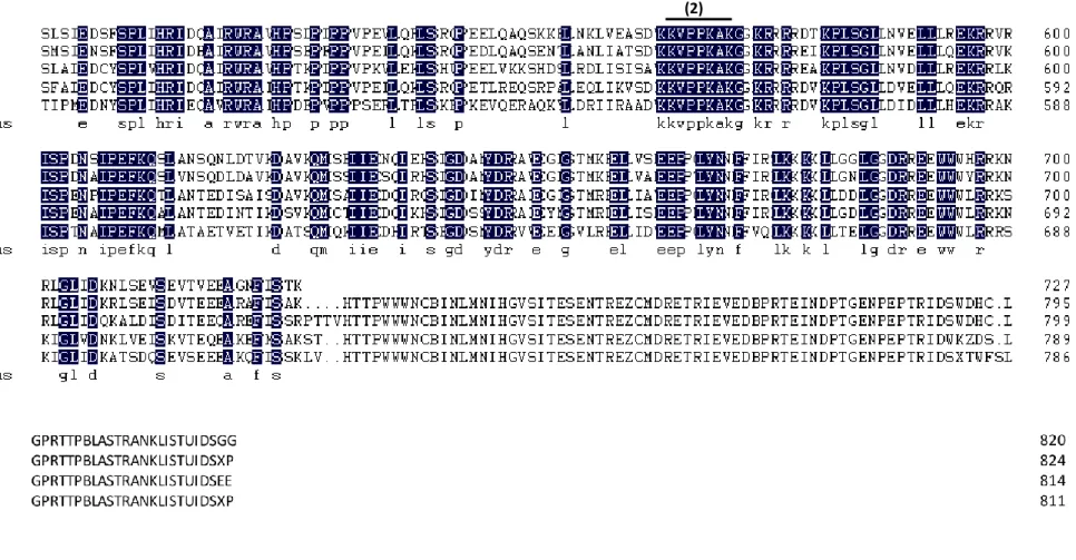
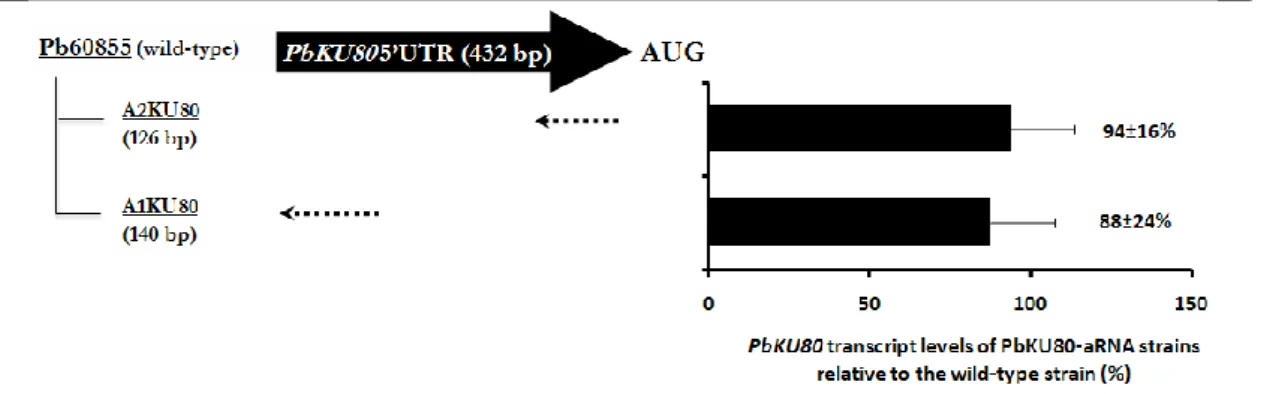
As formerly stated, the rate of homologous integration of exogenous DNA can be increased by blocking/downregulating NHEJ key players‟s function such as Ku80 (Ninomiya et al. 2004, Goins et al. 2006). In this sense, we established an antisense RNA (aRNA) system to downregulate the expression of PbKU80. Thus, to isolate P. brasiliensis KU80 (PbKU80) homolog, degenerate primers were designed from the highly conserved amino acid sequences GFKEEDK and KVPPKAK (positions 175-181 and 565-571 respectively) based on the alignment of counterparts from different organisms (Figure 1). Two DNA fragments of 888 and 675 bp were obtained by PCR on P. brasiliensis cDNA library. The DNA sequence analysis of these fragments revealed an incomplete open reading frame (ORF) with high degree of similarity to KU80 ORFs from other organisms (data not shown). The sequences of the isolated cDNA fragments revealed a 769-bp ORF flanked by a 342-bp 5‟ untranslated region (5‟UTR) and by a 186-bp 3‟ untranslated region (3‟UTR) together with a poly(A) tail (Figure 2). Then, with two sets of primers targeting the
PbKU80 5‟UTR (Figure 2A), two different aRNA molecules (A1KU80 and A2KU80) of
140 and 126-bp were amplified for independent downregulation of PbKU80 gene. To allow the construction of antisense molecules, specific restriction sites, namely XhoI and AscI were respectively added in the 5‟ and 3‟ ends of both sequences.
The repressing oligonucleotides were afterward individually cloned in pCR35 under the control of the Calcium-Binding Protein Promoter (CBP1) region from H. capsulatum (Rapleye et al. 2004).
Chapter 2
25
Figure 1. Multiple sequence alignment of the deduced amino acid sequence of P. brasiliensis KU80 (Pbku80p), Ajellomyces capsulatus KU80 (Acku80p), Coccidioides immitis KU80
(Ciku80p), Microsporum canis KU80 (Mcku80p) and Penicillium marneffei KU80 (Pmku80p). In black are underlined the degenerated primers (1) Deg2 and (2) Deg1.
Chapter 2
27
-342 TCAGGTTATG ATCAATTTTT CCTCCTCTTC TCCCTCTTCA TCTTTCCATG TCCAAGTGCA -282 GTACTAAGAA GAAAGATGGC GGACAAGGAA GCCACTGTGT ATATTGTGGA TGTCGGAAAA
-222 TCTATGGCCA ATTGTCACCA TGGTAGATCT ATATCAGACC TCGAGTGGGC GATGCGCTAT
–162 GTCTGGGACA AGATCACAAC CACGGTTGCC ACTGGCAGGA AAACCGCGAA TCTCGGAGTA -102 ATTGGTCTCA AAACAGATCG CTCTGACAAT CCATTATGGG AGAAAGAGGA AGAGAAAAGC
-42 TATGCCAATT TGACCGTTTT TCAAGACATA AGCCAGATTA AT
1 ATG GCG GAC AAG GAA GCC ACT GTG TAT ATT GTG GAT GTC GGA AAA TCT Met Ala Asp Lys Glu Ala Thr Val Tyr Ile Val Asp Val Gly Lys Ser 49 ATG GCC AAT TGT CAC CAT GGT AGA TCT ATA TCA GAC CTC GAG TGG GCG Met Ala Asn Cys His His Gly Arg Ser Ile Ser Asp Leu Glu Trp Ala 97 ATG CGC TAT GTC TGG GAC AAG ATC ACA ACC ACG GTT GCC ACT GGC AGG Met Arg Tyr Val Trp Asp Lys Ile Thr Thr Thr Val Ala Thr Gly Arg 145 AAA ACC GCG AAT CTC GGA GTA ATT GGT CTC AAA ACA GAT CGC TCT GAC Lys Thr Ala Asn Leu Gly Val Ile Gly Leu Lys Thr Asp Arg Ser Asp 193 AAT CCA TTA TGG GAG AAA GAG GAA GAG AAA AGC TAT GCC AAT TTG ACC Asn Pro Leu Trp Glu Lys Glu Glu Glu Lys Ser Tyr Ala Asn Leu Thr 241 GTT TTT CAA GAC ATA AGC CAG ATT AAT ATG CCT CAA ATC CGC GAA CTG
Val Phe Gln Asp Ile Ser Gln Ile Asn Met Pro Gln Ile Arg Glu Leu 289 CGT AAA GCG ATC AAA ATC AGC AAT ACA ACT GAA GGA GAC GCA ATA TCA Arg Lys Ala Ile Lys Ile Ser Asn Thr Thr Glu Gly Asp Ala Ile Ser 337 TCC CTT ATC TTG GCG ATT GAT ATG ATT GTA CGA TAC TGC AAG AAA TTG Ser Leu Ile Leu Ala Ile Asp Met Ile Val Arg Tyr Cys Lys Lys Leu 385 AAA TAC AAA AGG AAA GTC GTC CTT GTT ACG GAT GGA ACA GGT GCT ATG Lys Tyr Lys Arg Lys Val Val Leu Val Thr Asp Gly Thr Gly Ala Met 433 GAT ACG GAT GGG ATG GAG GGA ATT GTA TCC AAA ATA AAC GAG GAA TCG Asp Thr Asp Gly MET Glu Gly Ile Val Ser Lys Ile Asn Glu Glu Ser
481 ATT GAA CTT GTA GTC CTG GGT GTA GAT TTC GAT GAC CCA GAG TAC GGA Ile Glu Leu Val Val Leu Gly Val Asp Phe Asp Asp Pro Glu Tyr Gly 529 TTC AAG GAA GAG GAC AAG
Phe Lys Glu Glu Asp Lys
1 AAG GTA CCA CCC AAA GCC AAA GGT CTC AAA CGA GTC AGA GAC ACA GAA Lys Val Pro Pro Lys Ala Lys Gly Leu Lys Arg Val Arg Asp Thr Glu 49 AAG CCA CTT TCA GGC TTA AAC GTC GAA GAG CTT CTC CGG ACG GAG AAA Lys Pro Leu Ser Gly Leu Asn Val Glu Glu Leu Leu Arg Thr Glu Lys 97 CGT GTG AGG ATA TCC CCG GAC AAC TCC ATA CCC GAA TTC AAG CAG TCC Arg Val Arg Ile Ser Pro Asp Asn Ser Ile Pro Glu Phe Lys Gln Ser 145 CTG GCG AAT TCT CAG AAC CTT GAT ACA GTC AAG GAT GCC GTT AAG CAG Leu Ala Asn Ser Gln Asn Leu Asp Thr Val Lys Asp Ala Val Lys Gln 193 ATG TCC CCC ATC ATA GAA AAC CAA ATA GAG CAT AGC TTG GGC GAT GCC Met Ser Pro Ile Ile Glu Asn Gln Ile Glu His Ser Leu Gly Asp Ala 241 AAC TAT GAT CGG GCC GTT GAA GGC TTG GGC ACC ATG AAA GAA GAG CTG Asn Tyr Asp Arg Ala Val Glu Gly Leu Gly Thr Met Lys Glu Glu Leu
A
B
F1 F2 R2 R1289 GTT TCC TTT GAG GAG CCT GGT CTA TAT AAC GAT TTC ATC CGT AGC TTG Val Ser Phe Glu Glu Pro Gly Leu Tyr Asn Asp Phe Ile Arg Ser Leu 337 AAA GCG AAA CTG CTT GGG GGT GAA CTC GGT GGA GAT AGG CGC GAG ATG Lys Ala Lys Leu Leu Gly Gly Glu Leu Gly Gly Asp Arg Arg Glu Met 385 TGG TGG CAT GTG AGG AAG AAT AGA CTG GGG TTG ATT GAT AAG AAT CTC Trp Trp His Val Arg Lys Asn Arg Leu Gly Leu Ile Asp Lys Asn Leu 433 TCA GAA GTC TCT GAA GTG ACG GTG GAA GAA GCT GGG AAT TTT TTG TCC Ser Glu Val Ser Glu Val Thr Val Glu Glu Ala Gly Asn Phe Leu Ser 481 ACA AAA TGA
Thr Lys ***
+1 GTACACTAGT TTCGGTGAGA ATATTCCTGC CATGGTTTTG GAGTGTTCGA CATTATGAGC +61 GAATCCGTAT CCATACACGC CCCCTCAATG GATGATTGAT CATCAGGGTA GATTCCTTGT +121 CTTAAGTCTA ATAAAATTGG ATTATTCATA TTATCGCGAA AAAAAAAAAA AAAAAAAAAA +181 AAAAAA
Figure 2. Nucleotide and deduced amino acid sequence of the incomplete ORF of P. brasiliensis cDNA
encoding the KU80 homolog. The PbKU80 cDNA sequence contains an incomplete 1035-bp open reading frame flanked upstream by a 342-bp 5‟ untranslated region (A) and downstream by a 186-bp 3‟ untranslated region (B). In grey are highlighted the sequences used for the construction of A1KU80 aRNA and A2KU80 aRNA.
A1KU80 and A2KU80 aRNA constructs were amplified together with the flanking CBP-1 promoter and cat-B terminator, with KpnI restriction sites, and cloned into the binary vector pUR5750. This is a crucial and helpful tool for A. tumefaciens-mediated transformation, for the single copy insertion of the cassette (T-DNA) with the antisense oligonucleotide into P. brasiliensis genome.
2.3.2 Cloning strategy
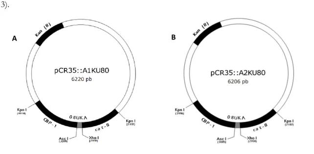
The strategy pursued in this study resulted in the construction of four plasmids, pCR35::A1KU80, pCR35::A2KU80, pUR5750::A1KU80 and pUR5750::A2KU80 (Figure 3).
Chapter 2
29
Figure 3. (A) pCR35::A1KU80, (B) pCR35::A2KU80, (C) pUR5750::A1KU80 and (D) pUR5750::A2KU80
vectors. For vector properties see Table 2, and for construction strategies see Material and Methods. Hph (R), hygromycin resistance selectable marker; Kan (R), kanamycin resistance selectable marker; CBP-1, calcium-binding protein promoter; 08UKA, antisense oligonucleotides of PbKU80 gene; cat-B, intergenic region downstream of the CATB gene as transcriptional termination signal; LB, left border; LR, right border.
In Figure 4 is represented the strategy used to clone the antisense oligonucleotides into the vectors to carry out gene transfer into P. brasiliensis.
Figure 4. Scheme of the strategy used for cloning the antisense molecules, as described in Materials and
Methods.