Article
*e-mail: rgsberlinck@iqsc.usp.br
A Method for Dextruxin Analysis by HPLC-PDA-ELSD-MS
Raquel P. Morais,a Simone P. Lira,b Mirna H. R. Seleghimc and Roberto G. S. Berlinck*,a
aInstituto de Química de São Carlos, Universidade de São Paulo,
CP 780, 13560-970 São Carlos-SP, Brazil
bEscola Superior de Agricultura “Luiz de Queiroz”, Universidade de São Paulo,
CP 9, 13418-900 Piracicaba-SP, Brazil
cDepartamento de Ecologia e Biologia Evolutiva, Universidade Federal de São Carlos,
CP 676, 13565-905 São Carlos-SP, Brazil
Destruxinas (Dtx) são ciclodepsipetídeos produzidos por fungos entomopatogênicos, que são utilizados como controle biológico em insetos pragas em diferentes agriculturas. A presente investigação reporta uma nova abordagem para análises de destruxinas produzidas por uma linhagem do fungo Beauveria felina, utilizando-se de LC-PDA-ELSD-MS. Em comparação
com os métodos anteriores, a nova abordagem utiliza-se de uma limpeza prévia da amostra em cartuchos C18 que removem efetivamente os constituintes do meio de cultura. Além disso, o uso dos solventes MeCN/MeOH 50:50, (v/v) como eluentes mais fortes no sistema de gradiente em 0,1% de H2O demonstrou fornecer a melhor resolução dos picos cromatográicos. Detecções simultâneas usando arranjo de fotodiodos (PDA), detector de espalhamento de luz evaporativa (ELSD) e espectrometria de massas (MS) indicaram praticamente uma resposta idêntica de todos os detectores na análise das destruxinas. Cinco amostras obtidas da cultura de B. felina foram analisadas, e indicaram a presença de 20 destruxinas conhecidas e de 6 ciclodepsipetídeos ainda não reportados. Considerando a redução do uso do MeCN, e a eicácia do ELSD como detector para destruxina, o método prova que pode ser de grande valia e de baixo custo operacional para controle de qualidade nas análises de destruxinas produzidas por linhagens de fungos.
Destruxins (Dtx) are cyclodepsipeptides produced by enthomopathogenic fungi, which are used in biological control of different agricultural insect plagues. The present investigation reports a new approach for analysis of destruxins produced by the fungal strain Beauveria felina, using LC-PDA-ELSD-MS. Compared to previous methods, the
new approach uses a clean-up on C18 cartridges, which effectively removes growth media constituents. Moreover, the use of 50:50 (v/v) MeCN/MeOH as the strongest eluting solvent in a gradient system over 0.1% H2O proved to give a better resolution of chromatographic peaks. Simultaneous detection using photodiode array (PDA), evaporative light scattering detector (ELSD) and mass spectrometry (MS) indicated a practically identical response of all detectors for destruxins analysis. Five samples obtained from the culture media of B. felina
were analysed, and indicated the presence of twenty known destruxins and six yet unreported cyclodepsipeptides. Considering the reduced use of MeCN, and the effectiveness of ELSD as a detector for destruxins, the method proved to be valuable and cost-effective for quality control analysis of destruxin-producing fungal strains.
Introduction
Destruxins (Dtx) are cyclodepsipeptides isolated from entomopathogenic fungal strains of economic importance. Dry spores of the fungus Metarhizium anisopliae are currently used in biological control of
agricultural plagues such as several Hemiptera and Lepidoptera species affecting sugar cane cultures and citrus plantations.1-3 Dry spores of Beauveria bassiana
are also used against infestations of banana plants by
Cosmopolites sordidus (Coleoptera: Curculionidae),
which causes death and a significative loss of fruit productivity.3 Both M. anisopliae and B. bassiana produce
destruxin cyclodepsipeptides and related metabolites as the active compounds against the insect plagues.4,5 Since
the cyclodepsipeptides production by these and related fungal strains is affected by growth and environmental conditions,6-10 the biological activity of the strains used
commercially can be inluenced if the compounds are not produced in suficient amount and chemical variety to maintain a high level of entomopathogenicity. Therefore, it is important to establish an analytical procedure to evaluate the composition of cyclodepsipeptides obtained from fungal cultures used in biological control on insect plagues.11
Although several approaches have been developed to detect and identify destruxins from different strains of
M. anisopliae,6,8,12-18 high cost instrumentation have been
used to identify destruxins, which are unaffordable for regular use in quality control of the culture media from entomopathogenic fungi used for biological control in several developing countries.
We have recently isolated a marine-derived strain of
B. felina fungus which is a source of new and known
destruxins.19,20 Therefore, we have been interested to
develop an universal and cost effective method for destruxins analysis, in order to detect a large variety of such compounds potentially useful for insect plague biological control. Herein we present and discuss a new method using HPLC-PDA-ELSD-MS for the analysis of destruxins mixtures obtained from B. felina, and provide
evidence that that this method is cost effective to analyse culture media of destruxin-producing fungi.
Experimental
Materials and reagents
Beauveria felina fungal strain has been previously
isolated from a marine alga of the genus Caulerpa.19,20
A voucher specimen has been deposited in the Brazilian collection CBMAI (reference # 738 and 739). Standards
used for monitoring destruxins were pseudodestruxin C, [Phe3, N-Me-Val5] destruxin B and roseotoxin B, previously
isolated from B. felina and identified by analysis of
NMR and MS data.19 The chemical structures of the
cyclodepsipeptides are shown in Figure 1. Water was purified using a double filtering system Rios/Milli-Q Gradient A 10 system (Millipore, Billerica, MA, USA). Acetonitrile, methanol and formic acid were of HPLC grade (J. T. Baker or Mallinkrodt).
Apparatus
HPLC analyses were performed using a Waters Alliance 2695 coupled online with a Waters 2996 photodiode array detector, followed by a Waters 2424 evaporative light scattering detector and a Micromass ZQ2000 mass spectrometry detector with an electrospray interface. Separations were performed on a C18 reversed-phase Waters X-terra (2.1 × 50 mm, 3.5 µm particle size) with a mobile phase low rate of 0.5 mL min–1. The mobile
phase consisted of (A) H2O containing 0.1% formic acid and (B) 1:1 (v/v) MeOH/MeCN containing 0.1% formic acid. A linear gradient elution program was applied as follows: 0-1.0 min hold on 10% B, 1.0-20.0 min linear gradient to 100% B, 20.0-25.0 min hold on 100% B, 25.0-30.0 min hold on 50% B for reequilibration. The total run time was of 30 min. The injection volume was of 20 µL. The pressure limits was established as follows: lowest 0 Psi, highest 5000 Psi; during elution, the highest pressure was 2200 Psi.
Determination was performed using three detectors online: a photodiode array UV detector, followed by an evaporative light scattering detector and a single quadrupole mass spectrometry detector. The photodiode array scanned the samples at λmax 205 and 254 nm. The evaporative light scattering detector condition was optimized to the following conditions: drift tube temperature 75 °C, gas pressure (N2) 50 Psi, Nebulizer 60%. The mass spectrometer detector was optimized to the following conditions: capillary voltage 3.00 kV, source block temperature 100 °C, desolvation temperature 350 °C, operating in electrospray positive mode, detection range 300-900 Da with total ion count extracting acquisition. The cone and desolvation gas low were 50 and 350 L h−1, respectively, and were obtained from
Preparation of standard solutions
Accurately weighed samples of each cyclodepsipeptide standard were dissolved in MeOH to prepare stock solutions of 1.0 mg mL−1. The stock solutions were kept closed in
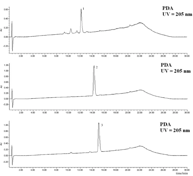
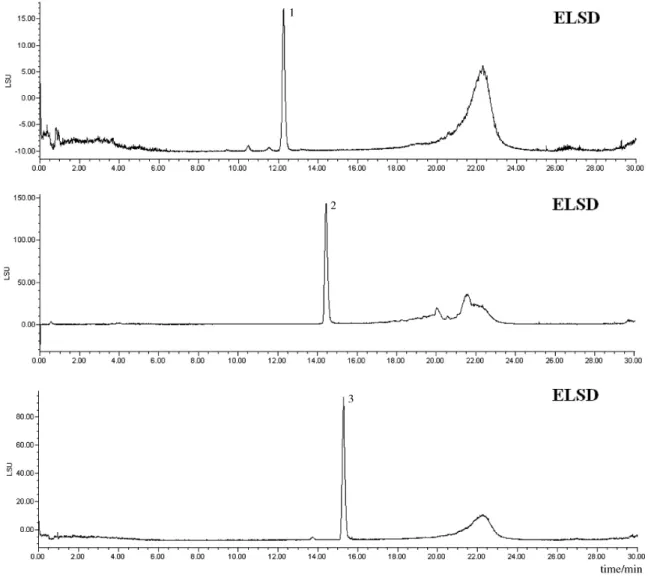
appropriate vials, at 4 °C until needed. The PDA, ELSD and MS chromatograms of the three cyclodepsipeptide standards are shown in Figures 2-5.
Sample preparation
B. felina was grown in 500 mL erlenmeyer flasks
containing 250 mL of MF broth as a culture medium. After 14 days of incubation at 28 °C, the culture medium and mycelia suspension were iltered through a 0.7 µm membrane. The iltered liquid medium was adsorbed into a solid-phase extraction (SPE) C18 Waters SepPak cartridge (5 g). Dessorption was performed using: 75:25 (fraction 1),
50:50 (fraction 2), 25:75 (fraction 3) H2O/MeOH and 100% MeOH (fraction 4). All fractions were dried in a Speedvac Savant high vacuum centrifuge. Dried samples were accurately weighed and solutions were prepared at a concentration of 1.0 mg mL−1.
Peak identiication
Isobaric cyclodepsipeptides were identiied by analysis of MS spectra and comparison with retention times of standards and literature data.5
Method validation
The validation protocol was performed following literature procedures.21,22 Acceptable values were deined
for the following parameters: selectivity, precision (repeatability and intermediate precision) and stability.
Compound Name MW (DA) R1 R2 R3 R4 R5 R6 R7 n
1 Dtx E diol 611 CH(Me)CH2Me Me H Me Me CHOHCH2OH H 1
2 Dtx Ed1 625 CH(Me)CH2Me Me H Me Me CHOHCH2OH H 2
3 Chlorohydrin Dtx A4 643 CH(Me)CH2Me Me Me Me Me CHOHCH2Cl H 1
4 Dtx A1 591 CH(Me)CH2Me Me H Me Me CH=CH2 H 2
5 Dtx A4 591 CH(Me)CH2Me Me Me Me Me CH=CH2 H 1
6 Roseotoxin B 591 CH(Me)CH2Me Me H Me Me CH=CH2 Me 1
7 Dtx B1 607 CH(Me)CH2Me Me H Me Me CHMe2 H 2
8 Dtx E1 607 CH(Me)CH2Me Me H Me Me oxirane H 2
9 HomoDtx B 607 CH(Me)CH2Me Me Me Me Me CHMe2 H 1
10 Roseotoxin A 607 CH(Me)CH2Me Me H Me Me CHMe2 Me 1
11 [Phe3, N-Me-Val5] Dtx B 655 CH
2Ph Me H Me CHMe2 CHMe2 H 1
12 PseudoDtx B 669 CH2Ph Me H Me CH2CHMe2 CHMe2 H 1
13 PseudoDtx C 669 CH2Ph Me H Me CHMe2 CHMe2 Me 1
14 Dtx C2 595 CHMe2 Me H Me Me CHMeCH2OH H 1
15 DesmethylDtx C 595 CH(Me)CH2Me H H Me Me CHMeCH2OH H 1
16 Dtx F 595 CH(Me)CH2Me Me H Me Me CHOHMe H 1
17 HydroxyDtx B 609 CH(Me)CH2Me Me H Me Me CMeMeOH H 1
18 Dtx C 609 CH(Me)CH2Me Me H Me Me CHMeCH2OH H 1
19 Dtx D2 609 CHMe2 Me H Me Me CHMeCO2H H 1
20 Dtx D1 637 CH(Me)CH2Me Me H Me Me CHMeCO2H H 2
Selectivity: The peak purity of the [Phe3, N-Me-Val5]
Dtx B (11), and of each chromatographic peak of samples C2-fr3-MF, C2-fr4-MF, C3-fr3-MF, C3-fr4-MF and C4-fr4-MF, was evaluated by comparison of the MS and UV spectra obtained at three points of each peak, using the MassLinx-Empower software (Waters Co.). Peaks were considered pure when their UV spectra similarity (230 to 400 nm) and mass spectra similarity was greater than 95%.
Precision: The precision was calculated by the evaluation of repeatability and intermediate precision at one concentration level (1.0 mg mL−1) for each analysis.
In order to measure the repeatability (intra-day precision), samples were analyzed in triplicate during the same day. For intermediate precision (inter-day precision), samples were analyzed in triplicate in three different days. The precision
was expressed as the relative standard deviation (RSD) of the concentration of [Phe3, N-Me-Val5] Dtx B (11).
Stability: For the internal and external standard methods, the standard [Phe3, N-Me-Val5] Dtx B (11) stability test
was performed at one concentration level (1.0 mg mL−1), in
triplicate, at regular intervals of 0, 24 and 48 h. The solution was stored at 20 °C during this period. The results are reported in the Supplementary Information section.
Results and Discussion
analysis reported the direct HPLC analysis of the crude culture media,6,7,18 HPLC analysis after extraction
of the culture media with organic solvents such as CH2Cl2/EtOAc,8,18 or with CH
2Cl2 followed by
pre-purification by silica-gel column chromatography,14,17
or by purification by ion-exchange and silica-gel chromatography,16 culture media centrifugation and
iltration through a size-exclusion membrane,12 or culture
media adsorption in XAD-16 followed by desorption and chromatography on Sephadex LH-20.15 We selected
a pre-purification method to remove most of culture media contaminants and avoid the use of environmentally unfriendly organic solvents. Indeed, after solid-phase extraction of B. felina culture media on C18 reversed-phase
cartridges, we observed that only apolar fractions 3 and 4 (see Experimental section) presented Dtx, but the most
polar fractions 1 and 2 were devoid of cyclodepsipeptides. Additionally, fractions 3 and 4 presented only destruxins and possibly lipids eluting later, but no media components such as sugars or amino acids were detected. Therefore, the clean-up procedure proved to be effective to remove culture media contaminants.
proportion between MeOH and MeCN remained constant (50:50, v/v) in increased amounts related to H2O (linear gradient), provided the best resolution for peaks separation. All previous HPLC methods for Dtx analysis consisted of MeCN/H2O as mobile phases.6,8,12-19 However, the number
of Dtx derivatives detected using these previous methods were much smaller than in the present investigation: one,7
two,6,16 three,8,12 four,14 and seven.18 Only one previous study
detected 24 Dtx derivatives, but with a poor separation resolution.17 Our method enabled us to detect up to twelve
Dtxs derivatives in a single run (Figure 5). Furthermore, due to the current high cost of acetonitrile, the amount of MeCN required in the present method is reduced by the half with a best separation resolution. Moreover, in using a short (2.1 × 50 mm) HPLC column and a smaller low rate (0.5 mL min−1), the analysis cost can be signiicantly
reduced, and very suitable for dried spores quality control
of M. anisopliae, Beauveria spp. or other Dtx producing
fungal strains.
Previous methods for Dtx analysis used detectors such as UV,6,7,16 UV-MS,8 PDA-MS,12,14 MS,17-18 and
MS/MS.15 Interest in using an evaporative light scattering
detector (ELSD) is based on the fact that in optimized and standardized conditions the signal intensity can be directly related to analyte amount, by plotting the peak area versus sample size in double logarithmic.23 Since
the response of light scattering is a function of the solute particle diameter, and that the structure of Dtx are closely related one to another (Figure 1), the signal intensity of each peak can be directly related to the amount of each Dtx in the sample under analysis. The same is not true for the UV detection, since some Dtx present a benzyl chromophore.19
Although the ELSD detection limit is lower than UV (one to two orders of magnitude),23-25 such is not the case for
analytes with a non-conjugated chromophore for which the UV response is much weaker. Since the large majority of Dtx present UV absorption at very low wavelengths (below λmax 210 nm), due to the absence of conjugated chromophores, the use of UV detection implies mobile phase absorption and a compromised detection. Moreover, the detection in such a high energy wavelength implies in a considerable reduction of expensive UV lamps lifetime. The laser LED of a light scattering detector is not only cheaper than deuterium UV lamps, but also has a considerably longer lifetime.26 More important,
in the case of Dtx analyses we observed an excellent relatedness of signal intensity for the three detectors used simultaneously, PDA-ELSD-MS. Therefore, it is possible to use either PDA-MS or ELSD-MS detection for Dtx analysis, without detection compromise. The parameters to be optimized for ELSD detection (drift tube
temperature, gas pressure and nebulizer temperature) are of easy operation and the detector can be easily cleaned as well. Therefore, Dtxs analysis using ELSD gave reliable results, with a cheapest and easy-to-operate detector.
Five fractions obtained from two distinct growth experiments of B. felina have been obtained and analyzed
by HPLC-PDA-ELSD-MS: C2-fr3-MF, C2-fr4-MF, C3-fr3-MF, C3-fr4-MF and C4-fr4-MF. Analysis of all five fractions indicated the presence of several Dtx. A typical analysis outset is shown in Figure 5, in which peaks are simultaneously detected by UV/ photodiode array, evaporative light scattering and mass spectrometry detectors. The chromatograms obtained using all three detectors showed an excellent agreement of signal intensity in each analysis (Figures 2-5). Since the optimized ionization conditions favored the observation of both [M+H]+ and [M+Na]+ quasi-molecular ion peaks,
Figure 5. Typical PDA-ELSD-MS chromatograms of Dtx detected in fractions obtained from B. felina. Chromatograms with PDA, ELSD and MS detection
Table 1. Dtxs in fractions obtained from growth experiments of B. felina
C3-fr3-MF
P4: unknown 10.841 630.0 651.9
P5: 3 11.512 644.0 666.0
P6: 4 or 5 or 6 12.460 592.0 614.0
P7: unknown 13.627 568.1 590.0
P8: 7 or 8 or 9 or 10 14.012 608.1 630.0
P9: 11 14.606 656.1 678.1
P10: unknown 15.973 557.1 and
628.1
579.0 and 650.0 C3-fr4-MF
P1: unknown 13.608 568.1 590.0
P2: 7 or 8 or 9 or 10 14.001 608.0 630.0
P3: 11 14.516 656.1 678.1
P4: 12 or 13 15.461 670.1 692.1
P5: 17 or 18 or 19 16.501 609.9 632.1
P6: 20 18.065 638.1 660.1
P7: unknown 19.218 601.5 624.5
C4-fr4-MF
P1: 4 or 5 or 6 12.492 592.0 614.0
P2: unknown 13.162 688.0 710.0
P3: unknown 13.572 568.0 590.0
P4: 7 or 8 or 9 or 10 14.010 608.1 630.0
P5: 11 14.575 656.1 678.1
P6: 12 or 13 and 14 or 15 or 16 15.428 670.1 and 596.0
692.0 and 618.1
P7: 20 18.078 638.1 660.1
P8: unknown 19.040 601.5 624.5
Fraction/peak assignments tR m/z
[M+H]+
m/z
[M+Na]+ C2-fr3-MF
P1: 1 9.160 612.0 634.0
P2: 2 9.958 626.0 648.0
P3: unknown 10.832 630.0 652.0
P4: 3 11.569 644.0 666.0
P5: 4 or 5 or 6 12.509 592.0 614.0
P6: unknown 13.649 568.0 590.0
P7: 7 or 8 or 9 or 10 14.074 608.0 630.0
P8: 11 14.636 656.2 678.1
P9: 14 or 15 or 16 15.390 596.0 618.0 C2-fr4-MF
P1: 2 9.923 626.0 648.0
P3: 3 11.494 644.0 666.0
P4: 4 or 5 or 6 12.446 592.0 614.0
P5: unknown 13.624 568.0 590.0
P6: 7 or 8 or 9 or 10 14.028 608.1 630.0
P7: 11 14.584 656.0 678.1
P8: 12 or 13 and 14 or 15 or 16
15.443 670.1 and 596.0
692.1 and 618.0 P9: 17 or 18 or 19 16.474 610.1 632.0
P10: 20 18.060 638.1 660.1
P11: unknown 19.153 601.6 624.6
C3-fr3-MF
P2: 1 9.331 612.0 634.0
P3: 2 9.905 626.0 648.0
the molecular mass assignment relied on the detection of both peaks for a single Dtx or isobaric Dtxs. Peak assignments are included in Table 1, while the structure of Dtxs detected in fractions obtained from B. felina are
represented in Figure 1. Fraction C2-fr3-MF showed the presence of ten peaks, seven of which could be assigned to known and two to unknown destruxin. The mass spectra recorded for the remaining peak in this fraction (P10) did not provide information to assign it to any Dtx. Fraction C2-fr4-MF showed the presence of twelve peaks (Figure 5), eight of which could be assigned for Dtxs, two of which are unknown. The mass spectra recorded for the remaining peak in this fraction (P2) did not provide information to assign it to any Dtx. Fraction C3-fr3-MF showed the presence of ten peaks, six of which could be assigned for Dtxs, but three are yet unknown. The mass spectra recorded for the remaining peak in this fraction (P1) did not provide information to assign it to any Dtx.
Fraction C3-fr3-MF showed the presence of ten peaks, nine of which could be assigned for Dtxs, but three are yet unknown. Fraction C3-fr4-MF presented seven Dtxs, two of which are unknown, while fraction C4fr4-MF presented eight Dtxs, three of which are unknown. Overall, twenty six distinct Dtx cyclohexadepsipeptides have been detected in fractions obtained from B. felina, six of which have not yet
been reported in the literature. The [M+H]+/[M+Na]+ values
recorded for the unknown Dtxs (630.0/652.0; 568.0/590.0; 601.6/624.6; 557.1/579.9; 628.1/650.0; 688.0/710.0) did not allow us to suggest possible structures for these compounds. Further investigations on semi-preparative scale production of Dtxs are currently underway in order to isolate and identify the unknown cyclodepsipeptides produced by B. felina.
As shown in Table 1, use of HPLC-PDA-ELSD-MS could be applied for the analysis of four fractions obtained from distinct growth experiments of B. felina, in order to
that all peaks in the chromatograms of the fractions analyzed were well separated, with exception of only three peaks, P8 of fraction C2-fr4-MF, P10 of fraction C3-fr3-MF and P6 of fraction C4-fr4-MF. These peaks presented two different Dtxs each one. Therefore, the separation conditions using 50:50 (v/v) MeOH/MeCN as the organic solvent in the gradient proved to be useful for the optimal separation of Dtx on a short reversed-phase C18 column. Moreover, as shown in Figure 2, the response of PDA, ELSD and MS detectors to Dtxs proved to be almost identical. Therefore, it is possible to use a cheapest HPLC-ELSD analysis for Dtxs detection and even quantiication, provided that calibration curves are constructed using different Dtx standards. Doubtless, the present method can be used for quality control of destruxin-producing fungal strains, whose dried mycelia is used in biological control of insect plagues.
As for the method validation, the selectivity was evaluated by measuring the peak purity of [Phe3,
N-Me-Val5] Dtx B (11) in fractions C2-fr3-MF, C2-fr4-MF, C3-fr3-MF, C3-fr4-MF and C4-fr4-MF. Selectivity was checked by carefully analyzing the UV spectra of all chromatographic peaks in three different regions of its chromatographic peak (smaller rt, center and longer rt), and no interference was observed. As for the precision, the relative standard deviation (RSD) to repeatability did not exceed 5.68% (UV detector), at the concentration level of 1.0 mg mL−1 for [Phe3, N-Me-Val5] Dtx B (11). As for the
intermediate precision, the RSD did not exceed 5.66% (UV detector) when it was calculated for measurements realized during three consecutive days (n = 9), at the concentration level of 1.0 mg mL−1 for [Phe3, N-Me-Val5] Dtx B (11) as
well. These precision values are slightly above the accepted values of high standard quality methods (ANVISA, RSD at 5%).22 In the case of the external standard method, the RSD
of repeatability (n = 3) did not exceed 5.57 % (MS detector), for 11 at 1.0 mg mL−1. Finally, the samples C2-fr3-MF,
C2-fr4-MF, C3-fr3-MF, C3-fr4-MF and C4-fr4-MF stability tests performed at one concentration level (1.0 mg mL−1)
showed no signiicant differences in the concentration values of [Phe3, N-Me-Val5] Dtx B (11) after 24 and 48 h
of samples preparation, using either the internal standard method or no standard. The overall results obtained showed very good agreement of selectivity, precision and stability, for samples analyzed with photodiode array, evaporative light scattering and mass spectrometry detectors. Therefore, our method could be conveniently validated.
Conclusions
HPLC-PDA-ELSD-MS analysis of Dtxs mixtures showed that HPLC-ELSD can be used for qualitative
analysis of such compounds. The use of a 50:50 (v/v) MeOH/MeCN mixture as the organic eluent showed a better resolution for Dtx separation than only MeOH or MeCN. The use of a short HPLC column coupled to a smaller low rate without resolution compromise could signiicantly reduce solvent consumption for the HPLC analyses. Sample clean-up using C18 SPE showed excellent results to obtain fractions largely enriched in Dtxs. Compared to methods previously reported, the present method proved to be among the best, including the advantage of using HPLC-ELSD as a cost effective alternative for destruxin analysis.
Supplementary Information
Supplementary data are available free of charge at http://jbcs.sbq.org.br, as PDF ile.
Acknowledgments
The authors thank to Dr. Álvaro Esteves Migotto, director of the Centro de Biologia Marinha of the Universidade de São Paulo, for providing facilities for
B. felina collection and to Aline Maria de Vita-Marques
for the isolation and preservation of B. felina. Financial
support provided by FAPESP BIOTA/BIOprospecTA research program (05/60175-2) is gratefully acknowledged. R. P. Morais also thanks FAPESP for a PhD scholarship (06/61694-6).
References
1. Fernandez, F. B.; Int. Sugar J.2000, 102, 482.
2. Acevedo, J. P. M.; Samuels, R. I.; Machado, I. R.; Dolinski, C.;
J.Invertebr. Pathol. 2007, 96,187.
3. Dolinski, C.; Lacey, L. A.; Neotropic. Entomol.2007, 36, 161. 4. Jegorov, A.; Hajduch, M.; Sulc, M.; Havlicek, V.; J.Mass
Spectrom.2006, 41, 563.
5. Pedras, M. S. C.; Zaharia, L. I.; Ward, D. E.; Phytochemistry
2002, 59, 579.
6. Hu, Q. B.; Ren, S. X.; Wu, J. H.; Chang, J. M.; Musa, P. D.;
Toxicon 2006, 48, 491.
7. Feng, K. C.; Rou, T. M.; Liu, B. L.; Tzeng,Y. M.; Chang, Y. N.;
Enzyme Microb.Technol.2004, 34, 22.
8. Wang, C.; Skrobek, A.; Butt, T. M.; J.Invertebr. Pathol.2004,
85, 168.
9. Amiri-Besheli, B.; Khambay, B.; Cameron, S.; Deadman, M. L.; Butt, T. M.; Mycol. Res.2000, 104, 447.
10. Liu, B. L.; Chen, J. W.; Tzeng, Y. M.; Biotechnol. Prog.2000,
16, 993.
11. Strasser, H.; Vey, A.; Butt, T. M.; Biocont. Sci. Technol.2000,
12. Seger, C.; Sturm, S.; Stuppner, H.; Butt, T. M.; Strasser, H.;
J.Cromatogr., A2004, 1061, 35.
13. Liu, C. M.; Huang, S. S.; Tzeng, Y. M.; J.Liq. Chromatogr. Relat. Technol.2004, 27, 1013.
14. Hsiao,Y. M.; Ko, J. L.; Toxicon2001, 39, 837.
15. Potterat, O.; Wagner, K.; Haag, H.; J. Chromatogr., A 2000, 872, 85.
16. Chen, J.-W.; Liu, B.-L.; Tzeng, Y.-M.; J. Chromatogr., A1999,
830, 115.
17. Jegorov, A.; Havlicek, V.; Sedmera, P.; J. Mass Spectrom. 1998,
33, 274.
18. Loutelier, C.; Cherton, J. C.; Lange, C.; Traris, M.; Vey, A.;
J. Chromatogr., A1996, 738, 181.
19. Lira, S. P.; Vita-Marques, A. M.; Seleghim, M. H. R.;Bugni, T. S.; La Barbera, D.; Sette, L. D.; Sponchiado, S. R. P.; Irleland, C. M.; Berlinck, R. G. S.; J. Antibiot.2006, 59, 553.
20. Vita-Marques, A. M.; Lira, S. P.; Berlinck, R. G. S.; Seleghim, M. H. R.; Sponchiado, S. R. P.; Tauk-Tornisielo, S. M.; Barata, M.; Pessoa, C.; Moraes, M. O.; Coelho, B.; Nascimento, G. G. F.; Souza, A. O.; Minarini, P. R. R.; Silva, C. L.; Silva, M.; Pimenta, E. F.; Thiemann, O.; Passarini, M. R. Z.; Sette, L. D.;
Quim. Nova2008, 31, 1099.
21. Bandeira, K. F.; Cavalheiro, A. J.; Chromatographia, 2009, 70, 1455.
22. National Agency of Sanitary Vigilance of Brazil-ANVISA. Resolução RE no. 889 de 29 de maio de 2003. Available at: http://www.anvisa.gov.br/legis/resol/2003/re/899_03re.htm, accessed in May 2010.
23. Kong, L.; Li, X.; Zou, H.; Wang, H.; Mao, X.; Zhang, Q.; Ni, J.; J. Chromatogr., A2001, 936, 111.
24. Megoulas, N. C.; Koupparis, M. A.; Crit. Rev. Anal. Chem.
2005, 35, 301.
25. Ganzera, M.; Stuppner, H.; Curr. Pharm. Anal.2005, 1, 135. 26. Seller representatives cost/lifetime estimation for a UV lamp is
approximately US$ 1.00/h (continuously working at λmax 200-210 nm), while for a ELSD LED is approximately US$ 0.32/h.
Submitted: December 2, 2009
Published online: September 8, 2010
Supplementary Information
*e-mail: rgsberlinck@iqsc.usp.br
A Method for Dextruxin Analysis by HPLC-PDA-ELSD-MS
Raquel P. Morais,a Simone P. Lira,b Mirna H. R. Seleghimc and Roberto G. S. Berlinck*,a
aInstituto de Química de São Carlos, Universidade de São Paulo, CP 780, 13560-970
São Carlos-SP, Brazil
bEscola Superior de Agricultura “Luiz de Queiroz”, Universidade de São Paulo, CP 9, 13418-900
Piracicaba-SP, Brazil
cDepartamento de Ecologia e Biologia Evolutiva, Universidade Federal de São Carlos, CP 676,
13565-905 São Carlos-SP, Brazil
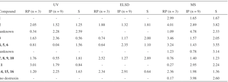
Table S1. Peak areas relative standard deviation (%) of repetibility (RP). Intermediate precision (IP) and stability (S) measured for peaks observed in
fraction C2-fr3-MF using UV; ELSD and MS HPLC detectors; peaks areas were measured relative to [Phe3, N-Me-Val5] Dtx B (11) as internal standard
UV ELSD MS
Compound RP (n = 3) IP (n = 9) S RP (n = 3) IP (n = 9) S RP (n = 3) IP (n = 9) S
1 - - - 3.09 1.57 2.72
2 3.92 5.09 1.51 - - - 3.79 1.43 5.25
unknown 2.73 4.50 1.85 - - - 1.17 3.74 3.90
3 3.62 3.13 0.43 - - - 3.20 2.27 1.19
4, 5, 6 2.48 1.17 2.40 - - - 3.44 1.06 4.18
unknown - - - 0.97 2.42 5.31
7, 8, 9, 10 2.99 3.18 2.65 - - - 0.84 2.09 3.13
11 0.00 0.00 0.00 - - - 0.00 0.00 0.00
14, 15, 16 2.43 2.01 2.05 - - - 2.34 2.93 3.26
no destruxin - - - 0.20 3.66 2.77
Table S2. Peak areas relative standard deviation (%) of repetibility (RP). Intermediate precision (IP) and stability (S) measured for peaks observed in fraction C2-fr3-MF using UV; ELSD and MS HPLC detectors. No internal standard was used
UV ELSD MS
Compound RP (n = 3) IP (n = 9) S RP (n = 3) IP (n = 9) S RP (n = 3) IP (n = 9) S
1 - - - - 2.99 1.65 1.67
2 2.05 1.52 1.25 1.88 1.32 1.81 4.01 2.89 3.82
unknown 0.34 2.28 2.59 - - - 1.09 4.78 2.33
3 1.63 2.36 0.56 0.74 1.17 2.00 3.46 1.57 2.05
4, 5, 6 0.81 0.04 1.56 0.64 2.35 1.10 3.24 1.43 3.55
unknown - - - - 1.23 0.78 3.14
7, 8, 9, 10 1.76 0.55 1.81 2.52 1.27 2.89 0.76 1.40 1.23
11 3.01 1.79 0.84 - - - 0.27 2.95 2.24
14, 15, 16 1.20 2.25 1.63 2.34 2.91 0.64 2.36 1.98 1.36
Table S3. Retention times relative standard deviation (%) of repetibility (RP). Intermediate precision (IP) and stability (S) measured for peaks observed in
fraction C2-fr3-MF using UV; ELSD and MS HPLC detectors. Retention times were measured relative to [Phe3, N-Me-Val5] Dtx B (11) as internal standard
UV ELSD MS
Compound RP (n = 3) IP (n = 9) S RP (n = 3) IP (n = 9) S RP (n = 3) IP (n = 9) S
1 - - - 1.24 0.51 0.12
2 0.24 0.22 0.06 - - - 1.52 0.49 0.04
unknown 0.22 0.17 0.23 - - - 1.46 0.42 0.18
3 0.16 0.19 0.03 - - - 1.34 0.30 0.05
4, 5, 6 0.09 0.74 0.06 - - - 1.28 0.14 0.03
unknown - - - 1.23 0.20 0.05
7, 8, 9, 10 0.01 0.05 0.01 - - - 1.19 0.11 0.07
11 0.00 0.00 0.00 - - - 0 0 0
14, 15, 16 0.09 0.04 0.06 - - - 1.11 0.12 0.12
no destruxin - - - 1.06 0.12 0.04
Table S4. Retention times relative standard deviation (%) of repetibility (RP). Intermediate precision (IP) and stability (S) measured for peaks observed in fraction C2-fr3-MF using UV; ELSD and MS HPLC detectors. No internal standard was used
UV ELSD MS
Compound RP (n = 3) IP (n = 9) S RP (n = 3) IP (n = 9) S RP (n = 3) IP (n = 9) S
1 - - - 0.18 1.07 0.14
2 0.43 0.36 0.06 0.48 0.39 0.14 0.46 0.42 0.04
unknown 0.24 0.38 0.16 - - - 0.41 0.35 0.14
3 0.35 0.26 0.08 0.27 0.27 0.09 0.28 0.36 0.04
4, 5, 6 0.26 0.54 0.03 0.27 0.30 0.18 0.22 0.21 0.09
unknown - - - 0.17 0.17 0.02
7, 8, 9, 10 0.19 0.14 0.08 0.11 0.10 0.16 0.13 0.15 0.09
11 0.20 0.13 0.07 - - - 1.06 0.16 0.07
14, 15, 16 0.12 0.10 0.12 0.11 0.11 0.08 0.16 0.08 0.16
fraction C2-fr4-MF using UV; ELSD and MS HPLC detectors. Peaks areas were measured relative to [Phe3, N-Me-Val5] Dtx B (11) as internal standard
UV ELSD MS
Compound RP (n = 3) IP (n = 9) S RP (n = 3) IP (n = 9) S RP (n = 3) IP (n = 9) S
2 2.72 3.55 2.78 3.30 1.22 4.29 3.81 1.86 1.34
no destruxin 0.70 4.71 0.74 - - - 0.71 0.87 2.16
3 2.34 1.58 3.85 1.17 2.03 4.09 3.57 1.38 2.41
4, 5, 6 2.29 2.33 1.49 2.88 2.44 4.95 2.31 1.01 0.50
unknown 2.91 5.66 2.63 2.69 3.80 1.95 1.08 1.51 3.58
7, 8, 9, 10 0.06 1.68 1.86 2.59 0.90 4.29 0.60 4.30 1.65
11 0.00 0.00 0.00 0.00 0.00 0.00 0.00 0.00 0.00
12, 13 and
14, 15, 16
0.20 3.58 3.40 1.20 1.82 3.11 1.02 1.57 2.00
17, 18, 19 - - - 1.12 3.37 1.66
20 - - - 1.13 2.70 1.75
unknown 1.11 3.31 2.36 2.44 0.80 2.57 0.46 0.10 1.42
Table S6. Peak areas relative standard deviation (%) of repetibility (RP). Intermediate precision (IP) and stability (S) measured for peaks observed in fraction C2-fr4-MF using UV; ELSD and MS HPLC detectors. No internal standard was used
UV ELSD MS
Compound RP (n = 3) IP (n = 9) S RP (n = 3) IP (n = 9) S RP (n = 3) IP (n = 9) S
2 2.27 0.37 1.24 2.14 0.60 1.65 4.04 1.42 0.62
no destruxin 3.20 1.37 2.33 - - - 1.65 2.51 2.27
3 2.49 1.94 1.60 0.30 0.53 0.57 3.17 1.35 3.74
4, 5, 6 0.26 1.66 1.34 2.73 0.40 3.64 3.09 2.24 1.10
unknown 1.14 1.01 0.88 2.01 1.72 1.66 1.11 1.37 3.67
7, 8, 9, 10 2.50 3.64 4.66 1.26 2.08 0.82 1.58 0.99 0.12
11 2.52 1.56 2.81 1.35 1.53 3.48 0.98 1.42 1.58
12, 13 and
14, 15, 16
2.47 1.03 0.97 2.42 1.24 2.66 1.37 1.50 0.87
17, 18, 19 - - - 0.35 0.63 1.48
20 - - - 0.17 2.42 2.27
Table S7. Retention times relative standard deviation (%) of repetibility (RP). Intermediate precision (IP) and stability (S) measured for peaks observed in
fraction C2-fr4-MF using UV; ELSD and MS HPLC detectors. Retention times were measured relative to [Phe3, N-Me-Val5] Dtx B (11) as internal standard
UV ELSD MS
Compound RP (n = 3) IP (n = 9) S RP (n = 3) IP (n = 9) S RP (n = 3) IP (n = 9) S
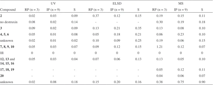
2 0.02 0.03 0.09 0.37 0.12 0.15 0.19 0.15 0.11
no destruxin 0.08 0.02 0.14 - - - 0.30 0.19 0.18
3 0.09 0.02 0.09 0.13 0.21 0.35 0.13 0.08 0.10
4, 5, 6 0.05 0.01 0.08 0.05 0.18 0.21 0.06 0.23 0.10
unknown 0.02 0.01 0.02 0.10 0.09 0.25 0.19 0.06 0.15
7, 8, 9, 10 0.05 0.03 0.07 0.09 0.12 0.15 1.21 0.12 0.07
11 0 0 0 0 0 0 0 0 0
12, 13 and
14, 15, 16
0.05 0.03 0.04 0.07 0.06 0.13 0.13 0.05 0.10
17, 18, 19 - - - 0.05 0.12 0.11
20 - - - 0.04 0.06 0.07
unknown 0.02 0.08 0.18 0.15 0.20 0.16 0.38 0.75 0.90
Table S8. Retention times relative standard deviation (%) of repetibility (RP). Intermediate precision (IP) and stability (S) measured for peaks observed in fraction C2-fr4-MF using UV; ELSD and MS HPLC detectors. No internal standard was used
UV ELSD MS
Compound RP (n = 3) IP (n = 9) S RP (n = 3) IP (n = 9) S RP (n = 3) IP (n = 9) S
2 0.03 0.09 0.14 0.34 0.07 0.07 0.13 0.04 0.06
no destruxin 0.04 0.11 0.13 - - - 0.29 0.30 0.17
3 0.08 0.10 0.10 0.15 0.05 0.20 0.05 0.04 0.07
4, 5, 6 0.03 0.11 0.12 0.04 0.07 0.03 0.06 0.21 0.19
unknown 0.03 0.11 0.11 0.08 0.08 0.09 0.13 0.07 0.02
7, 8, 9, 10 0.05 0.10 0.11 0.11 0.06 0.05 1.17 0.06 0.11
11 0.04 0.12 0.13 0.03 0.17 0.19 0.09 0.12 0.16
12, 13 and
14, 15, 16
0.06 0.09 0.13 0.06 0.12 0.05 0.07 0.08 0.09
17, 18, 19 - - - 0.05 0.01 0.06
20 - - - 0.05 0.06 0.10
fraction C3-fr3-MF using UV; ELSD and MS HPLC detectors. Peaks areas were measured relative to [Phe3, N-Me-Val5] Dtx B (11) as internal standard
UV ELSD MS
Compound RP (n = 3) IP (n = 9) S RP (n = 3) IP (n = 9) S RP (n = 3) IP (n = 9) S
no destruxin 1.78 1.58 3.50 - - - 0.44 0.51 1.19
1 1.77 2.79 4.29 - - - 1.76 0.58 0.89
2 0.66 3.42 4.67 3.34 3.30 1.88 1.12 0.66 3.12
unknown 2.55 5.65 2.98 - - - 3.21 0.60 4.45
3 1.03 2.73 2.61 3.97 1.48 3.18 2.91 0.92 2.19
4, 5, 6 0.23 1.08 1.77 4.97 1.88 2.41 1.43 0.29 2.77
unknown 3.93 3.65 5.32 - - - 4.26 2.18 2.01
7, 8, 9, 10 2.05 2.81 3.34 3.94 0.43 0.99 0.43 1.58 2.07
11 0.00 0.00 0.00 0.00 0.00 0.00 0.00 0.00 0.00
unknown 1.78 1.58 3.50 3.38 2.54 4.64 1.31 1.82 1.01
Table S10. Peak areas relative standard deviation (%) of repetibility (RP). Intermediate precision (IP) and stability (S) measured for peaks observed in fraction C3-fr3-MF using UV; ELSD and MS HPLC detectors. No internal standard was used
UV ELSD MS
Compound RP (n = 3) IP (n = 9) S RP (n = 3) IP (n = 9) S RP (n = 3) IP (n = 9) S
no destruxin 2.78 1.42 4.38 - - - 0.74 1.7 2.45
1 0.17 1.02 2.61 - - - 2.15 1.93 1.2
2 2.32 0.76 2.28 1.36 1.45 0.79 0.82 0.33 1.2
unknown 1.05 2.68 0.34 - - - 3.89 1.42 4.78
3 2.42 1.5 5.29 0.57 0.15 0.9 2.21 0.7 0.25
4, 5, 6 2.06 1.13 3.03 1.24 1.18 0.57 0.79 0.67 1.35
unknown 2.71 3.58 2.08 - - - 3.8 4.35 0.13
7, 8, 9, 10 1.12 4.87 1.46 1.14 3.34 1.86 0.88 1.38 1.71
11 1.84 0.47 3.34 3.66 1.67 2.69 0.69 0.39 1.93
Table S11. Retention times relative standard deviation (%) of repetibility (RP). Intermediate precision (IP) and stability (S) measured for peaks observed in
fraction C3-fr3-MF using UV; ELSD and MS HPLC detectors. Retention times were measured relative to [Phe3, N-Me-Val5] Dtx B (11) as internal standard
UV ELSD MS
Compound RP (n = 3) IP (n = 9) S RP (n = 3) IP (n = 9) S RP (n = 3) IP (n = 9) S
no destruxin 0.02 0.18 0.02 - - - 0.05 0.12 0.08
1 0.06 0.04 0.14 - - - 0.04 0.08 0.17
2 0.12 0.05 0.06 0.33 0.25 0.3 0.05 0.3 0.24
unknown 0.03 0.1 0.08 - - - 0.07 0.13 0.1
3 0.05 0.04 0.02 0.28 0.03 0 0.04 0.05 0.03
4, 5, 6 0.05 0.02 0.02 0.49 0.15 0.3 0.03 0.11 0.02
unknown 0.16 0.08 0.42 - - - 0.08 0.21 0.31
7, 8, 9, 10 0.07 0.03 0.03 0.33 0.28 0.15 0.12 0.15 0.15
11 0 0 0 0 0 0 0 0 0
unknown - - - 0.3 0.25 0.16 0.02 0.12 0.1
Table S12. Retention times relative standard deviation (%) of repetibility (RP). Intermediate precision (IP) and stability (S) measured for peaks observed in fraction C3-fr3-MF using UV; ELSD and MS HPLC detectors. No internal standard was used
UV ELSD MS
Compound RP (n = 3) IP (n = 9) S RP (n = 3) IP (n = 9) S RP (n = 3) IP (n = 9) S
no destruxin 0.01 0.14 0.07 - - - 0.03 0.12 0.11
1 0.04 0.03 0.06 - - - 0.07 0.01 0.15
2 0.12 0.11 0.1 0.11 0.21 0.24 0.06 0.23 0.13
unknown 0.03 0.04 0.04 - - - 0.09 0.05 0.16
3 0.04 0.05 0.09 0.08 0.05 0.08 0.06 0.05 0.11
4, 5, 6 0.04 0.06 0.09 0.24 0.17 0.09 0.06 0.06 0.11
unknown 0.15 0.12 0.39 - - - 0.07 0.15 0.18
7, 8, 9, 10 0.06 0.07 0.11 0.2 0.32 0.18 0.13 0.08 0.07
11 0.02 0.06 0.08 0.29 0.04 0.08 0.03 0.08 0.13
unknown - - - 0.07 0.28 0.12 0.03 0.04 0.11
Table S13. Peak areas relative standard deviation (%) of repetibility (RP). Intermediate precision (IP) and stability (S) measured for peaks observed in
fraction C3-fr4-MF using UV; ELSD and MS HPLC detectors. Peaks areas were measured relative to [Phe3, N-Me-Val5] Dtx B (11) as internal standard
UV ELSD MS
Compound RP (n = 3) IP (n = 9) S RP (n = 3) IP (n = 9) S RP (n = 3) IP (n = 9) S
unknown - - - 0.88 1.76 1.03
7, 8, 9, 10 2.01 2.05 5.32 3.54 2.46 3.48 0.99 1.35 4.47
11 0 0 0 0 0 0 0 0 0
12, 13 2.83 1.11 0.83 1.82 1.21 1.38 1.35 2.91 3.1
17, 18, 19 - - - 1.23 1.98 1.05
20 - - - 1.78 1.77 1.52
fraction C3-fr4-MF using UV; ELSD and MS HPLC detectors. No internal standard was used
UV ELSD MS
Compound RP (n = 3) IP (n = 9) S RP (n = 3) IP (n = 9) S RP (n = 3) IP (n = 9) S
unknown - - - 1.2 3.41 3.33
7, 8, 9, 10 0.72 2.84 2.76 1.19 2.48 2.93 1.5 0.76 1.72
11 1.29 0.35 3.76 2.34 1.71 1.97 0.56 0.64 2.93
12, 13 1.91 0.51 3.23 3.58 1.75 0.65 1.83 2.11 2.78
17, 18, 19 - - - 1.69 3.27 2.37
20 - - - 1.36 2.64 1.53
unknown 2.36 1.51 2.45 0.3 0.69 0.28 1.84 2.08 0.56
Table S15. Retention times relative standard deviation (%) of repetibility (RP). Intermediate precision (IP) and stability (S) measured for peaks observed in
fraction C3-fr4-MF using UV; ELSD and MS HPLC detectors. Retention times were measured relative to [Phe3, N-Me-Val5] Dtx B (11) as internal standard
UV ELSD MS
Compound RP (n = 3) IP (n = 9) S RP (n = 3) IP (n = 9) S RP (n = 3) IP (n = 9) S
unknown - - - 0.29 0.08 0.28
7, 8, 9, 10 0.03 0.16 0.26 0.06 0.08 0.11 0.2 0.05 0.19
11 0 0 0 0 0 0 0 0 0
12, 13 0.14 0.29 0.27 0.07 0.09 0.05 0.27 0.06 0.25
17, 18, 19 - - - 0.2 0.11 0.34
20 - - - 0.39 0.06 0.24
unknown 0.23 0.05 0.22 0.14 0.19 0.13 0.32 0.24 0.51
Table S16. Retention times relative standard deviation (%) of repetibility (RP). Intermediate precision (IP) and stability (S) measured for peaks observed in fraction C3-fr4-MF using UV; ELSD and MS HPLC detectors. No internal standard was used
UV ELSD MS
Compound RP (n = 3) IP (n = 9) S RP (n = 3) IP (n = 9) S RP (n = 3) IP (n = 9) S
unknown - - - 0.08 0.09 0.1
7, 8, 9, 10 0.05 0.2 0.12 0.01 0.22 0.2 0.02 0.11 0.13
11 0.07 0.3 0.13 0.07 0.14 0.1 0.22 0.11 0.32
12, 13 0.2 0.22 0.15 0.13 0.05 0.05 0.13 0.08 0.09
17, 18, 19 - - - 0.08 0.07 0.03
20 - - - 0.19 0.12 0.1
Table S17. Peak areas relative standard deviation (%) of repetibility (RP). Intermediate precision (IP) and stability (S) measured for peaks observed in
fraction C4-fr4-MF using UV; ELSD and MS HPLC detectors. Peaks areas were measured relative to [Phe3, N-Me-Val5] Dtx B (11) as internal standard
UV ELSD MS
Compound RP (n = 3) IP (n = 9) S RP (n = 3) IP (n = 9) S RP (n = 3) IP (n = 9) S
4, 5, 6 1.89 1.22 3.52 - - - 0.56 4.58 1.11
unknown 2.22 2.04 3.85 - - - 2.59 3.89 2.41
unknown 1.36 3.92 2.03 0.66 3.47 1.38 2.53 4.65 4.32
7, 8, 9, 10 2.78 1.68 5.31 1.44 1.09 2.9 1.54 4.72 2.45
11 0 0 0 0 0 0 0 0 0
12, 13 and
14, 15, 16
5.68 2.45 5.18 0.38 0.55 0.85 2.29 4.61 3.98
20 1.89 1.22 3.52 0.66 3.47 1.38 2.31 2.44 4.25
unknown 2.22 2.04 3.85 1.44 1.09 2.9 1.57 3.24 0.77
Table S18. Peak areas relative standard deviation (%) of repetibility (RP). Intermediate precision (IP) and stability (S) measured for peaks observed in fraction C4-fr4-MF using UV; ELSD and MS HPLC detectors. No internal standard was used
UV ELSD MS
Compound RP (n = 3) IP (n = 9) S RP (n = 3) IP (n = 9) S RP (n = 3) IP (n = 9) S
4, 5, 6 0.22 1.35 1.13 - - - 1.71 5.57 0.61
unknown 0.68 2.8 2.11 - - - 3.58 4.75 2.55
unknown 0.77 1.91 1.31 0.56 1.65 2.05 0.63 2.34 4.7
7, 8, 9, 10 2.03 3.75 5.04 1.02 3.43 0.59 1.13 2.04 1.75
11 1.66 1.14 2.71 0.48 1.69 3.44 2.16 2.52 0.79
12, 13 and
14, 15, 16
4.34 1.08 2.75 0.67 1.67 3.52 0.57 0.43 4.71
20 0.22 1.35 1.13 0.56 1.65 2.05 3.44 0.79 4.95
unknown 0.68 2.8 2.11 1.02 3.43 0.59 0.74 0.14 0.17
Table S19. Retention times relative standard deviation (%) of repetibility (RP). Intermediate precision (IP) and stability (S) measured for peaks observed in
fraction C4-fr4-MF using UV; ELSD and MS HPLC detectors. Retention times were measured relative to [Phe3, N-Me-Val5] Dtx B (11) as internal standard
UV ELSD MS
Compound RP (n = 3) IP (n = 9) S RP (n = 3) IP (n = 9) S RP (n = 3) IP (n = 9) S
4, 5, 6 0.28 0.04 0.06 - - - 0.16 0.27 0.15
unknown 0.05 0.09 0.17 - - - 0.45 0.05 0.74
unknown 0.05 0.12 0.19 0.22 0.05 0.14 0.23 0.25 0.09
7, 8, 9, 10 0.07 0.09 0.13 0.03 0.06 0.09 0.14 0.32 0.07
11 0 0 0 0 0 0 0 0 0
12, 13 and
14, 15, 16
0.07 0.12 0.1 0.01 0.08 0.13 0.39 0.14 0.09
20 - - - 0.23 0.22 0.12
in fraction C4-fr4-MF using UV; ELSD and MS HPLC detectors. No internal standard was used
UV ELSD MS
Compound RP (n = 3) IP (n = 9) S RP (n = 3) IP (n = 9) S RP (n = 3) IP (n = 9) S
4, 5, 6 0.41 0.09 0.05 - - - 0.07 0.15 0.19
unknown 0.1 0.09 0.13 - - - 0.61 0.11 0.76
unknown 0.11 0.03 0.08 0.15 0.05 0.23 0.05 0.09 0.11
7, 8, 9, 10 0.08 0.04 0.09 0.04 0.05 0.11 0.05 0.16 0.1
11 0.15 0.13 0.11 0.07 0.01 0.13 0.19 0.16 0.04
12, 13 and
14, 15, 16
0.13 0.06 0.05 0.06 0.07 0 0.2 0.18 0.1
20 - - - 0.06 0.06 0.16


