Ar
ti
cle
*e-mail: [email protected]; [email protected]
Kinetic and Equilibrium Mechanisms of Substrate Binding to
Mycobacterium tuberculosis
Enoyl Reductase: Implications to
Function-Based Antitubercular Agent Design
Igor B. Vasconcelos, Luiz A. Basso* and Diógenes S. Santos*
Centro de Pesquisas em Biologia Molecular e Funcional (CPBMF), Instituto Nacional de Ciência e Tecnologia em Tuberculose (INCT-TB), Pontifícia Universidade Católica do Rio Grande do Sul,
90619-900 Porto Alegre-RS, Brazil
A tuberculose (TB) continua sendo a principal causa de mortalidade devido a um único patógeno bacteriano, o Mycobacterium tuberculosis. Há, portanto, a necessidade de desenvolvimento de novos agentes antimicobacterianos. A 2-trans-enoil-ACP(CoA) redutase (InhA) de M. tuberculosis
é o principal alvo da isoniazida. Aqui nós apresentamos dados de equilíbrio e cinética em estado pré-estacionário da ligação do substrato 2-trans-dodecenoil-CoA à InhA. Os resultados demonstram cooperatividade homotrópica positiva da ligação deste substrato à InhA e um processo de associação bimolecular seguido por uma lenta isomerização do complexo binário enzima-substrato. Os dados aqui descritos devem auxiliar no desenho racional de novos inibidores de um alvo protéico validado e com potencial utilização no tratamento da TB.
Tuberculosis (TB) remains the leading cause of mortality due to a single bacterial pathogen,
Mycobacterium tuberculosis. There is a need for the development of new antimycobacterial agents.
M. tuberculosis 2-trans-enoyl-ACP(CoA) reductase (InhA) is the main target of isoniazid, the most prescribed anti-TB agent. Here we present pre-steady state kinetics and equilibrium data of 2-trans-dodecenoyl-CoA substrate binding to InhA. These results indicate both positive homotropic cooperativity upon substrate binding to InhA, and a bimolecular association process followed by a slow isomerization of the enzyme-substrate binary complex. The data here described should help the rational design of new agents against a validated and druggable protein target with potential anti-TB activity.
Keywords: tuberculosis, enoyl-ACP(CoA) reductase, mycolic acid, luorescence titration, pre-steady-state kinetics
Introduction
Tuberculosis (TB) remains the leading cause of mortality due to a bacterial pathogen, Mycobacterium tuberculosis.1 The World Health Organization (WHO)
reported that there were an estimated 9.27 million incident cases of TB in 2007, and that 1.37 million (15%) were HIV-positive patients, who are more likely to develop active TB than HIV-negative patients.2 In addition, there were
an estimated 0.5 million cases of multi-drug resistant TB (MDR-TB), which is deined as strains resistant to, at least, isoniazid and rifampicin.2 The emergence of extensively
drug-resistant (XDR) TB cases, deined as cases in persons with TB whose isolates are MDR that are also resistant to
a luoroquinolone and, at least, one second-line injectable agent (amikacin, kanamycin and/or capreomycin),2,3 its
widespread distribution,4 and unprecedented fatality rate,5
raise the prospect of incurable and deadly TB worldwide. There is thus is an urgent need for the development of new antimycobacterial agents.
Isoniazid (INH, isonicotinic acid hydrazide) is one of the oldest synthetic antitubercular, and the most prescribed drug for active infection and prophylaxis.6 The product of
the M. tuberculosis InHA structural gene (InhA) has been shown to be a major target for INH7 and to be an
NADH-dependent enoyl-ACP (acyl carrier protein) reductase enzyme speciic for long-chain enoyl thioester substrates.8
the meromycolate branch of mycolic acids, the hallmark of mycobacteria.9 However, it has recently been suggested that
the acyclic 4R isomer of INH-NADP inhibits M. tuberculosis dihydrofolate reductase, an enzyme essential for nucleic acid synthesis, prompting the authors to propose that this enzyme is also a target for isoniazid.10 In addition, results of
a proteome-wide proiling using INH adducts coupled to a solid support suggested that INH targets multiple enzymes in M. tuberculosis.11 However, determination of a clinically
relevant drug target requires the ability to transfer a single point mutation that causes drug resistance within a gene that putatively encodes a drug target and demonstrates that this transfer is suficient, by itself, to confer drug resistance. To the best of our knowledge, it has been shown for InhA,12
thereby indicating that mycobacterial enoyl reductase is the bona ide target for INH mode of action.
INH is a pro-drug that is activated by the mycobacterial katG-encoded catalase-peroxidase enzyme in the presence of manganese ions, NAD(H) and oxygen.13-16 The kat
G-produced acylpyridine fragment of INH is covalently attached to the C4 position of NADH that forms a binary complex with the wild-type (WT) enoyl reductase of M. tuberculosis.17 This isonicotinyl-NAD+ adduct has been
shown to be a slow, tight-binding competitive inhibitor of WT InhA with an overall dissociation constant value of 0.75 nmol L-1.18 Consistent with InhA as the primary target
of INH mode of action, INH-resistant clinical isolates of M. tuberculosis harboring InHA-structural gene missense mutations have higher dissociation constant values for NADH than INH-sensitive WT InhA.19 Pre-steady-state
kinetics studies on NADH binding to InhA showed that the limiting rate constant values for NADH dissociation from the InhA-NADH binary complexes were larger for INH-resistant InhA mutants as compared to INH-sensitive WT InhA,20 which was borned out by structural studies.20,21
Steady-state kinetic studies have indicated that WT InhA follows a sequential kinetic mechanism with preferred binding of NADH followed by enoyl-Coenzyme A (enoyl-CoA) substrate binding, and that hydride transfer occurs from the 4S hydrogen to the C3 position of 2-trans -enoyl-CoA(ACP) substrate.8 However, results of primary kinetic
deuterium isotope effects were consistent with a random order of addition of substrates to WT InhA.22 The kinetics
of NADH binding to WT and INH-resistant InhA proteins has also been investigated by luorescence spectroscopy in a stopped-low equipment.20 However, there has been no
report on binding of 2-trans-enoyl-CoA substrate to InhA. Here we present equilibrium and pre-steady-state data on 2-trans-dodecenoyl-CoA (DD-CoA) binding to WT InhA. A mechanism of binary complex formation between DD-CoA and WT InhA is proposed. Moreover, the results here
presented support a random order kinetic mechanism of substrate addition to InhA.
Experimental
Materials
All chemicals used were of analytical or reagent grade and required no further purification. Complete protease inhibitor cocktail tablets were from Boehringer (Mannheim, Germany). Amicon stirred ultrailtration cell and regenerated cellulose ultrailtration membranes were from Millipore. Fast performance liquid chromatography (FPLC) protein puriication (4 ºC) was carried out in an Äkta puriier (GE Helthcare).
Puriication of WT InhA
WT InhA was expressed and puriied to homogeneity as described elsewhere.19 In short, six liters of 1.5 × Luria broth
medium containing 50 mg mL-1 carbenicillin were inoculated
with a single colony, grown to an A600 of 0.8-1.0, induced with 1 mmol L-1 isopropyl-b-D-thiogalactopyranoside
(IPTG), and grown for 2 h. Cells were harvested by centrifugation, the pellet resuspended in 20 mmol L-1 PIPES
pH 7.3 (1 g cells per 3 mL buffer) containing lysozyme (0.2 mg mL-1) and protease inhibitor cocktail tablets,
disrupted by sonication, treated with streptomycin sulfate (1% m/v final), and centrifuged. The supernatant was dialysed against 20 mmol L-1 PIPES pH 7.3, centrifuged, and
the soluble fraction loaded on Q Sepharose Fast Flow anion exchange column, and eluted using a linear 0-0.5 mol L-1
NaCl gradient. Fractions containing WT InhA were pooled, concentrated by ultrailtration (molecular weight cut-off of 10,000 Da), loaded on Sephacryl S-200 gel iltration column, and eluted using isocratic 20 mmol L-1 PIPES pH 7.3.
Fractions containing WT InhA were pooled, loaded on Mono Q HR 16/10 anion exchange column, and eluted using a linear 0-0.5 mol L-1 NaCl gradient. Fractions containing
WT InhA were pooled and stored in 65% ammonium sulfate solution. Total protein concentration was determined by Bradford’s method using bovine serum albumin as standard (Bio-Rad Laboratories).
Synthesis and puriication of 2-trans-dodecenoyl-CoA (DD-CoA)
2-trans dodecenoyl-CoA was synthesized from 2-trans -dodecenoic acid and CoA by the mixed anhydride method as previously described.19 DD-CoA was puriied by
(Waters Associates, Milford, MA) as described elsewhere.22
In short, chromatography was performed using 20 mmol L-1
ammonium acetate/1.75% acetonitrile as buffer A and running a 0-100% gradient of 95% acetonitrile/5% H2O (buffer B) at 8 mL min-1. Elution was monitored at 260 nm
and 285 nm using an Äkta Puriier 10 (GE Healthcare) at constant room temperature (ca. 20 °C), and fractions containing DD-CoA were pooled and lyophilized. The retention time for DD-CoA was 63 min. The ratio of absorbance of puriied DD-CoA at 232 nm and 260 nm was 0.62, a value that meets the established criterion for pure thioesters (A232/A260≥ 0.52).23
Fluorescence spectroscopy
Protein luorescence titration at equilibrium was carried out at 25 ºC by making microliter additions of DD-CoA (1-28 mmol L-1) to 2 mL of 2 mmol L-1 WT InhA in
100 mmol L-1 Pipes, pH 7.0, keeping the dilution to a
maximum of 1.0%. Excitation and emission wavelenghts were, respectively, 299 and 335 nm; with excitation and emission slits of, respectively, 1.5 and 10 nm, using an RF-5301PC Spectrophotometer (Shimadzu). Control measurements were performed in the same conditions, except that DD-CoA was not added, and these values were subtracted from those obtained in the presence of the substrate. Control experiments were also employed to determine the maximum ligand concentrations to be used with no inner ilter effect on intrinsic protein luorescence.
Pre-steady-state kinetics of DD-CoA binding
The rate of WT InhA:DD-CoA binary complex formation was determined by monitoring the rate of quench in protein luorescence upon DD-CoA (2.5-40 µmol L-1)
binding to WT InhA (2 mmol L-1) in 100 mmol L-1 Pipes,
pH 7.0, using an Applied-Photophysics SX-18MV-R (Leatherhead, UK) stopped-low spectroluorimeter operated at 25 °C in luorescence mode (dead time = 1.37 ms). The excitation wavelength (299 nm) was selected by focusing a 150-W Xenon arc lamp onto a monochromator itted with a 250 nm holographic grating. The excitation slit width was set to 0.64 mm, corresponding to a spectral band width of 3 nm. The luorescence signal above 320 nm was collected using a WG320 Scott ilter, positioned between the photomultiplier and the sample cell as previously described.24,25 Data acquisition was carried out using a
split time base (1 and 10 s), with the irst half of data acquired over 10% of the time period monitored for the second half of the split time base. This procedure allows for more accurate determination of the rate and amplitude
of an exponentially changing signal.26 Data were stored
on an Acorn A5000 computer, and analyzed by non-linear regression. All concentrations in stopped-low experiments are given for mixing chamber.
Data analysis
Data from equilibrium fluorescence spectroscopy were itted to equation 1, the Hill equation,27 in which F
is the observed luorescence signal, Fmax is the maximal luorescence, n represents the minimum number of binding sites, and K’ is a constant comprising interaction factors and the intrinsic dissociation constant.
(1)
The rate of WT InhA:DD-CoA binary complex formation were characterized by a biphasic quench in protein luorescence, and thus all traces were itted to equation 2, a double exponential function equation with loating end point, yielding values for the observed apparent rate of WT InhA:DD-CoA binary complex formation for the fast (kobs1) and slow (kobs2) phases of binding.28 A
1 and
A2 are the signal amplitudes for, respectively, fast and slow phases.
(2)
A plot of kobs1 versus DD-CoA concentration was linear, the data were thus itted to equation 3. This equation describes a single-step reversible bimolecular association process, in which k1 is the association rate constant for WT InhA:DD-CoA binary complex formation, and k-1 represents the dissociation rate constant of DD-CoA from the WT InhA:DD-CoA binary complex.28,29
(3)
A plot of kobs2 against increasing DD-CoA concentration displayed a hyperbolic increase, and the data were thus best itted to equation 4 for a bimolecular association process followed by a slow isomerization of the binary complex. Data itting to equation 4 yields values for the forward rate constant (k2) and reverse rate constant (k-2) for the binary complex isomerization process, and dissociation constant at equilibrium for binary complex formation (Kd).25,28
(4)
InhA:DD-CoA binary complex formation was obtained from itting the data to equation 5.25
(5)
Results and Discussion
Equilibrium binding of DD-CoA to WT InhA
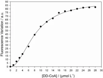
A plot of DD-CoA concentration versus quench in intrinsic protein luorescence upon WT InhA:DD-CoA binary complex formation at equilibrium (Figure 1) was sigmoidal. Accordingly, the data were itted to equation 1, yielding values of K’ = 8.2 ± 0.3 mmol L-1 and n = 2 ± 0.1.
These results clearly show that DD-CoA binds to free WT InhA, and that there is positive cooperativity in the binding of DD-CoA to WT InhA. Steady-state kinetic studies suggested that WT InhA follows a sequential kinetic mechanism with preferred binding of NADH.8 On the other
hand, a random order mechanism has been proposed based on primary kinetic deuterium isotope effects.22 The results
presented here showing DD-CoA binding to free WT InhA, and data previously published showing NADH binding to this enzyme,8,19,20 clearly demonstrate that the kinetic
mechanism is random order of addition of substrates. These results have a significant impact on rational-based drug design because the random order addition of substrates shown here demonstrates that analogues of any of the two substrates may be designed as potential InhA enzyme inhibitors. In the case of a sequential mechanism of substrate binding, analogues should be designed as mimicks of the irst substrate to bind to WT InhA, or as chemical compounds that are capable of binding to WT InhA:NADH binary complex.
Kinetics of WT InhA-DD-CoA binary complex formation
Steady-state kinetics and equilibrium binding are powerful tools for distinguishing mechanisms, in the sense that they can show which substrate binds to the enzyme irst, and which product dissociates irst. However, steady-state kinetics and equilibrium binding cannot inform us anything about isomerization of central complexes or individual rates of substrate binding to an enzyme. These limitations can be removed by studying directly partial reactions or elementary steps, instead of the overall reaction, providing that a way of detecting these interactions is available. This is the signiicance of transient kinetics, which aims to observe the changes occurring in the enzyme molecule itself to clarify the elementary steps of the enzyme reaction. It should be pointed out that the kinetics of NADH binding to WT and INH-resistant InhA proteins have been previously reported.20 However, there has been no report on binding
of 2-trans-enoyl-CoA substrate to InhA. Accordingly, the kinetics of DD-CoA binding to WT InhA was investigated by luorescence spectroscopy in a stopped-low equipment. All traces for DD-CoA binding were characterized by a biphasic quench in protein luorescence (Figure 2 - Inset), and the data were thus best itted to equation 2, a double exponential function, yielding values for the observed rate of WT InhA:DD-CoA binary complex formation for the fast (kobs1) and slow (kobs2) phases of binding. It should be pointed out that these two phases remained well separated from each other over the whole range of DD-CoA concentrations tested. A plot of kobs1 against DD-CoA concentration increased linearly (Figure 2), and thus the data were itted to equation 3, which describes a single-step reversible bimolecular association process, yielding values of 3.1 (± 0.5) × 103 L mol-1 s-1 for k
1, the association rate constant
of DD-CoA binding to WT InhA, and 53 (± 7) × 10-3 s-1 for k -1,
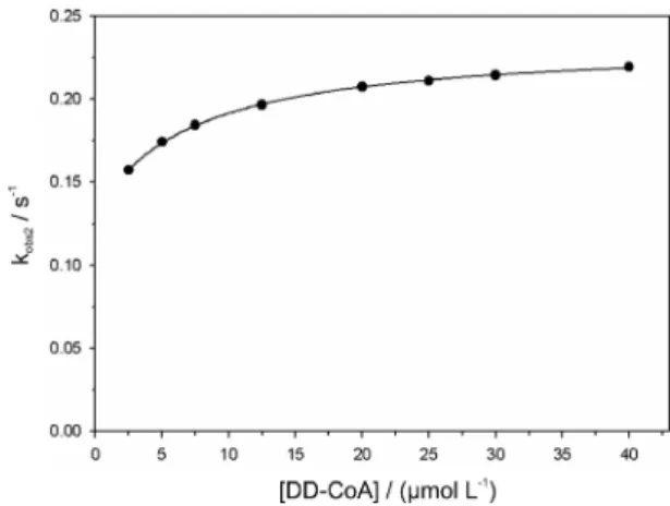
the dissociation rate constant of DD-CoA from WT InhA:DD-CoA binary complex. On the other hand, a plot ofkobs2 against increasing DD-CoA concentration showed a hyperbolic increase (Figure 3), which is consistent with a bimolecular association process followed by a slow isomerization of WT InhA:DD-CoA binary complex. Fitting these data to equation 4 yielded values for k2 = 0.104 (± 0.001) s-1,
k-2 = 0.130 (± 0.002) s-1 (the rate constants for, respectively,
the forward and reverse unimolecular isomerization step), and Kd = 6.9 (± 0.4) µmol L-1 (the dissociation constant for
the WT InhA:DD-CoA binary complex).
These results suggest that binding of DD-CoA to WT InhA is comprised of a bimolecular binding event followed by a slow isomerization of WT InhA:DD-CoA binary complex (Scheme 1). The overall dissociation constant for this mechanism (Kd(overall)) can be obtained from itting the
Figure 1. Fluorescence spectroscopy on the equilibrium binding of
data to equation 5, yielding a value of 3.8 (± 0.2) mmol L-1.
This value is in reasonably good agreement with the value of K’ determined from the equilibrium luorescence titration experiment.
The total signal amplitude changes derived from the biphasic quench in protein luorescence upon DD-CoA binding to WT InhA are given as the algebraic sum of the amplitudes of the fast (kobs1) and slow (kobs2) phases. Moreover, since no stopped-low signal was lost in the dead-time of the equipment (Figure 2), no correction was needed.24,25,28 A plot of total signal amplitude (A
1+A2)
against increasing DD-CoA concentrations was sigmoidal (Figure 4), and data itting to equation 1 yielded values of 14.1 (± 0.4) µmol L-1 and 2.5 (± 0.2) for, respectively, K’ and n.
These results are in good agreement with the equilibrium luorescence titration ones, thereby supporting the proposed mechanism for DD-CoA binding to WT InhA (Scheme 1).
Concerted or symmetry model
Equilibrium data supporting positive cooperativity as described are consistent with two major mechanisms involving interdependent binding sites. The first is the concerted mechanism or symmetry model proposed by Monod et al.,30 which predicts that there are two isomers of
free enzyme in equilibrium, E and E*, with substrate binding
effectively to E and negligibly to E*. The second is the
sequential mechanism proposed by Koshland et al.,31 which
predicts only one form of free enzyme in solution, substrate binding to one subunit, and a subsequent isomerization step that increases the afinity of the second subunit for the next
substrate molecule. In both mechanisms, isomerization steps are slower than binding steps. Measurements of binding rate constants can distinguish the symmetry and sequential mechanisms, since the dependence of the apparent rate constants on substrate concentration can be utilized to infer whether or not two forms of free InhA are present in solution.28,29 If free enzyme exists in equilibrium between
two forms, E and E*, one expects a hyperbolic decrease in
kobs values as the substrate concentration increases. On the other hand, if there is one form of free enzyme in solution, increasing substrate levels are accompanied by a hyperbolic increase in apparent rate constant values.29
The results presented here indicate that the binding of DD-CoA to WT InhA follows the sequential or induced-it model,31 with only one form of free WT InhA and
a slow isomerization of the enzyme-ligand complex following DD-CoA binding (Scheme 1). Interestingly, two forms in equilibrium were observed for M. tuberculosis
β-ketoacyl-ACP reductase,32 another member of the
mycobacterial FASII system. The equilibrium constant for the conformational change between WT InhA:DD-CoA
Figure 2. Linear dependence of kobs1 values on DD-CoA concentration.
Data were itted to equation 3. Inset: Representative biphasic stopped-low trace of DD-CoA (20 mmol L-1) binding to WT InhA (2 mmol L-1).
The top trace shows the control performed in the absence of DD-CoA.
Figure 3. Hyperbolic dependence of kobs2 values on DD-CoA
concentration. Data were itted to equation 4.
Figure 4. Sigmoidal dependence of total signal amplitude of stopped-low
data upon DD-CoA concentration. The solid curve represents the best it of the data to equation 1.
E+DD-CoA E–DD-CoA k1
k-1
E–DD-CoA* k2
k-2
and WT InhA:DD-CoA* binary complexes is near unity (k2/k-2 = 0.8), thereby indicating that the DD-CoA overall binding hinges on the bimolecular recognition process.
Conclusions
Enzyme inhibitors make up roughly 25% of the drugs marketed, and are thus important promising drug targets;33
however, mechanistic analysis should always be a top priority for enzyme-targeted drug programs since effective enzyme inhibitors take advantage of enzyme chemistry to achieve inhibitory activity.34 Moreover, it has recently been
pointed out that allostericity is a factor in pharmacological drug design because allosteric inhibitors may be more selective across species.35 To the best of our knowledge,
this is the irst report on enoyl-CoA substrate mode of binding to WT InhA. The results here presented support a random order kinetic mechanism of addition of substrates, and indicate that DD-CoA binding to free WT InhA follows the sequential model, with only one form of free enzyme in solution and isomerization of binary complex. These results are expected to be useful in the function-based design of inhibitors of a druggable and validated protein target that can be tested as new anti-tubercular agents.
Acknowledgments
This work was supported by National Institute of Science and Technology on Tuberculosis (Decit/SCTIE/MS-MCT-CNPq-FNDCT-CAPES) and Millennium Initiative Program (CNPq), Brazil, to D. S. S. L. A. B., D. S. S. (304051/1975-06) and L. A. B. (520182/99-5) are research career awardees from the National Council for Scientiic and Technological Development of Brazil (CNPq).
References
1. Harries, A. D.; Dye, C.; Ann. Trop. Med. Parasitol.2006, 100, 415. 2. World Health Organization, Geneva, Switzerland, 2009; www.who. int/tb/publications/global_report/2009, accessed on December 4, 2009.
3. Centers of Disease Control and Prevention; Morb. Mortal. Wkly. Rep.2006, 55, 301.
4. Dorman, S. E.; Chaisson, R. E.; Nat. Med. 2007, 13, 295. 5. Singh, J. A.; Upshur, R.; Padayatchi, N.; PLoS Med. 2007, 4, e50. 6. Basso, L. A.; Blanchard, J. S.; Adv. Exp. Med. Biol.1998, 456, 115. 7. Banerjee, A.; Dubnau, E.; Quémard, A.; Balasubramanian, V.;
Um, K. S.; Wilson, T.; Collins, D.; de Lisle, G.; Jacobs, W. R. Jr.; Science1994, 263, 227.
8. Quémard, A.; Sacchettini, J. C.; Dessen, A.; Vilchéze, C.; Bittman, R.; Jacobs, W. R. Jr.; Blanchard, J.S.; Biochemistry1995, 34, 8235.
9. Schroeder, E. K.; de Souza, O. N.; Santos, D. S.; Blanchard, J. S.; Basso, L. A.; Curr. Pharm. Biotechnol.2002, 3, 197.
10. Argyrou, A.; Vetting, M. W.; Aladegami, B.; Blanchard, J. S.; Nat.
Struct. Mol. Biol.2006, 13, 408.
11. Argyrou, A.; Jin, L.; Siconili-Baez, L.; Angeletti, R. H.;Blanchard, J. S.; Biochemistry2006, 45, 13947.
12. Vilchèze, C.; Wang, F.; Arai, M.; Hazbón, M. H.; Colangeli, R.; Kremer, L.; Weisbrod, T. R.; Alland, D.; Sacchettini, J. C.; Jacobs, W. R. Jr.; Nat. Med.2006, 12, 1027.
13. Johnson, K.; Schultz, P. G.; J. Am. Chem. Soc.1994, 116, 7425. 14. Johnson, K.; King, D. S.; Schultz, P. G.; J. Am. Chem. Soc.1995,
117, 5009.
15. Basso, L. A.; Zheng, R.; Blanchard, J. S.; J. Am. Chem. Soc.1996, 118, 11301.
16. Zabinski, R. F.; Blanchard, J. S.; J. Am. Chem. Soc.1997, 119, 2331.
17. Rozwarski, D. A.; Grant, G. A.; Barton, D. H. R.; Jacobs W. R. Jr.; Sacchettini, J. C.; Science1998, 279, 98.
18. Rawat, R.; Whitty, A.; and Tonge, P. J.; Proc. Natl. Acad. Sci.
U. S. A. 2003, 100, 13881.
19. Basso, L. A.; Zheng, R.; Musser, J. M.; Jacobs W. R. Jr.; Blanchard, J. S.; J. Infect. Dis.1998, 178, 769.
20. Oliveira, J. S.; Pereira, J. H.; Canduri, F.; Rodrigues, N. C.; de Souza, O. N.; Azevedo W. F. Jr.; Basso, L. A.; Santos, D. S.; J. Mol. Biol.2006, 359, 646.
21. Dias, M. V. B.; Vasconcelos, I. B.; Prado, A. M. X.; Fadel, V.; Basso, L. A.; Azevedo W. F. Jr.; Santos, D. S.; J. Struct. Biol.2007, 159, 369.
22. Parikh, S.; Moynihan, D. P.; Xiao, G.; Tonge, P. J.; Biochemistry
1999, 38, 13623.
23. Constantinides, P. P.; Steim, J. M.; J. Biol. Chem.1985, 260, 7573. 24. Basso, L. A.; Engel, P. C.; Walmsley, A.R.; Eur. J. Biochem.1993,
213, 935.
25. Basso, L. A.; Engel, P. C.; Walmsley, A. R.; Eur. J. Biochem.1995, 234, 603.
26. Walmsley, A. R.; Bagshaw, C. R.; Anal. Biochem.1989, 176, 313. 27. Hill, A. V.; Biochem. J.1913, 7, 471.
28. Nakatani, H.; Hiromi, K.; J. Biochem.1980, 87, 1805.
29. Basso, L. A.; Engel, P. C.; Walmsley, A. R.; Biochim. Biophys.
Acta1998, 1382, 345.
30. Monod, J.; Wyman, J.; Changeux, J. P.; J. Mol. Biol.1965, 12, 88. 31. Koshland, D. E.; Némethy, G.; Filmer, D.; Biochemistry1966, 5,
365.
32. Silva, R. G.; Rosado, L. A.; Santos, D. S.; Basso, L. A.; Arch. Biochem. Biophys.2008, 471, 1.
33. Robertson, J. G.; Biochemistry2005, 44, 5561. 34. Robertson, J. G.; Curr. Opin. Struct. Biol.2007, 17, 674. 35. Goodey, N. M.; Benkovic, S. J.; Nat. Chem. Biol. 2008, 4, 474.
Received: December 9, 2009