From the Disciplines of Surgery of the Digestive Tract and Coloproctology, Hospital das Clínicas, Faculty of Medicine, University of São Paulo - São Paulo/SP, Brazil. E-mail: [email protected]
Received for publication on January 29, 2004.
ORIGINAL RESEARCH
MICROSATELLITE INSTABILITY IN SOLITARY AND
SPORADIC GASTRIC CANCER
Rodrigo Oliva Perez, Carlos Eduardo Jacob, Fabricio L’ofreddo D’Ottaviano, Conrado Alvarenga, Adriana Safatle Ribeiro, Ulysses Ribeiro Jr., Cláudio José Caldas Bresciani, Bruno Zilberstein, José Eduardo Krieger, Angelita Habr-Gama and Joaquim José Gama-Rodrigues
Perez RO et al. Microsatellite instability in solitary and sporadic gastric cancer. Rev. Hosp. Clín. Fac. Med. S. Paulo 59(5):279-285, 2004.
Recently, the presence of microsatellite instability (MSI) has been reported in gastric cancer and associated with older age of presentation, distal tumor location, early disease staging, and better overall prognosis. Different characteristics in presentation and in tumor behavior may be explained by different genetic alterations during carcinogenesis of gastric cancer. Identification of specific genetic pathways in gastric cancer may have direct impact on prognosis and selection of treatment strategies.
PATIENTS AND METHODS: All 24 patients were treated by radical surgery. Fragments of normal and tumor tissues were extracted from the specimen and stored at -80oC before DNA purification and extraction. PCR amplification utilizing
microsatellite markers was performed. Tumors presenting PCR products of abnormal sizes were considered positive for microsatellite instability (MSI+).
RESULTS: Five patients (21%) had tumors that were MSI+ in at least 1 marker. In the group of patients with Lauren’s intestinal-type gastric carcinoma, 3 had tumors that were MSI+ (23%), while in the group of diffuse-type gastric cancer, 2 patients had tumors that were MSI+ (19%). The mean age of presentation and the male:female ratio was similar in both groups. Tumors that were MSI+ were more frequently located in proximal portion of the stomach compared to microsatellite-stable (MSS) tumors (40% vs. 16%). Although there was a trend of patients with MSI+ tumors towards a proximal gastric tumor location, early staging, and negative lymph node metastasis, there was no statistical significance compared to those with MSS tumors (P >.1). Comparison of overall and disease-free survival between gastric tumors that were MSI+ and those that were MSS found no statistically significant differences (P >.1).
CONCLUSIONS: Microsatellite instability is a frequent event in gastric carcinogenesis and shows a trend towards distinct clinical and pathological characteristics of gastric cancer.
KEY WORDS: Gastric cancer. Genetics. Microsatellite instability.
Gastric adenocarcinoma is one of the most common malignancies world-wide, but genetic steps involved in its carcinogenesis remain uncertain. Mul-tiple genetic alterations affecting proto-oncogenes, tumor suppressor genes, and mismatch repair genes (MMR) appear to be associated with the development of various human cancers including colorectal and gas-tric cancers.
Identification of MMR genes, which are responsible for hereditary nonpolyposis colon cancer, led to the hypothesis of a distinct genetic path-way in colorectal cancer associated
with microsatellite instability (MSI) or replication error phenotype (RER) by alterations in 1 or more of these genes, more frequently in Msh2 and Mlh1.17,7
presentation and in tumor behavior may be explained by different genetic alterations during carcinogenesis of gastric cancer. Identification of specific genetic pathways in gastric cancer may have direct impact on prognosis and selection of treatment strategies.
We report in this prospective study involving 24 Brazilian patients with solitary and sporadic gastric cancer, the incidence of MSI, its correlation with epidemiological, clinical and patho-logical characteristics, and impact on overall and disease-free survival.
PATIENTS AND METHODS
Twenty-four patients with a preoperative diagnosis of gastric ad-enocarcinoma treated at Hospital das Clínicas, Faculty of Medicine, Univer-sity of São Paulo (Department of Gas-troenterology) and “Hospital Alemão Osvaldo Cruz” entered the study. All patients were treated by radical surgery including gastric resection with free margins and D2 lymphadenectomy ac-cording to the JSRGC anatomical clas-sification. All patients were eligible for the study and had their resected speci-mens available for pathologic exami-nation and MSI determiexami-nation.
Tumors were characterized accord-ing to Lauren´s histologic classifica-tion into intestinal - and diffuse-type gastric cancer and staged according to JGCA recomendations.19 The same
sur-geon and surgical team performed all operations.
Of the 24 patients, 15 were male (62.5%) and 9 female (37.5%). The mean age was 63.6 years (range, 26 to 86 years). Eleven patients (46%) had Lauren’s intestinal-type adenocarcinoma while 13 patients (54%) had diffuse-type adenocarcinoma. Twelve patients (50%) had tumors located in lower third of the stomach, 7 in the middle third (29%), and 5 in the upper third (21%). Total gast-rectomy was performed in 11 patients
(46%), and subtotal gastrectomy in the remaining 13 patients (54%). Associated resection of the spleen was performed in 8 patients who were treated by total gas-trectomy (84.5%). Pathological exami-nation revealed a mean number of lymph nodes resected of 33.6 (range, 17 to 76). Patient demographics are summarized in Table 1.
Nine patients (37.5%) had T1 (mu-cosa and submu(mu-cosa) tumors, 2 pa-tients (8.3%) had T2 (muscular layer), 12 patients (50%) had T3 (serosal or subserosal), and 1 (4.2%) had a T4 (adjacent organs). Nine patients (37.5%) had no positive lymph nodes (N0), 9 (37.5%) were classified as N1, and 6 (25%) were classified as N2.
All patients had preoperative en-doscopic biopsies of primary tumors. However, these preoperative studies did not include normal adjacent tissue biopsies for Helicobacter pylori infec-tion determinainfec-tion. Since the surfaces of all resected specimens were washed with saline solution in order to avoid DNA contamination, determination of
Helicobacter pylori infection would be seriously compromised and
inaccu-rate in this setting. For this reason, pathologic examination of the resected specimen did not include H. pylori in-fection determination.
Only 1 patient (4.2%) had a distant metastasis in the left ovary and was classified as stage IV. Eight patients (33.3%) had stage I disease (stage Ia + Ib), 5 (21%) had stage II, and 10 (41.5%) had stage III (IIIa + IIIb) dis-ease. Stage classification and patient distribution are summarized in Tables 2 and 3.
Table 1 - Patient Demographics (N=24).
Age 26-86
Mean 63.6 years
Gender
Male 15 (62.5%)
Female 9 (37.5%)
Histologic type (Lauren)
Intestinal 13 (54%)
Diffuse 11 (46%)
Tumor location
L (distal third) 12 (50%) M (intermediate) 7 (29%) U (proximal third) 5 (21%) Types of gastrectomy
Total 11 (46%)
Subtotal 13 (54%)
Lymphadenectomy (17-76)
Mean 34 lymph nodes
Table 2 - Stage and histologic type (Lauren´s classification) (N=24).
(JGCA-1998) Intestinal Diffuse Total
Ia 5 (38.5%) 1 ( 9%) 6 (25%)
I b 0 2 (18%) 2 ( 8%)
II 4 (30%) 1 ( 9%) 5 (21%)
IIIa 1 ( 8%) 6 (55%) 7 (29%)
IIIb 2 (15.5%) 1 ( 9%) 3 (13%)
IV 1 ( 8%) 0 1 ( 4%)
(TNM) Intestinal Diffuse Total
T
T1 5 (38.5%) 4 (36%) 9 (37.5%)
T2 1 ( 8%) 1 ( 9.5%) 2 ( 8%)
T3 7 (54%) 5 (45%) 12 (50%)
T4 0 1 ( 9.5%) 1 ( 4%)
N
N0 8 (61.5%) 1 ( 9%) 9 (37.5%)
N1 2 (15.5%) 7 (63%) 9 (37.5%)
N2 3 (23%) 3 (27%) 6 (25%)
M
M0 12 (91%) 11 (100%) 23 (96%)
M1 1 ( 9%) 0 (0%) 1 ( 4%)
Tissue and DNA preparation
Fragments of normal and tumor tissues were extracted from the speci-men immediately after resection. The specimen surface was washed with sa-line fluid prior to fragment extraction to avoid DNA contamination of tissues sent for MSI determination. Areas of tissue extraction from the specimen were demarcated for routine pathologic examination. Only tissue fragments containing adenocarcinoma were in-cluded for MSI analysis. Normal areas were used as controls. Tissues were stored at -80oC before DNA
purifica-tion and extracpurifica-tion.
Tissues were incubated overnight at 550C in a buffer containing 100 mM
TRIS-HCl (pH 8.5), 5 mM EDTA, 200 µg of proteinase K/mL, and 0.2% so-dium dodecyl sulfate. The samples were cooled to room temperature, and DNA was precipitated with isopropa-nol and dissolved in 500 mL of buffer containing 10 mM TRIS (pH 8.2) and 1 mM EDTA.1
Microsatellite markers and PCR am-plification
Oligonucleotide primers for microsatellite markers from the long arm of chromosome 18 were designed
on the basis of published sequences (D18S55, D18S58, D18S61, D18S64, and D18S69)8. PCR-based dinucleotide
repeat assays were carried out in 96-well plates for 30 cycles; each cycle was car-ried out at 950C for 30 seconds, 500C
for 1 minute, and 700C for 1 minute.
Two volumes of stop buffer (95% of formamide, 20 uM sodium hydroxide, and 0.05% bromophenol blue and xy-lene cyanate) were added at the end of the amplification, plates were boiled in a water bath for 10 minutes at 1000C,
and the samples were loaded onto 7% polyacrylamide gels containing 32% formamide and 5.6 M urea.4
Determination of microsatellite in-stability (MSI)
Tumors presenting PCR products of abnormal sizes were considered posi-tive for MSI (MSI+). Tumors that were MSI+ for 1 marker were considered low-MSI, and tumors that were MSI+ for 2 or more markers were considered high-MSI. Detection of PCR products was made by Sybr-Green staining of the polyacrylamide gels.
Statistical analysis
Statistical analysis was performed using the Student t test and chi-square test. Overall survival and disease-free Kaplan-Meier curves were compared using the log-rank test.
RESULTS
Five patients (21%) had tumors that were MSI+ in at least 1 marker (MSI+ group), and 19 patients (79%) had tumors that were microsatellite-stable (MSS) (MSS group). Of the patients having tumors that were MSI+ 3 (12.5%) were considered high-MSI, and 2 (8.3%) were considered low-MSI.
In the group of patients with Lauren’s intestinal-type gastric
carci-noma, 3 (23%) had tumors that were MSI+, while in the group of diffuse-type gastric cancer, 2 patients (19%) were MSI+. These differences were not statistically significant (P = .58).
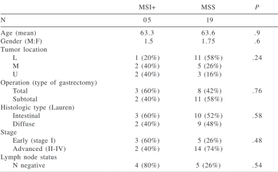
In the MSI+ group, the mean age was 63.6 years and the male:female ra-tio was 1.5; in the MSS group, the mean age was also 63.6 and the male:female ratio was 1.75. Tumor location in pa-tients having MSI+ tumors was 1 (20%) in the distal third, 2 (40%) in the mid-dle third, and 2 (40%) in the upper third of the stomach. In the MSS group, 11 patients (58%) had tumors in the lower third, 5 (26%) in the middle, and 3 (16%) in the upper third of the stom-ach. Three patients in the MSI+ group (60%) and 5 patients in the MSS group (26%) had Stage I disease. Lymph node metastasis was present in 1 patient in the MSI+ group (20%) and in 14 pa-tients in the MSS group (74%). There were no statistically significant differ-ences between groups in terms of age and gender distribution (P = .9 and P = .6, respectively). Although there was a trend for patients in MSI+ group to-wards proximal tumor location, early staging, and negative lymph node me-tastasis, there was no statistical signifi-cance, P >.1. (Table 4)
In the group of patients with Lauren’s intestinal-type gastric carci-noma, the mean overall survival was 33.6 months for patients with MSI+ tumors and 33.1 months for patients with MSS tumors. In this subgroup, none of the patients with MSI+ tumors had recurrence of the disease, while 2 patients with MSS tumors (20%) had systemic recurrent disease. In this group of patients, when patients with MSI+ and MSS tumors are compared within stages, mean overall survival and dis-ease recurrence rates are identical.
In the group of patients with Lauren’s diffuse-type gastric carci-noma, mean overall survival was 45.5 months for patients with tumors that were MSI+ and 30.9 months for
pa-Table 3 - Japanese Gastric Cancer Association Staging System-199819.
Staging JGCA (1998)
Stage T N M
IA T 1 N0 M 0
IB T 1 N1 M 0
T 2 N0 M 0
II T 1 N2 M 0
T 2 N1 M 0
T 3 N0 M 0
IIIA T 2 N2 M 0
T 3 N1 M 0
T 4 N0 M 0
IIIB T 3 N2 M 0
T 4 N1 M 0
IV T 4 N2 M 0
Any T N3 Any M
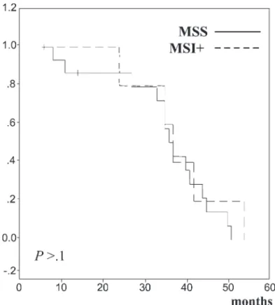
tients with tumors that were MSS. In this subgroup, none of the patients with MSI+ tumors had recurrence of the disease, while 3 patients with MSS tumors (20%) had systemic recurrent disease. These differences were not sta-tistically significant. In patients with Lauren´s diffuse-type gastric cancer, the difference between mean overall survival and disease-free survival was seen only in the stage III subgroup of patients, where the mean survival time of patients with MSI+ tumors was 54 months with no recurrence and for pa-tients with MSS tumors was 31.5 months with 1 recurrence (25%). (Figs. 1 and 2). However, the small number of patients in this subgroup (stage III) may account for such differences.
DISCUSSION
We found microsatellite instability (MSI) in 21% of 24 sporadic and soli-tary gastric adenocarcinomas in Brazil-ian patients, which is in accordance with other reports in the literature, which cite incidences of 9% to 33%.10,13,8,16,2,18,6 Furthermore, we also
found similar results concerning the frequent early disease staging
encoun-tered by others, since 60% of MSI+ tumors were stage I (Ia + Ib), while only 26% of the MSS tumors were stage I. Other characteristics have been associated with MSI+ gastric tumors, such as advanced age of patients, distally located tumors, and better overall prognosis.13.16,2,18,6 The
associa-tion with these latter characteristics was not observed in this study. Al-though not statistically significant, MSI+ tumors in our study were more frequently located in the proximal
third (40%) of the stomach when com-pared to MSS tumors (16%). Also, mean age was identical between both groups at 63.6 years.
We compared overall and disease-free survival between patients with MSI+ and MSS gastric tumors and found no statistically significant dif-ferences. These results also differ from earlier reports in the literature.13,16,2,18,6
The presence of the replication er-ror (RER) phenotype in gastric cancer appears to represent an independent genetic pathway in gastric carcinogen-esis.9,12,15 The incidence of this
pheno-type has been reported to be even higher (50%) in multiple or synchronic gastric tumors.9 A recent study has
shown that tumors that exhibit RER rarely exhibit loss of heterozygosity, considered to be the other major ge-netic pathway in gastric carcinogen-esis.12,11 Additionally, multiple tumors
in the same stomach usually present the same phenotype more frequently with MSI.12 These synchronic or
mul-tiple gastric tumors may be further di-vided in 2 subgroups, the first when there is an adenoma-associated adeno-carcinoma (type I) and the second when there is no adenoma associated (type II). These authors report an
im-Table 4 - Clinicopathological differences between microsatellite instability (MSI+) and microsatellite-stable (MSS).
MSI+ MSS P
N 0 5 19
Age (mean) 63.3 63.6 . 9
Gender (M:F) 1.5 1.75 . 6
Tumor location
L 1 (20%) 11 (58%) .24
M 2 (40%) 5 (26%)
U 2 (40%) 3 (16%)
Operation (type of gastrectomy)
Total 3 (60%) 8 (42%) .76
Subtotal 2 (40%) 11 (58%)
Histologic type (Lauren)
Intestinal 3 (60%) 10 (52%) .58
Diffuse 2 (40%) 9 (48%)
Stage
Early (stage I) 3 (60%) 5 (26%) .48
Advanced (II-IV) 2 (40%) 14 (74%)
Lymph node status
N negative 4 (80%) 5 (26%) .54
portant difference in the incidence of MSI between these subgroups, being more frequent in type I (56%) and in-frequent in type II (8%).9 The presence
of MSI may be associated with muta-tions in specific target genes such as TGFb RII, IGFII R, and BAX. 13,9 The
combination of mutation in target genes may be responsible for differ-ences in biological behavior and clini-cal-pathological characteristics within MSI+ tumors.
Association of genetic alterations and dietary factors have also been studied. Recently, an Italian study has suggested a correlation between red meat ingestion and MSI+ gastric can-cer, suggesting an environmental fac-tor as a causative event in susceptible individuals or increased tolerance to DNA damage associated with reduced MMR activity.14
MLH1 and MSH2 appear to be re-sponsible for the majority of the MSI both in colorectal cancer and gastric cancer.3,4 The mechanism involved
more frequently is hypermethylation of the MLH1 promoter region resulting in underexpression of the protein as
ob-served by immunohistochemistry.3,4,1
Recent observations have indicated that even adjacent noncancerous tis-sue may present hypermethylation of MLH1 promoter region and may be as-sociated with increased risk for the de-velopment of gastric cancer.15,1
These results suggest that MSI may be an important and early event in a subset of gastric adenocarcinomas. In our study, although not statistically significant, MSI+ tumors showed a trend towards early disease staging, proximal gastric location, and lower risk for lymph node metastasis. Fur-thermore, we found no difference in the mean age of these patients, over-all survival, and disease-free survival, in contrast with observations of oth-ers.13,16,2,18,6 We believe that tumor
ag-gressiveness and biological behavior may be determined by target gene mu-tations and thus these should be iden-tified in order to compare different subsets of gastric tumors all exhibiting the RER or MSI+ phenotype. Further-more, MSI+ gastric cancer may be the end-result of environmental or dietary factors in a susceptible stomach. These
two arguments may account for the clinical-pathologic and survival differ-ences observed in our study.
Identification of genetic pathways in gastric cancer may be of great portance and have direct practical im-plications in the management of gas-tric cancer. Tumor biological behavior may be directly associated with spe-cific genetic alterations during car-cinogenesis of gastric cancer. These differences may account for the hetero-geneity of presentation and prognosis of this neoplasm. Thus, patients may have a distinct prognosis, presentation characteristics, and response to therapy according to specific genetic alterations and pathway.
Treatment of gastric cancer is based on radical surgical resection. Extent of gastric resection is deter-mined by tumor histology and loca-tion. Some genetic alterations, such as MSI and mutations in MMR genes, may be associated with increased risk of multiple synchronous tumors and therefore may require total gastrec-tomy as an initial treatment strategy, once it can be diagnosed preoperatively. An environmental fac-tor may also influence these aspects, since dietary factors and specific car-cinogenic agents may influence in a variable manner the manifestation in a common genetic background. Our results differ from other reports in sev-eral aspects of tumor presentation and behavior of MSI+ gastric tumors, which may be partially explained by a different environmental factor not yet identified.
early gastric cancer diagnosis, improve overall survival, and revive the discus-sion about prophylactic treatment of gastric cancer.
Adjuvant therapy has not yet shown consistent improvement in overall survival for gastric cancer. Characterization of specific genetic al-terations and pathways may allow identification of a subset of patients with gastric cancer that is susceptible to specific antineoplastic drugs. Fur-thermore, neoadjuvant treatment with chemotherapy and radiotherapy may prove to be beneficial in selected cases of advanced or even unresectable dis-ease. Alterations in specific genes such as p53 and other genes associated with apoptosis may be responsible for some of these differences observed in adjuvant treatment responses. In theory, a genetic pathway that does not include frequent alterations in such genes may be associated with better
response to chemotherapy and radio-therapy with adjuvant or neoadjuvant intent. As mentioned earlier, MSI may represent a distinct genetic pathway of gastric cancer and does not include frequent alterations in p53 gene and others. One could expect better re-sponse rates with specific chemothera-peutic regimens for adjuvant treatment in MSI+ tumors.
Finally, identification of these spe-cific genetic alterations and pathways may aid in the development of new drugs involving gene therapy, such as viral vectors directed to specific target genes causing tumor cell death with minimum toxicity.
The importance of the study of ge-netic alterations in gastric cancer devel-opment cannot be overemphasized, but new management strategies based on these characteristics should await fur-ther consistent results. Caution should be taken with interpretation of these
re-sults with special consideration to dif-ferent geographical areas and other en-vironmental factors. These factors may be responsible for different influences in gastric carcinogenesis, giving rise to tumors with distinct biological behaviors and presentation over simi-lar genetic alterations or pathways.
In conclusion, MSI is a frequent event in gastric carcinogenesis and can be easily detected. The presence of MSI may be associated with distinct clinical, epidemiological, and patho-logical characteristics, such as primary tumor location, disease stage, and pres-ence of lymph node metastases. Differ-ent geographical areas and environ-mental factors may further influence the association between MSI and these features in gastric cancer. Identifica-tion of genetic and molecular events in gastric carcinogenesis may result in improvement of disease management and overall survival.
RESUMO
Perez RO e col. Instabilidade de microsatelites no cancer gástrico solitário e esporádico. Rev. Hosp. Clín. Fac. Med. S. Paulo 59(5): 279-285, 2004.
A presença de Instabilidade de microsatellites (IMS) tem sido relata-da no cancer gastrico e associarelata-da a pa-cientes com idade mais avançada, lo-calização mais distal do tumor, estadios mais precoces e melhor prog-nostico. Relatamos neste prospectivo estudo envolvendo 24 pacientes com cancer gastrico solitario e esporadico, a incidencia de IMS, sua correlação com parametros epidemiologicos, clinicos e anatomo patológicos e o seu impacto sobre a sobrevida geral e li-vre de doença.
PACIENTES E MÉTODOS: To-dos os pacientes haviam sido trataTo-dos com cirurgia radical. Fragmentos de tecido normal e tumoral eram extraidos das peças e armazenados a – 80oC antes da extração e purificação
DNA. Realizava-se então a amplifica-ção com PCR utilizando marcadores específicos de microsatelites. Os tumo-res que aptumo-resentavam produtos de am-plificação anormais foram considera-dos positivos para IMS.
RESULTADOS: Cinco pacientes (21%) apresentaram Instabilidade de microsatelites (IMS+) com pelo menos um marcador (primer) No grupo de pa-cientes com adenocarcinomas gástri-cos do tipo histológico de Lauren, três apresentavam IMS (23%) enquanto no grupo portador de cancar gástrico
difuso, dois pacientes mostraram IMS (19%).. Embora haja uma tendência dos pacientes IMS+ apresentarem tu-mores de localização mais proximal, estadios mais precoces e ausência de metástases linfonodais, não se obser-vou diferenças estatisticamente signi-ficativas (p > 0,1). A comparação en-tre as taxas de sobrevida geral e livre de doença não mostrou significância estatistica (p > 0,1).
CONCLUSÕES: IMS é um even-to frequente na carcinogese gástrica e pode estar associado a caracteristicas clinicas e anátomo-patológicas do cân-cer gástrico.
REFERENCES
1 . Baek MJ, Kang H, Kim SE, Park JH, Lee JS, Paik YK, Kim H. Expression of hMLH1 is inactivated in the gastric adenomas with enhanced microsatellite instability. Br J Cancer 2001;85:1147-52.
2 . Chung YJ, Park SW, Song JM, Lee KY, Seo EJ, Choi SW, et al. Evidence of genetic progression in human gastric carcinomas with microsatellite instability. Oncogene1997;15:1719-26. 3 . Cunningham JM, Christensen ER, Tester DJ, Kim CY, Roche PC,
Burgart LJ, et al. Hypermethylation of the hMLH1 promoter in colon cancer with microsatellite instability. Cancer Res 1998;58:3455-60.
4 . Fleisher AS, Esteller M, Wang S, Tamura G, Suzuki H, Yin J, et al. Hypermethylation of the hMLH1 promoter in human gastric cancer with microsatellite instability. Cancer Res 1999;59:1090-5 .
5 . Han HJ, Yanagisawa A, Kato Y, Park JG, Nakamura Y. Genetic instability in pancreatic cancer and poorly differentiated type of gastric cancer. Cancer Res 1993;53:5087-9.
6 . Hayden JD, Cawkwell l, Quirke P, Dixon MF, Goldstone AR, Sue-Ling H, et al. Prognostic significance of microsatellite instability in patients with gastric carcinoma. Eur J Cancer 1997;33:2342-6 .
7 . Ionov Y, Peinado MA, Malkhosyan S, Shibata D, Perucho M. Ubiquitous somatic mutations in simple repeated sequences reveals a new mechanism for colonic carcinogenesis. Nature 1993;363:558-61.
8 . Jen J, Kim H, Piantadosi S, Liu ZF, Levitt RC, Sistonen P, et al. Allelic loss of chromosome 18q and prognosis in colorectal cancer. N Engl J Med 1994);331(4):213-21.
9 . Lee HS, Lee BL, Kim SH, Woo DK, Kim HS, Kim WH. Microsatellite instability in synchronous gastric carcinomas. Int J Cancer 2001);91:619-24.
10. Nakashima H, Inoue H, Mori M, Ueo H, Ikeda M, Akiyoshi T. Microsatellite instability in Japanese gastric cancer. Cancer 1995;75(3)supp:1503-7.
11. Nishizuka S, Tamura G, Terashima M, Satodate R. Loss of heterozygosity during the development and progression of differentiated adenocarcinoma of the stomach. J Pathol 1998);185:38-43.
12. Ogata S, Tamura G, Endoh Y, Sakata K, Ohmura K, Motoyama T. Microsatellite alterations and target gene mutations in the early stages of multiple gastric cancer. J Pathol 2001;194:334-40. 13. Oliveira C, Seruca R, Sobrinho-Simões M, Seixas M. The
clinicopathological features of gastric carcinoma with microsatellite instability may be mediated by mutations of different “target genes”. Am J Pathol1998;153(4):1211-9. 14. Palli D, Russo A, Ottini L, Masala G, Saieva C, Amorosi A, et al.
Red meat, family history, and increased risk of gastric cancer with of gastric cancer with microsatellite instability. Cancer Res2001;61:5415-9.
15. Sakata K, Tamura G, En Y, Ohmura K, Ogata S, Motoyama T. Hypermethylation of the hMLH1 gene promoter in solitary and multiple gastric cancers with microsatellite instability. Br J Cancer 2002;86:564-67.
16. Seruca R, Santos NR, David L, Constancia M, Barroca H, Carneiro F, et al. Sporadic gastric carcinomas with microsatellite instability display a particular clinicopathologic profile. Int J Cancer 1995;64:32-6.
17. Thibodeau SN, Bren G, Schaid D. Microsatellite instability in cancer of the proximal colon. Science 1993;260:816-9. 18. Wu MS, Lee CW, Shunt CT, Wang HP, Lee WJ, Sheu JC, Lin JT
Clinico-pathological significance of altered loci of replication error and microsatellite instability-associated mutations in gastric cancer. Cancer Res 1998;58:1494-7.