From the Department of Pediatrics, Children’s Institute, Hospital das Clínicas, Faculty of Medicine, University of São Paulo – São Paulo/SP, Brazil.
E-mail: [email protected] Received for publication on
November 15, 2003.
ORIGINAL RESEARCH
OROPHARYNGEAL COLONIZATION BY
HAEMOPHILUS INFLUENZAE IN HEALTHY
CHILDREN FROM TAUBATÉ (SÃO PAULO), PRIOR TO
THE
HAEMOPHILUS INFLUENZAE TYPE B
VACCINATION PROGRAM IN BRAZIL
Lucia Ferro Bricks, Caio Márcio Figueredo Mendes, Bianca Rezende Lucarevschi, Carmem Paz Oplustil, Rosemeire C. Zanella, Adriana Bori and Ciro João Bertoli
BRICKS LF et al. Oropharyngeal colonization by Haemophilus influenzae in healthy children from Taubaté (São Paulo), prior to the Haemophilus influenzae type b vaccination program in Brazil. Rev. Hosp. Clin. Fac. Med. S. Paulo 59(5): 236-243, 2004.
Haemophilus influenzae is one of the most important bacterial agents of otitis and sinusitis. H. influenzae type b (Hib) is one of the main causes of meningitis, pneumonia, and septicemia in nonvaccinated children under 6 years of age.
The aims of this study were to determine the prevalence of H. influenzae and Hib oropharyngeal colonization prior to the onset of the Hib vaccination program in Brazil in previously healthy children and to assess the susceptibility profile of this microorganism to a selected group of antimicrobials that are used to treat acute respiratory infections.
METHOD: Cultures of Haemophilus influenzae were made from oropharynx swabs from 987 children under 6 years of
age who were enrolled in 29 day-care centers in Taubaté (a city of São Paulo state, Brazil) between July and December 1998.
RESULTS: The prevalence of H. influenzae carriers was 17.4%, and only 5.5% of the strains were beta-lactamase
producers. The prevalence of Hib carriers was high, 7.3% on average (range, 0.0 – 33.3%).
CONCLUSIONS: The low prevalence of colonization by penicillin-resistant strains indicates that it is not necessary to substitute ampicilin or amoxicilin to effectively treat otitis and sinusitis caused by H. influenzae in Taubaté.
KEY WORDS: Haemophilus influenzae. Haemophilus influenzae type b. Children. Antimicrobial resistance. Oropharynx colonization.
Haemophilus influenzae may cause many events, which vary from an asymptomatic infection to a severe in-vasive disease with high mortality rates. The colonization of the upper respiratory tract by nonidentified strains and encapsulated strains of H. influenzae (serotypes a through f) can be influenced by personal and envi-ronmental factors.1-6
Upper respiratory tract coloniza-tion occurs earlier in children who go to day-care centers than in those who stay home. Consequently, the exposure and risk of these children for
acquir-ing invasive disease is higher for chil-dren attending day-care centers.1-6
During the past few decades, anti-biotic-resistant H. influenzae strains have appeared, and the major resist-ance mechanism has been production of beta-lactamase. The patterns of an-tibiotic resistance vary in different
parts of the world and have been grow-ing, hampering the therapy for diseases caused by H. influenzae.7-12
The increasing prevalence of beta-lactamase–producing strains is one of the main reasons for the prescription of wide-spectrum antibiotics to treat children with acute respiratory infec-tion; on the other hand, the use of an-tibiotics is considered one of the main factors associated with the dissemina-tion of antibiotic-resistant strains, be-cause of alterations induced in oropha-ryngeal bacterial flora.13-17
microorganisms through prevalence studies of resistant strains in the oropharynx or nasopharynx of previ-ously healthy children may be an im-portant instrument in establishing a reference guide for acute respiratory infection therapy.7,13-18
In nonvaccinated populations, most invasive diseases caused by H. influenzae are associated with serotype b, particularly among children under 5 who attend day-care centers.1-6
The relationship between being a H. influenzae type b (Hib) carrier and the risk of disease has not yet been es-tablished, because there is disease dis-semination in populations with low colonization rates and absence of dis-ease in populations with high coloni-zation rates by Hib. However, asymp-tomatic carriers play an important role in microorganism dissemination. Al-though few subjects colonized by Hib develop the disease, airway coloniza-tion by this microorganism is the first step to the development of invasive disease.1,3,18,19
In developing countries, there have been few studies concerning oropha-ryngeal colonization in children and the resistance profile of H. influenzae in nonhospitalized children.6,20,24
In Brazil, vaccination against Hib was only introduced at the end of 1999, and until that year, no study of Hib carriers had been published.23 The choice to perform this study in chil-dren who attend day-care centers is due to the higher Hib colonization rates and antibiotic exposure in this population.
The purposes of this study were to identify the prevalence of H. influenzae colonization and the prevalence of strains resistant to antibiotics in the oropharynx of healthy children prior to the onset of the Hib vaccination pro-gram in Brazil; to analyze risk factors for the acquisition of antibiotic-resist-ant strains; to identify the prevalence of asymptomatic Hib carriers; and to
verify the main risk factors associated to Hib oropharynx colonization.
SUBJECTS AND METHOD
Study Population
Children under 6 years of age who were attending municipal day-care centers in 1998 in Taubaté (SP, Brazil) were included in the study. The city has approximately 244,000 inhabit-ants and 31 day-care centers that ac-cept children under 7. Two day-care centers are specifically for children be-tween 6 and 7; the other 29 are distrib-uted, with 1 in each district.
The exclusion criteria were: age above 6, children whose parents or le-gally responsible guardians did not authorize participation in the study, those who did not attend the day-care center at the collection date or who had not been fasting on the collection occasion.
Method
After the approval of the project by the Ethical Committee of the involved Institutions, the legally responsible person for each child was informed about the study objectives and signed an Informed Consent Form. A stand-ardized questionnaire containing in-formation about the child, socio-demographic characteristics, environ-mental conditions, previous and cur-rent morbidity, and antimicrobial us-age was filled out.
The following variables were analyzed: gender, age, race, period of time attending the day-care center, how much time the child spends at the day-care center, number of people who live in the same house, number of siblings who share the same room and who are under 5 years of age, parent’s educa-tion, and number of smokers in the domicile. There were questions about
respiratory symptoms and antimicro-bial use on the collection date, within the last 4 weeks, and within the 3 months prior to the collection.
Sample collection and microbiologi-cal analysis
Oropharynx sample collection was carried out weekly from July through December 1998 (winter and spring) by only 1 investigator, who also inter-viewed the parents and filled in the forms.
A sample from each child was col-lected using a sterile swab that was in-troduced through the mouth to the oropharynx. Immediately after sample collection, the swab was placed in an adequate transportation media (Amies with charcoal, StarSwab™). The sam-ples received a code number and were sent to the microbiology laboratory within 4 hours after sample collection. The sample was plated in the same day of collection in the following culture media: blood agar (BA), blood agar containing gentamycin (BA-G), and chocolate agar (CHOC). All the media were incubated at 35°C in a 5% CO2 atmosphere for at least 48 hours. After incubation, plates were analyzed, and microorganisms were isolated and identified through a standard metho-dology.25-27
Haemophilus isolation was done from characteristic colonies growing on chocolate agar. These colonies were identified using X and V growth fac-tor discs (Cefar).25-27
Identified H. influenzae colonies were preserved in lamb blood and fro-zen at -70°C. Subsequently, they were referred to the bacteriological section, where serotyping was performed by conventional slide agglutination method using polyclonal sera against the 6 serotypes (Difco).27
nitrocefin, BBL) and susceptibility to some antimicrobials, using disc diffu-sion methodology in HTM (Haemophilus Test Medium) agar plates.25,26
The tested antibiotics were ampi-cillin, amoxicillin/clavulanic acid, azithromycin, ceftriaxone, cefuroxime, chloramphenicol, and levofloxacine.
Statistical analysis
Data from a standard questionnaire containing information about the child and laboratory results were entered in a computer and analyzed by the sta-tistical program, Epi Info 6.04. Analy-sis of variables was performed by us-ing the χ2 or Fisher exact test, when-ever appropriate. To compare means, the Student t test was used; a P value < . 05 was considered significant.
Results were expressed in simple frequencies, and the coefficients for Hib colonization risk factors were ex-pressed as odds ratios (OR) with a 95% confidence interval.
RESULTS
Of the total of 1200 children un-der 6 years of age that attended the 29 day-care centers in Taubaté, 213 did not participate in the study.
Nine hundred eighty-seven chil-dren, aged 8 to 71 months (median = 53 mo) participated in the study. Twenty children (2.0%) were under 24 months of age.
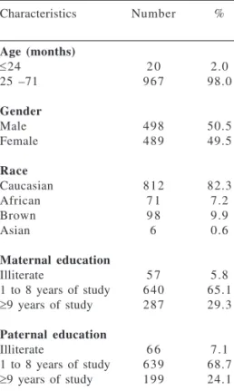
The children’s main sociode-mographic characteristics are listed in Table 1. Children attending day-care centers in Taubaté are, in general, from low-income families, and more than 65.0% of parents were poorly edu-cated.
The environmental conditions are summarized in Table 2. The period of time that children had been attending the day-care center ranged from 0 to
48 months (median = 8 months), and the majority of children (68.0%) stayed at the day-care center for 8 hours per day.
The number of people living in the house ranged from 2 to 15 (median = 4), and the number of people sleeping in the same child’s room ranged from
0 to 7 (median = 2). Children’s expo-sure to tobacco smoke at home was high (51.5%); 24.3% of the mothers and 39.1% of the fathers were smok-ers. Both parents of 131 children were smokers (13.2%). No child presented meningitis, epiglottitis, or contact with people presenting those diseases during the previous year.
Two hundred forty-six children (24.9%) presented respiratory symp-toms at the collection date. The most common problems were cough or wheezing (14.3%), common cold, in-fluenza, or watery nose (10.0%).
Only 32 children (3.2%) were re-ceiving antibiotics on the day of swab collection; 18.9% (n = 187) had re-ceived antibiotics during the previous month, and 30.1% (n = 297) had re-ceived antibiotics during the previous 3 months. The most commonly used antibiotics are listed in Table 3.
H. influenzae colonization and sus-ceptibility profile to antibiotics
Of the 987 children who were swabbed, 172 (17.4%) had a positive culture for H. influenzae; 91 of these were tested for antimicrobial suscepti-bility. Five of these tested isolates (5.5%) were beta-lactamase producers (4 were type b). Resistance to other Table 1 - Sociodemographic
charac-teristics of the study population: number and percentage of children, according to age, gender, race, and parents’ education.
Characteristics Number %
Age (months)
≤24 2 0 2.0
25 –71 9 6 7 98.0
Gender
Male 4 9 8 50.5
Female 4 8 9 49.5
Race
Caucasian 8 1 2 82.3
African 7 1 7.2
Brown 9 8 9.9
Asian 6 0.6
Maternal education
Illiterate 5 7 5.8
1 to 8 years of study 6 4 0 65.1
≥9 years of study 2 8 7 29.3
Paternal education
Illiterate 6 6 7.1
1 to 8 years of study 6 3 9 68.7
≥9 years of study 1 9 9 24.1
Table 2 - Distribution of number and percentage of studied children according environmental conditions.
Environmental conditions Number %
Period of time attending the day-care center (months)
≤6 3 8 1 38.6
>6 6 0 6 61.3
Time at the day-care center
Part time (4 hours/day) 3 1 6 32.0
Full time (8 hours/day) 6 7 1 68.0
Presence of smokers at domicile 5 0 9 51.5
Number of people who live with the child
<4 2 5 2 25.5
≥4 7 3 5 74.5
Children with siblings <5 years 3 8 2 38.7
Number of people sharing the same room with the child
<2 3 0 9 31.3
2 – 3 5 6 9 57.7
tested antimicrobials was nil (Table 4). There was no relationship between microorganism resistance and the analyzed variables. The P value was greater than .05 for gender, age, anti-biotics usage, number of siblings, pres-ence of smokers, history of respiratory diseases, and vaccine status.
Hib colonization
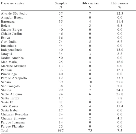
From the 172 isolated cultures of H. influenzae, 73 (collected from 7.3% of the test population) were of type b.
Hib colonization ranged from 0.0% to 33.3% in the different day-care centers (Table 5), and except for the fact that a child attended a particular day-care center, none of the analyzed variables was significantly associated with the risk of Hib colonization (Ta-ble 6).
In order to assess whether exposure to larger numbers of children was re-lated to the number of Hib carriers at a particular center, we made crossings between the total number of children in each day-care center (including children older than 6) and the number of Hib carriers under 5; the P value was not significant (OR = 0.69 [95% CI: 0.42-1.16), P = 0.13).
DISCUSSION
Oropharynx colonization by en-capsulated strains of H. influenzae is very common and varies between 6.0% and 90.0%, in different populations.2,4,6,7,13,14,16,18 In this study, the prevalence of children colonized by H. influenzae was 17%, similar to rates found by other authors.20,23,28
It is possible that some strains may not have survived the transport to the lab; however, the elevated rate of Hib colonization found (7.3%) suggests that this biasing factor did not occur.
Longitudinal studies conducted in healthy children and in children with
Table 3 - Most common antibiotics used by children who attended day-care centers in Taubaté on the day of swab collection and 30 and 90 days prior to the sample collection.
Antibiotics At collection date Last month Previous 90 days
Amoxicillin 47.0% 49.0% 49.5%
Penicillin benzathine 12.5% 16.6% 15.3%
TMP-SMZ* 12.5% 9.7% 10.5%
*TMP-SMZ: trimethoprim-sulphamethoxazole
Table 4 - Prevalence of H. influenzae strains, according to the pattern of susceptibility to different antibiotics.
Sensitive Intermediate Complete Number of
Antibiotics (%) resistance resistance tested
(%) (%) strains
Ampicillin * 94.4 0.0 5.5 9 1
Amoxicillin/clavulanate 100.0 0.0 0.0 9 1
Azithromycin 100.0 0.0 0.0 9 1
Cefuroxime sodium 100.0 0.0 0.0 9 1
Ceftriaxone 100.0 0.0 0.0 9 1
Chloramphenicol 100.0 0.0 0.0 9 1
Levofloxacin 100.0 0.0 0.0 6 7
* Total of beta-lactamase–producing strains = 5
Table 5 - Number of children who participated in study, number swabbed (samples), number of Haemophilus influenzae type b (Hib) carriers, and percentage of Hib carriers by day-care center.
Day-care center Samples Hib carriers Hib carriers
N N %
Alto de São Pedro 5 7 7 12.3
Amador Bueno 4 7 0 0.0
Baroneza 3 5 4 11.4
Belém 4 4 3 6.8
Canuto Borges 4 0 0 0.0
Cidade Jardim 4 6 0 0.0
Estiva 1 6 0 0.0
Gurilândia 7 4 5 6.7
Imaculada 4 4 0 0.0
Independência 4 0 6 15.0
Jaraguá 3 4 3 8.8
Jardim América 5 0 0 0.0
Mãe Maria 2 5 4 16.0
Marlene Miranda 1 3 0 0.0
Paduan 3 3 4 12.1
Piratininga 4 0 0 0.0
Parque Aeroporto 1 2 4 33.3
Sabará 3 9 1 0 25.6
São Gonçalo 3 8 3 7.8
Shalon 2 9 7 24.1
Santo Antonio 2 4 6 25.0
Santa Tereza 1 7 1 5.8
Santa Fé 3 1 0 0.0
Três Marias 3 5 4 11.4
Santa Isabel 1 7 0 0.0
Chácaras Reunidas 2 4 0 0.0
Chácara Silvestre 4 4 2 4.5
Parque Ipanema 1 4 0 0.0
Parque Planalto 2 5 0 0.0
recurrent otitis media have demon-strated that the status of a nasophar-ynx carrier of some pathogens like H. influenzae, S. pneumoniae, and M. catarrhalis is maintained in an almost constant proportion (21.0% to 37.0%) during the first 3 years of life; however, during high respiratory airway infec-tions, there is an increase in those pro-portions.20 In the present study, H. influenzae colonization was not asso-ciated with the presence of acute res-piratory infections.
The profile of microorganism resist-ance varies from region to region; there-fore, it is essential to know the local susceptibility patterns to antibiotics to appropriately guide the best therapeu-tic option. Most of the studies concern-ing microorganism resistance are re-lated to strains isore-lated from body flu-ids of children with invasive disease, and their data are not always useful for evaluating the microorganism-resist-ance profile in the community.7-17
In many countries, the proportion of H. influenzae beta-lactamase–pro-ducingstrains in children with invasive disease varies between 6.0% and 60.0% and has been increasing yearly.9-11
In Brazil between 1990 and 1999, from a total of 3204 strains of Haemophilus influenzae isolated from
patients with invasive disease (91.0% under 4 years of age), it was noted that 18.1% of strains were resistant to ampi-cillin, 19.1% to chloramphenicol, and 13.9% to ampicillin and chloram-phenicol, a very high prevalence com-pared to the results of this study. In 1996, 15.0% of the strains were beta-lactamase producers, increasing to 21.0% in 1999; during the same pe-riod, a significant increase in chloram-phenicol resistance was not detected (18.8% vs. 19.9%). All strains were sen-sitive to rifampicin (used for chemo-prophylaxis) and cephalosporin.10
In the present study, we found that only 5.5% of the isolated strains were beta-lactamase producers, results that are very similar to the rates found in healthy children who attended day-care centers in United Kingdom in the 1980s (5.4%)8 and in Italian children with se-rous otitis.16 In other regions of Europe, the United States, and Israel, the preva-lence of strains causing serous otitis that are resistant to ampicillin and other antibiotics is much higher.12-15
Acute respiratory infections, par-ticularly otitis media, are the main reason for antibiotics usage in chil-dren.12-17 There are controversies about the value of oropharyngeal cultures for predicting the etiology of acute respiratory infections. Some studies
have shown that the positive predic-tive value is low (50.0% for H. influenzae, 40.0% for S. pneumoniae, and 20.0% for M. catarrhalis)12; how-ever, the negative predictive value is excellent (95%).12,14
The low prevalence of beta-lactamase–producing strains found in the study suggests that in Taubaté, ampicillin should be the antibiotic of choice for the therapy of otitis and si-nusitis that is presumably caused by Haemophilus influenzae.
Although the previous use of anti-biotics is related to the colonization of respiratory airways by resistant strains,14,17 in the present study, previ-ous usage of antibiotics was not asso-ciated with an increase in the preva-lence of beta-lactamase–producing strains.
In the present study, Hib coloniza-tion rate was 7.3%, and in 11 day-care centers was greater than 10.0% (Table 5).
Of the many factors associated with Hib colonization, young age, contact with people who had invasive diseases caused by Hib, attending day-care centers, and vaccination status have been found to be the most important. Results concerning other risk factors such as gender, number of siblings un-der 5 years of age, and prior respira-tory diseases are controversial.1-7,20-24
In the Dominican Republic, a higher colonization rate was found among children who had siblings un-der the age of 522; in the present study, as in others,2,23 there were no signifi-cant differences among Hib-colonized children regarding the different analyzed variables.
Although a quarter of the children were presenting respiratory symptoms at the time of the swab collection, this variable was not associated with an in-crease in Hib colonization rates. Since acute respiratory infections are risk fac-tors for Hib invasive disease, high rates of colonization by Hib are worrisome.1 Table 6 - Possible risk factors for Haemophilus influenzae type b (Hib) oropharynx
colonization.
Risk factors Hib Carriers Odds ratio (95% IC) P value
Gender
Female 3 9 1.18 (0.73 – 1.97) 0.57
Male 3 4
Siblings under age of 5
Yes 4 3 0.9 (0.54 – 1.51) 0.75
No 3 0
Smokers at domicile
Yes 3 7 0.91 (0.55 – 1.51) 0.78
No 3 6
Hib Vaccination
Yes 1 1 1.26 (0.60 – 2.57) 0.65
In 1998, the vaccine against Hib had not yet been incorporated into the pediatric vaccination calendar in Bra-zil. The rates of Hib carriers were simi-lar to those found in preschool chil-dren living in developing countries such as the Dominican Republic (7.7%)22 and Turkey (9.0%)6 as well as Alaska (5.0% to 7.0%),24 and they were much higher than in children living in industrialized nations in the pre-vac-cine era.1
In populations vaccinated against Hib, the prevalence of airway carriers has significantly decreased19,23,28-32; however, the use of conjugated vaccines protects only partially against the carrier status and does not com-pletely eliminate the risk of infection by this microorganism.1,24,31,32
In Brazil, vaccination against Hib was introduced into the routine pediatric vaccination calendar by the end of 1999. In Curitiba (PR), vacci-nation was introduced in 1996, and the following year the prevalence of Hib colonization in 657 vaccinated chil-dren was 1.2%, lower than that found in 643 nonvaccinated children (4.8%) living in Porto Alegre (RS).23
In the present study, we did not find significant differences in Hib coloni-zation rates in vaccinated and nonvaccinated children; it is worth-while to mention that few children had been vaccinated against Hib and that the vaccine is effective in preventing colonization in the community only when the vaccine coverage is high.1
In Alaska, after the introduction of conjugated vaccines against Hib, inva-sive disease caused by this microorgan-ism has drastically decreased; however, a significant increase in cases of inva-sive disease caused by Hib has been recently detected.24,32 It is believed that high rates of colonization by Hib in
the Alaska population played an im-portant role in the re-emergence of Hib invasive disease.24,31,32
In different regions in Alaska, the rates of Hib carriers were variable. In Barrow, 4.4% of the children were colonized by Hib, with higher preva-lence between 6 and 7 years of age (7.8%), dropping to 0.0% in adoles-cents between 14 and 16 years of age. In Delta YK, they found a 6.6% rate of carriers among 365 children under the age of 4, this rate being much greater than that found in 383 children less than 4 years of age living in Anchor-age (1.0%). The persistence of a great number of children who were carriers of Hib in some communities in Alaska after the introduction of conjugated vaccine against Hib suggests that chil-dren of school age (who have not been vaccinated) may constitute an impor-tant reservoir of Hib. It seems that the elevated rate of carriers among nonvaccinated children was responsi-ble, at least in part, to the re-emer-gence of invasive disease in Alaska.24,31 These are very troubling data be-cause in Taubaté the rates of coloni-zation by Hib in children under 6 were as high as those found in Alaska in the prevaccine era. Unfortunately, we did not evaluate the prevalence of carriers among children older than 6 years in this city.
Preschool and school children colonized by Hib constitute the main reservoir of this microorganism, and the colonization may persist for pro-longed periods of time—1 or more years. Since the status of Hib carrier, in general, does not produce an in-crease in the titres of protecting anti-bodies against Hib, children carrying this microorganism may develop inva-sive disease caused by this agent.1,19,24
The incubation period of invasive and noninvasive disease caused by Haemophilus influenzae is unknown; however, high rates of colonization of the respiratory airways are associated with a higher risk of disease that at-tacks mucous tissue as well as invasive disease. Without a doubt, vaccination against Hib is the most effective meas-ure for reducing the impact of Hib in-vasive disease, although it does not play a significant role in reducing mu-cous tissue disease.1,30 After this study was completed, all children between 2 and 60 months of age who attended day-care centers in Taubaté were vac-cinated against Hib.
CONCLUSION
The prevalence of strains of beta-lactamase–producing Haemophilus influenzae (5.5%) was low among chil-dren from Taubaté, indicating that ampicillin or amoxicillin can be used as the antibiotic of choice to treat oti-tis and sinusioti-tis presumably caused by this microorganism.
The prevalence of Hib carriers was high (7.3%) in this study. Since the human being is the only reservoir of Hib and colonization has a relevant role in the transmission cycle of inva-sive disease caused by this microorgan-ism, it is essential to vaccinate children under 5 years of age against Hib and to monitor the impact of such a meas-ure.
ACKNOWLEDGMENT
RESUMO
orofaringe de crianças previamente saudáveis por H. influenzae e Hib e avaliar o perfil de suscetibilidade des-ses microorganismos a um grupo sele-to de antimicrobianos, que habitual-mente são utilizados para tratar as in-fecções respiratórias agudas.
MÉTODO: Foram colhidos swabs da orofaringe de 987 crianças menores de seis anos de idade que freqüentavam 29 creches da cidade de Taubaté (São Paulo, Brasil), entre julho e dezembro de 1998, para realização de culturas de H. influenzae e antibiograma.
RESULTADOS: A prevalência de portadores do H. influenzae foi de 17,4% e somente 5,5% das cepas
iso-ladas eram produtoras de beta-lacta-mase. A prevalência de portadores do Hib foi alta, com média de 7,3% (vari-ando entre 0.0 e 33,3%).
CONCLUSÕES: A baixa preva-lência da colonização por cepas resis-tentes às penicilinas indica que não é necessário substituir esses antibióticos para tratar empiricamente as otites e si-nusites causadas por H. influenzae em Taubaté.
UNITERMOS: Haemophilus influenzae. Haemophilus influenzae type b (Hib). Crianças. Resistência antimicrobiana. Colonização da orofaringe.
BRICK LF e col. Colonização da orofaringe de crianças saudáveis de Taubaté (São Paulo) por Haemophilus influenzae, antes da introdução da vacina contra Haemophilus influenzae do tipo b no Brasil. Rev. Hosp. Clin. Fac. Med. S. Paulo 59(5):236-243, 2004.
Haemophilus influenzae é um dos mais importantes agentes bacterianos de otites e sinusites. Em crianças menores de seis anos de idade não vacinadas con-tra o H. influenzae do tipo b (Hib), essa bactéria é uma das principais causado-ras de meningite, pneumonia e sepse.
O objetivo deste estudo foi deter-minar a prevalência da colonização da
REFERENCES
1 . Barbour ML. Conjugate vaccines and the carriage of Haemophilus influenzae type b. Emerg Infect Dis 1996;2:1-9.
2 . Michaels RH, Poziviak CS, Stonebraker FE, Norden CW. Factors affecting pharyngeal Haemophilus influenzae type b colonization rates in children. J Clin Microbiol 1976;4:413-7. 3 . Granoff DM, Daum RS. Spread of Haemophilus influenzae type b: recent epidemiologic and therapeutic considerations. Pediatrics 1980;97:854-860.
4 . Murphy TV, Pastor P, Medley F, Osterholm MT, Granoff DM. Pharyngeal colonization with Haemophilus influenzae type b in children in a day care center without invasive disease. J Pediatr 1985;106:712-6.
5 . Li KI, Dashefsky B, Wald ER. Haemophilus influenzae type b colonization in household contacts of infected and colonized children enrolled in day care. Pediatrics 1986;78:15-20. 6 . Bakir M, Yagci A, Ulger N, Akbenlioglu C, Ilki A, Soyletir G, et
al. Pharyngeal colonization with Haemophilus influenzae type b among healthy Turkish infants and children. Pediatr Intern 2002;44:381-6.
7 . Lerman SJ, Kucera JC, Brunken JM. Nasopharyngeal carriage of antibiotic resistant Haemophilus influenzae in health children. Pediatrics 1979;64:287-90.
8 . Howard AF, Dunkin KT, Millar GW. Nasopharyngeal carriage and antibiotic resistance of Haemophilus influenzae in healthy children. Epidemiol Infect 1988;100:193-203.
9 . Thornsberry C, Ogilvie PT, Holley HP Jr, Sahm DF. Survey of susceptibilities of Streptococcus pneumoniae, Haemophilus influenzae, and Moraxella catarrhalis. Isolates to 26 antimicrobial agents: a prospective US Study, Antim Agent Chem 1999;43:2612-23.
10. Zanella RC, Casagrande ST, Bokermann S, Almeida SC, Brandileone MC. Characterization of Haemophilus influenzae
isolated from invasive disease in Brazil from 1990 to 1999. Microb Drug Resist 2002;8;67-72.
11. Blosser-Middleton R, Sahm DF, Thornsberry C, Jones ME, Hogan PA, Critchley IA, et al. Antimicrobial susceptibility of 840 clinical isolates of Haemophilus influenzae collected in four European countries in 2000-2001. Clin Microbiol Infect 2003; 9:431-6.
12. Gehanno P, Lenoir G, Barry B, Bons J, Boucot I, Berche P. Evaluation of nasopharyngeal cultures for bacteriologic assessment of acute otitis media in children. Pediatr Infect Dis 1996;15:329-32.
13. Klein J. Role of nontypeable Haemopilus influenzae in pediatric respiratory tract infections. Pediatric Infect Dis J 1997;16:S5-8 .
14. Harper M. Nasopharyngeal colonization with pathogens causing otitis media: how does this information help us? Pediatr Infect Dis 1999;181:1120-24.
15. Leibovitz E, Satran R, Piglansky L, Raiz S, Press J, Leiberman A, et al. Can acute otitis media caused by Haemophilus influenzae
be distinguished from that caused by Streptococcus pneumoniae? Pediatr Infect Dis 2003;22:509-14.
16. Marchisio P, Claut L, Rognoni A, Esposito S, Passali D, Bellussi L, et al. Differences in nasopharyngeal bacterial flora in children with nonsevere recurrent acute otitis media and chronic otitis media with effusion: implications for management. Pediatr Infect Dis 2003;22:262-8.
18. Rapola S, Salo E, Leinonen M et al. Comparison of four different methods for detecting pharyngeal carriage of Streptococcus pneumoniae and Haemohilus influenzae in children. J Clin Microbiol 1997; 35:1077-9.
19. CDC. Progress toward elimination of Haemophilus influenzae
type b invasive disease among infants and children—United States, 1998-2000. MMWR 2002;51:234-7.
20. Villaseñor-Sierra A, Herrera-Basto E, Vázquez-Salazar P et al. Prevalencia de estado de portador de Haemophilus influenzae
in niños de Ciudad Nezahualcóyotl, estado de México, México. Salud Publica Mex 1996;38:87-93.
21. Vives M, Garcia ME, Saenz P, Mora MA, Mata L, Sabharwal H, et al. Nasopharyngeal colonization in Costa Rican children during the first year of life.. Pediatr Infect Dis 1997;16:852-8. 22. Gomez E, Moore A, Sanchez J, Kool J, Castellanos PL, Feris JM,
et al. The epidemiology of Haemophilus influenzae type b carriage among infants and young children in Santo Domingo, Dominican Republic. Pediatr Infect Dis 1998;17:782-6. 23. Forleo-Neto E, de Oliveira CF, Maluf EM, Bataglin C, Araujo JM,
Kunz LF Jr, et al. Decreased point prevalence of Haemophilus influenzae type b (Hib) oropharyngeal colonization by mass immunization of Brazilian children less then 5 years old with Hib polyribosylribitol phosphate polysaccharide-tetanus toxoid conjugate vaccine in combination with diphtheria-tetanus toxoid-pertussis vaccine. J Infec Dis 1999;180:1153-8.
24. Galil K, Singleton R, Levine OS, Fitzgerald MA, Bulkow L, Getty M, et al. Reemergence of invasive Haemophilus influenzae
type b disease in a well-vaccinated population in remote Alaska. J Infect Dis 1999;179:101-6.
25. Isenberg HD. Clinical Microbiology Procedures Handbook. ASM, Washington, v1, 1992.
26. National Committee for Clinical Laboratory Standards. 1998, Performance standards for antimicrobial disk susceptibility tests. Approved standard M2-A5. Wayne, Pa.
27. Campos JM. Haemophilus. In: Murray PR, Baron EJ, Pfaller MA, et al. Manual of Clinical Microbiology, 7th ed. American Society
for Microbiology, Washington, 1999, p.605-13.
28. Takala AK, Eskola J, Leinonen M, Kayhty H, Nissinen A, Pekkanen E, et al. Reduction of oropharyngeal carriage of Haemophilus influenzae type b (Hib) in children immunized with a Hib conjugate vaccine. J Infect Dis 1991;164:982-6.
29. Mohle-Boetani JC, Ajello G, Breneman E, Deaver KA, Harvey C, Plikaytis BD, et al. Carriage of Haemophilus influenzae type b in children after widespread vaccination with conjugate
Haemophilus influenzae type b vaccines. Pediatr Infect Dis 1993;12:589-93.
30. Peltola H. Worldwide Haemophilus influenzae type b disease at the beginning of the 21st century: global analysis of the disease
burden 25 years after the use of polysaccharide vaccine and a decade after the advent of conjugates. Clin Micro Rev 2001;13:302-17.
31. Singleton R, Bulkow LR, Levine OS, Butler JC, Hennessy TW, Parkinson A. Experience with the prevention of invasive
Haemophilus influenzae type b disease vaccination in Alaska: impact of persistent oropharyngeal carriage. J Pediatr 2000;137:313-32.
32. Trotter CL, Ramsay ME, Slack MPE. The epidemiology of