194
Celso Marcelo da Cunha
1, Renato José Bett Correia
2, Jéssica Teixeira Cunha
3A
BSTRACTObjective: To compare changes in refraction and ocular biometric parameters in hyperopic children with and without full optical cor-rection. Methods: Non-randomized prospecting study with 41 subjects (21 males and 20 females) aged 4 to 6 years with accommodative esotropia and or hyperopia between 3 to 6 diopters, select in Hospital Geral Universitário and Oftalmocenter Santa Rosa. The patients were divided in two groups: group 1 for hyperopic patients that did not need to use optical correction or could use partial correction, and group 2 for patients with accommodative esotropia or hyperopia who needed to use full optical correction all the time. The patients were examined to a complete ophthalmological examination, including objective cycloplegic refraction with auto refractometer, optical biometry and corneal topography, in baseline measurements and 3 years after that. Refraction and ocular biometric parameters were compared using T student test. Results: The mean initial age was 5.23 ± 0.81 and 5.36 ± 0.74 years; the initial refractive error in average was +3.99 ± 0.92 e +4.27 ± 0.85 D, the initial axial length was 21.42 ± 0.84 and 21.22 ± 0.86 mm, and initial keratometry was 42.55 ± 1.24 e 42.39 ± 1.22 D for group 1 and 2, respectively. In relation to refractive error, there was a significant decrease in group 1 and there was not in group 2 (p < 0.05). In relation to axial length, there was significant increase in group1 and there was not in group 2 (p<0.05). The 3-year comparison showed no statistically significant differences in keratometry for both groups. Conclusion: This study suggests that full optical correction of hyperopia may inhibit natural emmetropization during early and late childhood.
Keywords: Refraction errors; Hyperopia; Eye/growth & development.
R
ESUMOObjetivo: Comparar as alterações da refração e da biometria ocular na população infantil hipermetrópica com e sem correção óptica total. Métodos: Realizou-se estudo prospectivo longitudinal não randomizado em 41 pacientes com hipermetropia, entre 3 e 6 dioptrias ou/e com esotropia acomodativa pura nos ambulatórios do Hospital Geral Universitário e Oftalmocenter Santa Rosa, com idade inicial entre 4 e 6 anos. Os pacientes foram divididos em dois grupos, em que o Grupo 1 compôs-se pelos pacientes hipermétropes que não necessitavam usar sua correção óptica ou poderiam usá-la parcialmente, e o Grupo 2 por pacientes com esotropia acomodativa pura e pelos hipermétropes que necessitavam usar toda sua correção óptica. Os pacientes submeteram-se a exame oftalmológico completo, incluindo refração objetiva em autorrefrator com cicloplegia, biometria óptica e topografia corneana em uma medida inicial e outra 3 anos mais tarde. Comparou-se a refração e parâmetros biométricos com teste T student. Resultados: A média da idade inicial foi de 5,23 ± 0,81 e 5,36 ± 0,74 anos, a refração inicial foi +3,99 ± 0,92 e +4,27 ± 0,85 D, o diâmetro anteroposterior do globo ocular foi de 21,42 ± 0,84 e 21,22 ± 0,86 mm, e a ceratometria foi de 42,55 ± 1,24 e 42,39 ± 1,22 D, para os Grupos 1 e 2, respectivamente. Em rela-ção à refrarela-ção, houve redurela-ção significativa do poder esférico no Grupo 1, em 3 anos; e não houve no Grupo 2 (p<0,05). Com relarela-ção ao diâmetro anteroposterior do globo ocular, ocorreu aumento significativo no Grupo 1 e não houve no Grupo 2 (p<0,05 ). Não se verificou diferença significativa na comparação das ceratometrias em 3 anos nos Grupos 1 e 2. Conclusão: Estes dados permitiram concluir que a correção total da hipermetropia pode prejudicar a emetropização natural em crianças.
Descritores: Erros de refração; Hipermetropia; Olho/crescimento & desenvolvimento.
1 Oftalmocenter Santa Rosa. Cuiabá, MT, Brazil. 2 Hospital Geral Universitário. Cuiabá, MT, Brazil.
3 Academic Course in Medicine, Centro Universitário da UNIVAG. Várzea Grande, MT, Brazil.
Received for publication 13/07/2017 - Accepted for publication 18/07/2017.
The authors declare no conflicts of interestss.
O
RIGINALA
RTICLERev Bras Oftalmol. 2017; 76 (4): 194-7
Optical correction and evolution of hipermetropy
Correção óptica e evolução da hipermetropia
195
I
NTRODUCTIONH
ypermetropia is the most common ametropia in chil-dhood. Pre-verbal children have a high initial accom-modative capacity (12 diopters of accommodation am-plitude) that slowly decreases for decades. However, they usually tolerate only low and medium hypermetropias without decreased visual acuity or asthenopia, as long as it is within the limits of their accommodative tolerance.(1,2) Accommodative tolerance is theability of the child to be able to exert long-term accommodation without asthenopia or other clinical manifestations. The formula to estimate this value is: TA = 4.5 – 0.1 x Y (where Y is the age of the child in years).
Because they have such active accommodation, often hyper-metropic children who are within their accommodative tolerance wear their glasses irregularly. In turn, children with accommodative esotropia regularly wear their glasses, often due to keeping their binocular parallelism this way.
The emmetropization of the eyeball occurs by changes in the corneal curvature, by growth of the anteroposterior diameter of the eyeball, and by diminution of the dioptric power of the crystalline. The most important changes in the corneal curvature usually occur in the first two years of life.(3)
Previous studies have not been conclusive on the influence of total hypermetropia correction on its development. However, it is believed that hypermetropic children aged under 10 years and who have used their total optical correction for many years do not na-turally regress their hypermetropia. This fact could occur nana-turally with hyperopic children who do not use their optical correction.
This study was designed to compare hypermetropic child patients with the total optical correction to uncorrected or partially corrected.
M
ETHODSA non-randomized, longitudinal prospective study was performed at the Ophthalmology ambulatories of Hospital Geral Universitário de Cuiabá and Oftalmocenter Santa Rosa. Patients with initial age between 4 and 6 years were included.
Forty-one patients were selected during the care period of the authors, in a consecutive way, after their decision-makers approved the informed consent term, and met the inclusion and exclusion criteria. The patients were selected for two groups, Group 1 (23 patients) comprising hypermetropic patients who did not use their optical correction or could use partial optical correction (8 patients) within their accommodative tolerance. Group 2 (18 patients) comprised patients with pure accommo-dative esotropia or who needed continuous use of their hyper-metropic optical correction (4 patients).(4) Only the right eye of
every patient was assessed for the study.
Patients with visual acuity of 0.07 or better in both eyes, with hypermetropia between 3 and 6 diopters, with or without pure accommodative esotropia, and with other normal ophthalmologic examinations were included in this study. We excluded candidates with astigmatism greater than 1.00 DC, anisometropia greater than 1.00 R, accommodative esotropia with high CA/A, partially accommodative esotropia, those who did not return for two con-secutive reviews or the last one, those who were cyclopleghed outside the planned period, those who no longer wear glasses for over 30 days (of patients who should wear glasses continuously), with neurological change, and/or those who did not cooperate nor understood the carrying out of the examinations.
The children were examined by the first author, who mea-sured their visual acuity, refraction in autorefractor (five measu-rements; the prevalent average power was used) with cycloplegia (cyclopentolate 1% and tropicamide 1% twice, preceded by 1 dro of proximetacaine 0.5%) held 40 min after the last drop of eyedrops, coverage test with and without prisms, ocular motility, biomicroscopy of the anterior segment, fundoscopy, computed to-mography and optical biometry as to measure the anteroposterior diameter of the eyeball (DAP). A second complete evaluation was held 3 years later. Other half-yearly evaluations in this interval did not have cycloplegia, computerized keratoscopy or biometry.
The statistical analysis was done using the software SPSS for Windows (Statistical Package for Social Sciences, version 9.0, SPSS Inc., Chicago, USA). Continuous variables were compared using the Student t test, and the categorical variables using the Chi-square test. The longitudinal changes for each parameter (re-fraction, keratometry and DAP) were analyzed with the Student’s t-test. P <0.05 was considered statistically significant.
Record in Brazil Platform: 09123713.9.0000.5165 . May 2013
R
ESULTSOf the 41 patients, 21 (51.21%) were male and 20 (48.79%) female. The average age was 5.23 ± 0.81 and 5.36 ± 0.74 years, and the average initial hypermetropia was 3.99 ± 0.92 and 4.27 ± 0.85 D, the average initial was 21.42 ± 0.84 e 21.22 ± 0.86 mm, the average keratometry was 42.55 ± 1.24 and 42.39 ± 1.22 D for groups 1 and 2, respectively.
Of the 23 patients from Group 1, one was excluded for not having returned to the final evaluation, and of the 18 from Group 2, four were excluded, one for not wearing glasses continuously, two for not returning for the final evaluation, and one for having refrac-tion with cycloplegia outside the period determined in this study.
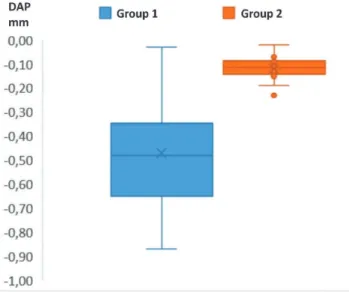
Regarding the data collected at the study onset and 3 years later, there was significant reduction of hypermetropia in Group 1 (from 3.99 to 3.00 D, p<0.05), and there was no significant reduc-tion of hypermetropia in Group 2 (from 4.27 to 4.16 D, p<0.05). Regarding DAP, there was an increase in Group 1 (from 21.42 to 21.89 mm, p<0.05), and there was no significant increase in Group 2 (from 21.22 to 21.34 mm, p<0.05). There was no difference in the K of the two groups. Compared to the average ages, there were no significant differences in the refractions and DAP between male and female.
Figure 1 shows the negative influence of the total optical correction of hypermetropia on emmetropization.
Figure 2 shows the negative influence of the total optical correction of hypermetropia on the increase of DAP.
D
ISCUSSIONThe growth of the eyeball is bigger in myopic, lower in emmetropic, and even lower in hypermetropic patients. The compensation of this increase in this population is made by the crystalline. Therefore, for the emmetropization to occur in hyper-metropic patients of this age group studied, it would be necessary to have a higher growth of the eyeball and/or a lower rectification of the curves of the lens.(5)
For decades it has been discussed how much to prescribe hypermetropia to orthophoric children with asthenopia, since it has been considered only how much of the correction of hyper-metropia would be satisfactory to ameliorate the symptoms, and
196
it had not been considered if the total correction may or may not influence the ocular emmetropization. There are very few studies in the literature on this topic, and some point out controversial results. Two of them have demonstrated the interference of total correction in emmetropization of the child, and this study cor-roborated these findings.(6,7) We believe that children can reduce
from 0.25 to 0.37 D/year, from 4 to 15 years. Another study showed that there is no difference in younger children. However, this last study carried out exams under cycloplegia quarterly, which seems to interfere with the process of modeling the crystalline, one of the pillars of emmetropization in the child-juvenile population.(5,8)
Not all hypocorrected or uncorrected patients show im-portant reduction of hypermetropia. In this study, it was found that patients who had eyes smaller than 20.0 mm did not reduce their hypermetropia. These eyes may have thicker sclerae, which
would prevent the natural growth of the eyes so that emmetro-pization occurs.
Some authors consider that the non-correction of hyperopia manifested in children could be a risk factor for the development of accommodative esotropia. However, an extensive review of Cochrane in 2014 found no significant association.(9)
Another factor that could interfere with emetropization is amblyopia. Studies indicate that it may interfere with this process, but other authors have not found this fact.(10)
Recently, it has been proposed that the non-correction of hypermetropia manifested above the accommodative tolerance may interfere with school learning.(11)
Some studies have observed an increase in hyperopia in the first years of life. This may be related to the non-use of cy-clopentolate for cycloplegia in children younger than 2 years, or even greater changes in the corneal curvature that occurs in the first years of life.(3)
This study presents some limitations, one of which is that it did not objectively evaluate the accommodation amplitude, near point, and curvature of the crystalline, due to the difficul-ties of measuring methods in children of about 4 years, and the lack of an apparatus that accurately measures the curvature of the crystalline. Secondly, it would be preferable to compare the group with accommodative esotropia treated with total correction of hypermetropia to a group with the same pathology, but not using their total correction. However, this did not seem ethical. An alternative would be not to include the group with accom-modative esotropia, and consider only orthophoric patients. This would leave a very small number of participants with a high bias of children who would have to wear their glasses continuously, which this group does not do so often. Finally, the number of participants and follow-up would be preferable to better clarify the emmetropization in the child-juvenile population.
C
ONCLUSIONThe results showed that the constant use of total optical correction of hypermetropia inhibits the natural process of emmetropization in greater hypermetropia during childhood. Non or partial optical correction, when necessary, helped in the process of the child’s emmetropization, with the exception of infants with minor PAD. Other larger studies are required for data from other regions in Brazil to be analyzed, and the reference value for DAP to be more accurate, which could interfere with emmetropization, is referred.
A
CKNOWLEDGEMENTSTo Prof. Gilmar J. de Oliveira Júnior, by the support in the statistical analysis.
R
EFERENCES1. Uras R. Temas de refração. Arq Bras Oftalmol. 1993; 56(1):7-12. 2. Alves MR. Óptica, Refração e Visão Subnormal. 2ª ed. Rio de Janeiro:
Editora Cultura Médica; 2011.
3. Gordon RA, Donzis PB. Refractive development of human eye. ArchOphthalmol.1985;103(6):785-9.
4. Prieto-Díaz, J. Souza-Dias, C. Estrabismo. 5ª ed. Buenos Aires: Edi-ciones Científicas Argentinas; 2005.
5. Shih YF, Chiang TH, Lin LLK. Lens Thickness Changes among Scho-olchildren in Taiwan. Invest Ophthalmol Vis Sci. 2009; 50(6):2337-44.
Figure 1. Difference between refraction measurements in 3 years
(in Diopters)
Rev Bras Oftalmol. 2017; 76 (4): 194-7
Figure 2. Difference between measurements of the anteroposterior
197
6. Yang HK, Choi JY, Kim DH, Hwang JM. Changes in Refractive Errors Related To Spectacle Correction of Hyperopia. Plos One. 2014; 9: e110663.
7. Ingram RM, Gill LE, Lambert TW. Effect of spectacles on changes of spherical hypermetropia in infants who did, and did not, have strabismus. Br J Ophthalmol. 2000; 84(3):324-6.
8. Atkinson J, Anker S, Bobier W, Braddick O, Nardini M. Normal emmetropization in Infants with spectacle correction for hyperopia. Invest Ophthalmol Vis Sci. 2000; 41(12):3726-31.
9. Jones-Jordan L, Wang X, Scherer RW, Mutti DO. Spectacle correc-tion versus no spectacles for prevencorrec-tion of strabismus in hyperopic children. Cochrane Database Syst Rev. 2014;18;(8):CD007738. 10. Kulp MT, Foster NC, Holmes JM, Kraker RT, Melia BM, Repka MX,
Tien DR; Pediatric Eye Disease Investigator Group. Effect of ocular alignment on emmetropization in children <10 years with amblyopia. Am J Ophthalmol. 2012; 154 (2):297-302.
11. Kulp MT, Ciner E, Maguire M, Moore B, Pentimonti J, Pistilli M, Cyert L, Candy TR, Quinn G, Ying GS. VIP-HIP Study Group. Un-corrected Hyperopia and Preschool Early Literacy. Results of the Vision in Preschoolers-Hyperopia in Preschoolers (VIP-HIP) Study. Ophthalmology. 2016; 123(4):681-9.
Rev Bras Oftalmol. 2017; 76 (4): 194-7
Corresponding Author:
Celso Marcelo da Cunha Oftalmocenter Santa Rosa
Av.: Miguel Sutil, 8000. Sala 208. Edifício Santa Rosa Tower. Bairro Jardim Mariana,
ZIP Code 78040400. Cuiabá, MT. Phone No.: 65 36266757
Email: [email protected]