Cardiac Development in Zebrafish and
Human Embryonic Stem Cells Is Inhibited by
Exposure to Tobacco Cigarettes and
E-Cigarettes
Nathan J. Palpant1,4,5, Peter Hofsteen1,4,5, Lil Pabon1,4,5, Hans Reinecke1,4,5, Charles E. Murry1,2,3,4,5*
1Department of Pathology, University of Washington School of Medicine, Seattle, Washington, United States of America,2Department of Bioengineering, University of Washington School of Medicine, Seattle, Washington, United States of America,3Department of Medicine/Cardiology, University of Washington School of Medicine, Seattle, Washington, United States of America,4Center for Cardiovascular Biology, University of Washington School of Medicine, Seattle, Washington, United States of America,5Institute for Stem Cell and Regenerative Medicine, University of Washington School of Medicine, Seattle, Washington, United States of America
Abstract
Background
Maternal smoking is a risk factor for low birth weight and other adverse developmental outcomes.
Objective
We sought to determine the impact of standard tobacco cigarettes and e-cigarettes on heart developmentin vitroandin vivo.
Methods
Zebrafish (Danio rerio) were used to assess developmental effectsin vivoand cardiac
differ-entiation of human embryonic stem cells (hESCs) was used as a model forin vitro
cardiac development.
Results
In zebrafish, exposure to both types of cigarettes results in broad, dose-dependent develop-mental defects coupled with severe heart malformation, pericardial edema and reduced heart function. Tobacco cigarettes are more toxic than e-cigarettes at comparable nicotine concentrations. During cardiac differentiation of hESCs, tobacco smoke exposure results in a delayed transition through mesoderm. Both types of cigarettes decrease expression of cardiac transcription factors in cardiac progenitor cells, suggesting a persistent delay in dif-ferentiation. In definitive human cardiomyocytes, both e- and tobacco
cigarette-a11111
OPEN ACCESS
Citation:Palpant NJ, Hofsteen P, Pabon L, Reinecke H, Murry CE (2015) Cardiac Development in Zebrafish and Human Embryonic Stem Cells Is Inhibited by Exposure to Tobacco Cigarettes and E-Cigarettes. PLoS ONE 10(5): e0126259. doi:10.1371/ journal.pone.0126259
Academic Editor:Robert W Dettman, Northwestern University, UNITED STATES
Received:November 7, 2014
Accepted:March 31, 2015
Published:May 15, 2015
Copyright:© 2015 Palpant et al. This is an open access article distributed under the terms of the Creative Commons Attribution License, which permits unrestricted use, distribution, and reproduction in any medium, provided the original author and source are credited.
Data Availability Statement:All relevant data are within the paper and its Supporting Information files.
Funding:This work was supported by National Institutes of Health grants P01 HL094374- 04S1, R01 HL084642, P01 GM81619, and U01 HL100405 (CEM), and 5T32HL007312-35 (NJP and PH).
treated samples showed reduced expression of sarcomeric genes such as MLC2v and MYL6. Furthermore, tobacco cigarette-treated samples had delayed onset of beating and showed low levels and aberrant localization of N-cadherin, reduced myofilament content with significantly reduced sarcomere length, and increased expression of the immature car-diac marker smooth muscle alpha-actin.
Conclusion
These data indicate a negative effect of both tobacco cigarettes and e-cigarettes on heart developmentin vitroandin vivo. Tobacco cigarettes are more toxic than E-cigarettes and
exhibit a broader spectrum of cardiac developmental defects.
Introduction
Maternal smoking is one of the most significant causes of preventable perinatal morbidity and mortality worldwide, accounting for nearly $200 billion in health care costs each year within
the United States alone[1]. Smoking increases the risk of a wide range of pregnancy
complica-tions including miscarriage. Studies have shown that maternal smoking is strongly associated with placental developmental deficits, ectopic implantation, stillbirth, reduced birth weight, structural malformations as well as systemic disorders including postnatal neurobehavioral and cardiovascular problems and increased risk of Sudden Infant Death Syndrome (SIDS) [2–4].
Tobacco cigarette smoke has over 7,000 chemicals, including at least 69 known carcinogens such as tar, heavy metals, polycyclic aromatic hydrocarbons, phenol, benzene, carbon monox-ide, nitrosamines, and hydrogen cyanide distributed throughout the gas and particulate phases
[4]. The complexity of these chemical components and their interactions with each other in the
context of human physiology are not well understood. Among the chemicals in tobacco smoke,
nicotine is without question the most widely studied and understood bioactive agent [4].
Dur-ing pregnancy, nicotine and other metabolites diffuse across the chorioamnionic membrane into fetal serum, where the low pH of amniotic fluid favors the accumulation of basic metabo-lites like nicotine in the fetus [5]. In accordance with this, studies have shown that fetal nicotine levels are higher than maternal serum and tend to accumulate in amniotic fluid, fetal serum and meconium [6,7].
Electronic cigarettes (e-cigarettes) are a recent technology that allows for nicotine delivery without the complex components of tobacco cigarette. E-cigarettes contain a cartridge with a humectant mixed with a variety of artificial flavorings and nicotine that is aerosolized by heat generated by an atomizer. Based on the chemical composition of e-cigarettes (containing fewer chemicals including the wide range of known carcinogens found in traditional cigarettes), they are gaining significant popularity in recent years [8–10], and currently there is little oversight
or regulation over their use and sale[11]. More research is necessary to determine the
conse-quences of e-cigarettes on pregnancy or development.
A variety of cell culture and animal model systems have been used to study the effects of cig-arette smoke on development and physiology. These have helped to identify genetic and molec-ular mechanisms underlying issues of addiction and the wide variety of complications that
arise from smoking both during development and in the adult [4,12–14]. In recent years,
human embryonic stem cells (hESCs) have been used to study the effects of smoking on
The current study builds on these important findings that have set the groundwork for com-parative analysis of tobacco cigarette smoke and e-cigarette aerosol on developmental events. Despite the advance of protocols for directed differentiation of hESCs into definitive cell types, there have been no studies to our knowledge that have employed these protocols to provide fo-cused insights into the impact of cigarette smoke on cardiac development. Here we utilize a high efficiency directed cardiac differentiation method as a disease modeling platform to ad-dress unanswered questions about the impact of tobacco smoke and e-cigarette aerosol on car-diac lineage specification. This approach is coupled with studies in zebrafish, a well-known
medium-throughput animal model for heart development and toxicology studies [16–21], to
assess toxicity as well as functional and morphological defects caused by exposure to cigarette
smoke during developmentin vivo. The rationale for these studies was to determine the impact
of tobacco smoke and e-cigarettes on cardiac development.
Materials and Methods
Smoke and Aerosol Extracts
Smoke and aerosol extracts were generated with an approach adapted from previous
publica-tions [6,22]. To generate smoke extracts, 85 mL of DMEM-F12 medium was placed in a gas
washing bottle. A vacuum was used to draw tobacco smoke or e-cigarette aerosol into the media through a gas diffuser to generate media containing the full contents of either 1 aerosol-ized e-cigarette cartridge (South Beach Smoke, Tobacco Classic, Full Flavored, 16 mg nicotine/ cartridge) or the smoke generated from 22 tobacco cigarettes (University of Kentucky, 3R4F Research grade cigarettes). Given that e-cigarettes can have a wide range of effects on cell
phys-iology [13], we opted to study a single common brand e-cigarette in greater detail rather than a
sampling of many types in lesser detail. Tobacco cigarettes were purchased from the University of Kentucky. These tobacco cigarettes are fully defined and are established for research grade
studies (http://www2.ca.uky.edu/refcig/). Purified nicotine (Sigma) in media was used as a
con-trol. Tobacco cigarette extracts, e-cigarette aerosol extracts, and purified nicotine samples were aliquoted into 1 mL volume and frozen at -80C. To quantify the nicotine concentration, all samples were analyzed using gas chromatography (GC) by the UCSF Clinical Pharmacology Laboratory. Concentrations of nicotine were determined by gas chromatography with
nitro-gen-phosphorus detection [23], using 5-methylnicotine as an internal standard. This method
has been modified for simultaneous extraction of nicotine and cotinine with determination using capillary GC [24]. The limits of quantitation are 1 ng/ml for nicotine.
Cigarette extract exposure
Cleavage stage zebrafish (Danio rerio) embryos were continuously exposed for 3 days in
non-tissue culture plates (1 embryo per 0.5 mL) containing purified nicotine, e-cigarette or tobacco
extracts (6.8, 13.7 and 34μM nicotine) in fish water containing 0.1% dimethyl sulfoxide
(DMSO; sigma) or vehicle control (0.1% DMSO). DMSO was used to increase permeability of
the chorion [25] and exposures were replenished every 24 h. Cigarette smoke extracts were
di-luted in either culture medium or fish water and nicotine concentration was used as a common reference point. Forin vitroassays, cells were treated with varying doses (1.7, 3.4, 6.8 or
13.7μM) of nicotine from tobacco smoke extract, e-cigarette aerosol extracts, or media
Zebrafish husbandry and assays
Wild-type (AB; Zebrafish International Resource Center, Eugene, OR, USA) zebrafish were
bred and embryos were raised following procedures previously described [26]. Adult
zebra-fish were housed in 10 liter (L) aquaria at a density of ~5 zebra-fish per 1L with a 14 h/10 h light/ dark cycle. Fish were fed Zeigler Adult Zebrafish Diet (Pentair Aquatic Eco-Systems) twice
daily and recordings of water temperature (~27.5°C), pH (7.5), conductivity (800μS) were
collected daily. Single embryos were cultured in individual wells of multiwall plates to permit individual dosing and phenotyping. To assess vitality and growth following extract expo-sures: survival, hatching from chorion and pigment formation (Full, partial or none) were as-sessed every 24 h. At approximately 72 hours post exposure (hpe), incidence and severity of heart malformation was scored. Heart rate was determined by counting ventricular contrac-tions over a period of one minute from randomly selected zebrafish larvae at 27°C. For qRT-PCR, zebrafish embryos (cleavage stage) were exposed to either control, e-cigarette or
tobacco extracts at 13.7μM and embryos were collected at 24 hpe for RNA isolation as
de-scribed below. Bright field images were obtained with a Nikon SMZ1000 microscope using a Canon Rebel T3i camera. All experimental procedures involving animals were approved by the Institutional Animal Care and Use Committee at the University of Washington, Seattle. All assays consist of a minimum of three independent breeding trials and data were collected in a blinded fashion.
Human ESC Directed Differentiation
Undifferentiated RUES2 hESCs (Female line, Rockefeller University, NIH registry number
0013) were plated at 1.6x10⁵cells/cm2on Matrigel (BD) coated plates and maintained in an
undifferentiated state with mouse embryonic fibroblast (MEF) conditioned media containing 5 ng/mL hbFGF (Peprotech, 100-18B). Directed differentiations using a monolayer platform
were performed based on previous reports [27] with a modified protocol. Undifferentiated
hESCs were plated as single cells as described previously and upon reaching appropriate
con-fluency, treated with the Wnt/β-catenin agonist CHIR-99021 (1μM, Cayman chemical, 13122)
for 24 hours. Cells were then exposed to Activin A (R&D SYSTEMS, 338-AC-050) (100 ng/ mL) in RPMI/B27 medium (day 0). After 17 hours, media was changed to RPMI/B27 medium
containing BMP4 (R&D SYSTEMS, 314-BP-050) (5 ng/mL) and CHIR-99021 (1μM, Cayman
chemical,13122). On day 3, media was changed to RPMI/B27 medium containing the Wnt/β
-catenin antagonist XAV-939 (1μM; Tocris, 3748). Media was then changed on day 5 to RPMI/
B27 medium. From day 0 to day 5, the B27 supplement utilized did not contain insulin
(Invi-trogen, 0050129SA). From day 7–14 a B27 supplement with insulin was used (Invitrogen,
17504044). For assays assessing the onset and rate of beating, cultures were analyzed indepen-dently during differentiation, with each well counted as n = 1.
qRT-PCR
Flow Cytometry
WT RUES2 differentiated cells were labeled for flow cytometry using the following antibodies:
cardiac troponin T (Thermo Scientific, Ab-1 (13–11)) or smooth muscle actin (Abcam, Ab
32575) or corresponding isotype controls. Cells were analyzed using a BD FACSCANTO II (Beckton Dickinson, San Jose, CA) with FACSDiva software (BD Biosciences). Instrument set-tings were adjusted to avoid spectral overlap. Data analysis was performed using FlowJo (Tree Star, Ashland, Oregon).
Immunofluorescence
Cells were prepared for immunofluorescence exactly as has been described previously for
stain-ing of hESC-derived cells [27]. In brief, cells were fixed with either 4% paraformaldehyde or
methanol, permeabilized in PBS containing 0.025% Triton-X, and blocked in PBS containing 1.5% normal goat serum. Cells were stained with NKX2.5 (R&D Systems, Cat.# AF2444, 1:400), Mouse monoclonal anti-PAN cadherin (Sigma C1821, 1:500), mouse monoclonal anti cardiac troponin T (Thermo Scientific MS-295-P1, 1:400), monoclonal mouse anti-human Smooth Muscle Actin (DAKO, Clone 1A4, 1:500), and mouse monoclonal anti-alpha-Actinin (Sigma, A7732 Clone EA-53, 1:800) followed by secondary staining with AlexaFlour-594 Don-key Anti-Goat (Invitrogen lot #1180089, 1:200) or AlexaFlour-594 Goat Anti-Mouse (Invitro-gen lot # 1219862, 1:200). Nuclei were counterstained with DAPI. For quantification of immunohistochemistry results, images were analyzed using ImageJ to quantify the pixel inten-sity of various proteins. NKX2-5 was normalized to DAPI and all other samples were normal-ized to phalloidin. Sarcomere length measurements were accomplished by measuring the
distance between ten sarcomeres inα-actinin stained samples and that distance divided by ten
to determine the length between each sarcomere. Measurements were generated from at least 3
different biological replicates with 40–90 sarcomeres measured per treatment group.
Cell Stress Array
Protein samples were isolated from control, e-cigarette aerosol extract (6.8μM), or tobacco
cig-arette smoke extract (6.8μM) treated samples on day 14 of differentiation using RIPA buffer
(Sigma). Samples were quantified for protein concentration by BCA protein assay analysis
(Thermo Scientific, 23227). A total of 300μg of protein (100μg of protein from each of 3
bio-logical replicates) was used to analyze abundance of cell stress proteins in each condition using the Proteome Profiler, Human cell stress array kit (R&D, ARY018).
Statistics
Single variable analysis between 2 samples was compared by Student’s t-test. Single and
multi-variable assays were analyzed by one or two way ANOVA. Results are presented as
mean ± SEM. For all results:P<0.05. All data were derived from at least six independent
ex-periments (biological replicates). Each figure legend describes the number of biological repli-cates (n) used to generate statistical comparisons within each experiment.
Results
This study was designed to determine the impact of tobacco cigarette smoke extract and
e-ciga-rette aerosol extracts on cardiac developmentin vivoandin vitro. As opposed to using percent
refill solution[13], puff equivalents (PE)[14] or concentrations of cigarette smoke condensate
(CSC)[28] as used in a wide range of toxicological studies, we chose to normalize our groups to
provides us with a quantifiable baseline with which to compare our results to other studies using a similar metric. Although supraphysiological with reference to doses measured in fetal
serum [29], these doses were established within the working range of previous studies using
embryonic stem cells and zebrafish for toxicology studies of nicotine (1.2–10μM)[6,15,16].
Further discussion of this dosing rationale is provided below.
The effects of cigarette exposure on zebrafish development
Zebrafish are an establishedin vivomedium-throughput vertebrate model for studying
chemi-cal toxicity and heart development [18,19,21,30]. We used this system to explore the effect of
cigarette exposure duringin vivodevelopment. To assess the effects of tobacco smoke and
e-cigarette aerosol extracts on vertebrate development, zebrafish were reared for 72 h in e-cigarette
extracts at 6.8, 13.7 and 34μM nicotine. As indicators of general growth and heart development
we collected data on survival, hatching from the chorion, pigment formation, incidence and se-verity of heart malformation and heart rate as described in the methods.
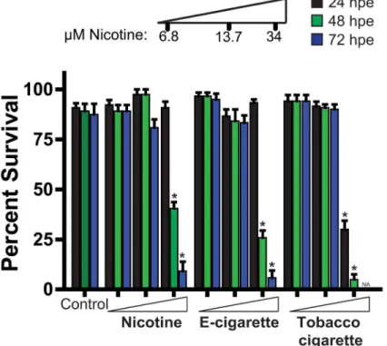
Treatment with nicotine, e-cigarette aerosol extract, and tobacco smoke extract showed no change in embryo survival over the initial 24 hours at all the doses examined, with the
excep-tion of reduced survival observed in the 34μM tobacco smoke extract treated cohort (Fig 1).
Exposure to 34μM nicotine, e-cigarette aerosol extract, and tobacco-cigarette smoke extract at
48 hrs resulted in markedly reduced survival when compared to controls. Although the 34μM
tobacco-exposed cohort had 0% survival by 72 hpe, exposure to the same dose with nicotine and e-cigarettes also had a significant impact in survival (nicotine: 9.2%, e-cigarette: 5.8%). (Fig
1). E-cigarette extract exposed zebrafish showed no striking differences in pigment formation
Fig 1. Zebrafish embryo-larval survival analysis following escalating doses of nicotine, e-cigarette and tobacco extracts.Assessment of zebrafish survival over 72 h with increasing concentrations of pure nicotine, e-cigarettes and tobacco cigarettes extracts or vehicle control (0.1% DMSO). n3 (independent experiments with each n containing between 24–48 animals per treatment).*P<0.05, hpe = hours post exposure.
and chorion hatching when exposed at 6.8 or 13.7μM nicotine when compared to control (S1
Fig). Although 6.8μM tobacco cigarette-exposed fish were similar to controls, decreased
hatch-ing and pigment formation was observed at the 13.7μM nicotine (S1 Fig).
To assay the effects on cardiac development, fish were scored for the incidence of heart ab-normalities and their severity at 72 hpe. Four phenotypes were observed and were classified as:
normal, looped heart with no pericardial edema [31];mild, slight pericardial edema with
looped heart;intermediate, unlooped, balloon shaped heart chambers coupled with pericardial
edema;severe, stretched unlooped heart, no directional blood flow with an unabsorbed yolk
(Fig 2a). Due to the overt lethality observed at 34μM nicotine across all groups we assessed
car-diac defects in the 0, 6.8 and 13.7μM treatment groups. Analysis of zebrafish exposed to
nico-tine alone showed no significant difference from controls regarding the frequency of heart defects (Fig 2b). However, fish exposed to e-cigarette aerosol extract or tobacco cigarette extract showed markedly increased heart defect incidence with tobacco cigarette treated cohorts
show-ing the most number and greatest severity of defects in a dose dependent manner (Fig2band
2c). To investigate heart function we quantified heart beating rate at 72 hpe. While 13.7μM
nicotine from tobacco cigarette exposed fish showed markedly decreased heart rate, e-cigarette aerosol extract exposure at an equivalent dose was not different from controls (control: 155 ± 1.7; e-cigarette: 152 ± 1.8; tobacco cigarette: 134 ± 11 bpm) (Fig 2d).
We also performed transcriptional profiling of 1 day old zebrafish embryos following a 24 h exposure to e-cigarette and tobacco extracts (13.7 uM). These data show a marked decrease in
contractile proteinscmlc2andtnnt2, the transcription factormef2ca, and the junctional protein
responsible for electromechanical conduction in the heart (cx43) and a significant increase in
the cardiac homeobox genenkx2.5only in tobacco cigarette extract treated fish (Fig 2e). These data indicate that both e-cigarette aerosol extract and tobacco cigarette smoke extract exposure affects heart development and function with a more severe impact in the context of tobacco cigarette extract.
Analysis of smoke exposure during cardiac directed differentiation of
hESCs
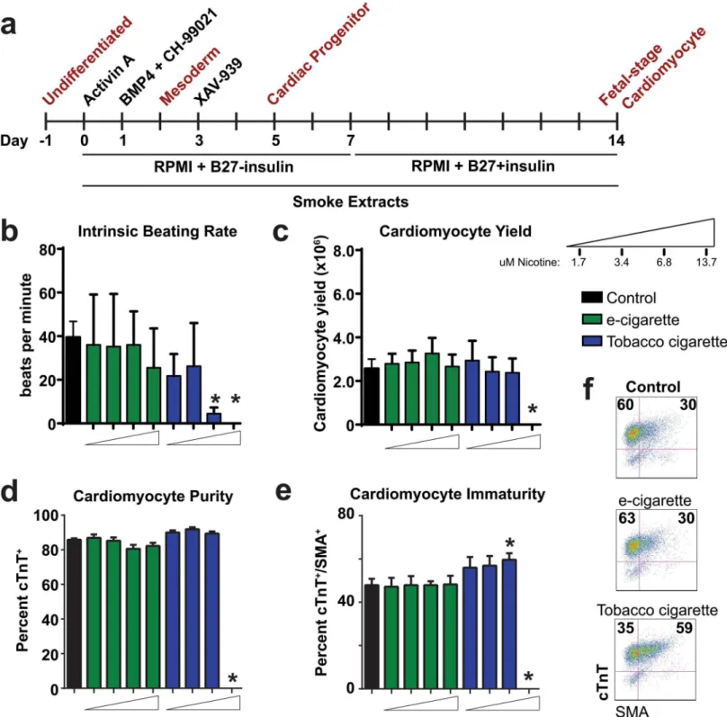
To study the effects of smoke exposure on hESC cardiac development, we used a monolayer-based directed hESC cardiac differentiation platform [27,32,33] (Fig 3a). The serial induction of differentiation with Activin A and BMP4 signaling, in combination with the small molecule
Wnt/β-catenin agonist CHIR-99021 leads to a transition from pluripotency (day 0) to lateral
plate mesoderm (day 2). Use of the small molecule Wnt/β-catenin inhibitor XAV-939 at
differ-entiation day 3 facilitates the transition from mesoderm to cardiac progenitors at day 5 of dif-ferentiation. Cells then progress to definitive fetal-stage cardiomyocytes by completion of the protocol (day 14). We utilized this protocol to assess the effects of smoke extracts on human cardiomyocyte differentiation (smoke extracts was added fresh at each media change througout the experimental time course) (Fig 3a).
In an initial dosescalation analysis, we compared control cohorts to those exposed to e-cigarette aerosol extractor tobacco smoke extract assayed at day 14 of differentiation. During the time course of differentiation, we first observed that tobacco cigarette smoke extract at a
dose of 13.7μM nicotine was cytotoxic, resulting in cell death within 2 days of differentiation
(Fig3b–3e). Among all other viable samples, we carried out a general assessment of
cardiomyo-cyte development at day 14 on the basis of intrinsic beating rate (beats/minute), cardiomyocardiomyo-cyte
yield (total number of cardiomyocytes harvested at day 14), cardiomyocyte purity (cTnT+cells
Fig 2. Cardiac developmental defects observed in zebrafish treated with cigarette smoke.(a) Representative whole mount images of zebrafish at 72 hpe showing normal, mild, intermediate, and severe cardiac developmental defects. v = ventricle, At = atrium. (b-c) Analysis of percent zebrafish with heart defects (b) severity of heart defects and (c). (d) Analysis of heart function in control, e-cigarette and tobacco treated groups at 72 hpe. (e) Quantitative RT-PCR analysis (fold change from control) of a panel of genes with critical roles in early heart development at 24 hpe. n3 (independent experiments with each n containing between 24–48 animals per treatment). For qRT-PCR, n = 3 with each n consisting of 28–35 embryos from independent breeding pools.* P<0.05, hpe = hours post exposure; N = Nicotine, E = E-cigarette, T = Tobacco.
Fig 3. Analysis of e-cigarette and tobacco cigarette on hESC cardiac differentiation.(a) Timeline of differentiation protocol for cardiac directed differentiation of hESCs. (b-e) Analysis of cardiac endpoints including intrinsic beating rate (b) cardiomyocyte yield (c), cardiomyocyte purity (d), and cardiomyocyte immaturity based on percent cTnT+/SMA+(e) and representative flow cytometry plots (f) on day 14 of differentiation with increasing doses of purified e-cigarette and tobacco cigarette extracts. n6 per group.*P<0.05.
per minute, we found no difference in the beating rate of samples treated with e-cigarette
aero-sol extract. However, tobacco cigarette smoke extract treatment at the 6.8μM nicotine dose
showed significant reduction in intrinsic beating rate (4.5 ± 1 beats per minute) (Fig 3b).
Anal-ysis of cardiomyocyte yield and purity (based on cTnT+cells) showed no difference between
groups with the exception of the highest dose of tobacco cigarette extract (Fig3cand3d).
Smooth muscle actin (SMA) is known to be expressed early in cardiac development but is
pro-gressively reduced as cells mature[34]. We therefore assessed cardiac immaturity on the basis
of cells co-expressing cTnT and SMA at day 14 and found that tobacco cigarettes showed
in-creased percent double-positive cells at the 6.8μM nicotine dose (Fig3eand3f). We found that
cells differentiated in purified nicotine were not significantly different on the basis of all these
endpoints compared to control samples (S2 Fig). These initial studies illustrated the cytotoxic
effect of tobacco cigarette extract exposure on developing hESCs and revealed its inhibitory ef-fect on cardiomyocyte differentiation.
Analysis of early cell state transitions during hESC cardiac directed
differentiation
Given results from initial studies looking at increasing doses of nicotine from different cigarette
sources, a dose of 6.8μM nicotine was chosen to compare the effects of e-cigarette aerosol
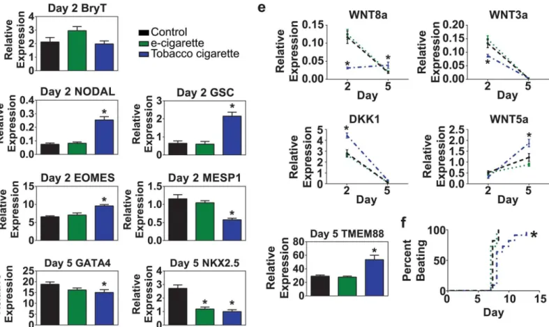
ex-tract and tobacco cigarette smoke exex-tract to control samples during a time course analysis. To determine the impact of cigarette smoke treatment on different stages of cardiac differentia-tion, RNA samples were harvested at day 2 (mesoderm), day 5 (cardiac progenitor cell), and day 14 (definitive fetal-stage cardiomyocytes). Quantitative RT-PCR analyses were performed to determine the transcript abundance of a panel of genes known to have critical roles in cell fate decisions or to participate in the functional development of the cardiomyocyte at each of these stages.
During the transition through mesoderm on day 2, we assessed expression of the pan mesendoderm marker Brachyury T and found no difference between control, e-cigarette
aero-sol extract and tobacco cigarette smoke extract treated samples (Fig 4a). However, genes
in-volved in patterning anterior primitive streak-derived mesendoderm development including the bicoid homeobox protein Goosecoid (GSC) and NODAL were significantly higher only in
cells treated with tobacco cigarette smoke extract (Fig 4b). Among the transcription factors
known to specify the early stages of cardiac development, eomesodermin (EOMES) is known to regulate MESP1 in an axis of signaling to directed pre-cardiac mesoderm fate specification
[35]. We found that cells treated with tobacco cigarette smoke extract had significantly higher
levels of EOMES and lower levels of MESP1 compared to control and e-cigarette aerosol
ex-tract treated samples (Fig 4c). We also analyzed a panel of genes involved in transition through
the cardiac progenitor cell stage (day 5). GATA4 and NKX2.5 are canonical cardiac transcrip-tion factors known to mediate expression of a broad range of cardiac developmental and
struc-tural genes [36,37]. Both e-cigarette aerosol extract and tobacco smoke extract treated samples
showed significantly reduced levels of NKX2.5 compared to control samples. Tobacco smoke
extract treated samples also showed significantly lower expression of GATA4 (Fig 4d).
Wnt/β-catenin has long been known to be a critical mediator of cardiac development where
stimulation of the pathway is required for transitioning through mesoderm and down-regula-tion of Wnts are required to mediate specificadown-regula-tion into the cardiac lineage [27,38]. Expression analyses of Wnt modulators found that samples treated with tobacco cigarette smoke extract had significantly lower expression of canonical Wnt ligands WNT3a and WNT8a as the cells
transitioned through mesoderm on day 2 of differentiation (Fig 4e). Expression of both ligands
progenitor (day 5) stage with the exception of WNT8a which was sustained in the tobacco
ciga-rette group. In contrast, the non-canonical ligand WNT5a (β-catenin-independent) was
up-reg-ulated in all groups during the transition to the cardiac progenitor cell, however, a significant difference in expression levels was observed between the tobacco treated samples over that
ob-served with control and e-cigarette aerosol extract treated samples (Fig 4e). Tobacco smoke
ex-tract treated samples showed significantly higher levels of DKK1 on day 2 of differentiation with down-regulation of this WNT inhibitor observed in all groups by day 5. Lastly, we analyzed
TMEM88, a transmembrane protein known to inhibit Wnt/β-catenin signaling by binding to
Disheveled [27,39]. We found that TMEM88 was significantly up-regulated in tobacco extract
treated samples. These data show significant dysregulation of a key signaling pathway required for fate specification in cardiac development following exposure to tobacco cigarette extracts.
Analysis of cigarette exposure on fetal stage myocytes generated from
hESCs
We assessed the onset of beating during the progression from cardiac progenitors at day 5 to
definitive cardiomyocyte development at day 14 (Fig 4f). These data indicate that control and
Fig 4. Impact of cigarette exposure on cardiogenic mesoderm development.(a-c) Quantitative RT-PCR analysis of expression levels on day 2 of differentiation for genes involved in mesoderm development following exposure to 6.8μM e-cigarette, or tobacco cigarette extract. Analysis included the pan mesendoderm marker Brachyury T (T) (b), mesendoderm genes involved in patterning anterior primitive streak, NODAL and goosecoid (GSC) (c), and cardiogenic mesoderm genes eomesodermin (EOMES) and MESP1. (d) Quantitative RT-PCR analysis of cardiac progenitor cell markers on day 5 of differentiation including GATA4 and NKX2.5. (e) Quantitative RT-PCR analysis of Wnt/β-catenin ligands WNT3a, WNT8a, WNT5a and Wnt/β-catenin signaling inhibitors DKK1 and TMEM88 between days 2 and 5 of differentiation. (f) Time course analysis of onset of beating during cardiac differentiation. n6 per group.*P<0.05.
e-cigarette aerosol extract treated samples showed active contraction around day 7–8 of differ-entiation. However, tobacco smoke extract treated samples were significantly delayed in the onset of beating with contraction occurring variably between days 7 and 13 of differentiation (Fig 4f).
Human ESC-derived cardiomyocyte samples were analyzed for a panel of transcription fac-tors, calcium handling proteins, and contractile proteins that have been shown to have important roles in cardiac development and function. In contrast to day 5 samples which exhibited a reduc-tion in cardiac transcripreduc-tion factors, we found that GATA4 and NKX2.5 mRNA levels were not
different between groups at day 14 (Fig 5a). Immunohistochemical analysis of Nkx2.5 protein
ex-pression corroborated this finding (Fig5cand5d). Furthermore, analysis of calcium handling
proteins such as the L-type calcium channel and SERCA2a showed no difference in expression between e-cigarette aerosol extract and tobacco smoke extract treated samples compared to
con-trol (Fig 5b). However, the junctional protein connexin 43 which is responsible for coordinating
electromechanical transduction in cardiomyocytes was significantly down-regulated in tobacco extract treated samples compared to control or e-cigarette treated cohorts (Fig 5b).
We also assessed the junction protein cadherin involved in signaling and structural integrity during cardiac development. During embryogenesis cadherins accumulate in the Golgi appara-tus and then shuttle to the membrane as cells mature. Immunohistochemical analysis showed uniform cadherin expression in control cardiomyocytes and e-cigarette aerosol extract treated samples with robust localization at the cell membrane and cell-cell junctions. In contrast, to-bacco cigarette smoke extract treated samples had very low levels of cadherin protein
expres-sion that was localized in the peri-nuclear domain as opposed to the cell membrane (Fig5eand
5fandS3 Fig).
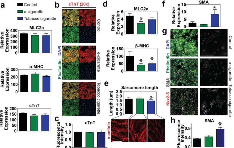
Lastly, we analyzed the expression levels of a variety of contractile proteins involved at vari-ous stages of maturation during cardiomyocyte development. Early developmental myofila-ment genes including the atrial isoform of myosin light chain (MLC2a), the fast ATPase alpha
isoform of myosin heavy chain (α-MHC/MYH6), and the thin filament protein cardiac
tropo-nin T (cTnT) were shown to be expressed equivalently in all groups (Fig 6a). These findings
were confirmed by immunohistochemical analysis of day 14 cells demonstrating similar
ex-pression of cTnT between all groups (Fig6band6c). During development isoform switching
of myofilament proteins is known to occur reflecting changes in the maturation state[40]. In
particular, myocyte development involves a switch from the atrial myosin light chain (MLC2a) to the ventricular specific myosin light chain (MLC2v) and similarly the early development
myosin isoform (α-MHC) is replaced with the adult slow ATPase myosin isoform (β-MHC).
Although all groups showed similar expression of the early developmental variants, both e-cig-arette aerosol extract and tobacco cige-cig-arette smoke extract treated samples had significantly
lower expression of mature developmental markerβ-MHC. Additionally, e-cigarette aerosol
extract treated samples exhibited significantly lower levels of MLC2v (Fig 6d).
Sarcomerogenesis was used as another marker of developmental maturity in which sarco-mere length was measured between control and cigarette treated samples. We found that con-trol and e-cigarette aerosol extract treated samples had myofilaments with longer, more
mature sarcomere lengths (Control: 1.68 ± 0.03μm; e-cigarette: 1.70 ± 0.03μm) compared to
tobacco cigarette extract treated samples which showed etiolated myofilaments with shorter
sarcomere lengths (1.47 ± 0.02μm) (Fig 6e). Lastly, we analyzed smooth muscle actin
expres-sion as a marker of myocyte immaturity. Much as we observed in flow cytometry outcomes
from initial dosing assays (Fig3eand3f), we found that transcript levels analyzed by RT-PCR
and protein abundance by IHC showed significantly higher SMA expression in tobacco extract treated samples compared to either control or e-cigarette aerosol extract treated samples (Fig
have significant developmental deficiencies with more modest defects observed in e-cigarette aerosol extract treated cohorts.
We also determined whether a broad-based cellular stress response was activated with ciga-rette smoke exposure. To address this question, we tested whether markers of stress-related sig-naling cascades were significantly up-regulated in hESC-derived cardiomyocytes treated with both types of cigarette extracts compared to control samples. Protein samples from day 14 fetal cardiomyocytes differentiated with continuous exposure to e-cigarette and tobacco extracts Fig 5. Analysis of hESC derived fetal cardiomyocyte transcription factor, calcium handling, and junction protein expression.(a) Expression level of cardiac transcription factors GATA4 and NKX2.5 (a) and calcium handling proteins including the L-type calcium channel and SERCA2a, and the junctional protein CNX43 (b) by quantitative RT-PCR in cells treated with 6.8μM e-cigarette or tobacco cigarette extracts vs. control. (c-d) Representative
immunocytochemistry (c) and quantification (e) for NKX2.5 in fetal cardiomyocytes with various cigarette treatments compared to control. (e-f) Representative immunohistochemistry (e) and quantification (f) for the junction protein cadherin in fetal hESC cardiomyocytes with various cigarette treatments compared to control. Inset shown to the right. Arrows indicate perinuclear expression of cadherin. n6 per group. Scale bar = 100μm.* P<0.05.
(6.8μM) were isolated and profiled for 26 different stress related proteins including redox
en-zymes, oxidative stress proteins, heat shock proteins, and proteins involved in NFκB and p53
signaling pathways. These results show that exposure to smoke resulted in no significant differ-ences in stress-related proteins between the tested conditions (S4 Fig).
Discussion
It is well established that smoking impacts development, causing a wide range of
pregnancy-re-lated problems including effects on heart development and function[2–4]. With the growing
popularity of electronic cigarettes, these studies establish an important basis for understanding the impact of e-cigarettes and tobacco cigarettes in various physiological contexts. Here we sought to understand the implications for e-cigarette and tobacco smoke extract on cardiac
de-velopment usingin vitrohuman embryonic stem cell cardiac differentiation as well as zebrafish
developmentin vivo. Although there are species dependent differences in the sensitivity to
to-bacco related chemicals such as aromatic hydrocarbons, our findings show a strong correlation Fig 6. Analysis of cardiac myofilament and structural protein expression.(a) Quantitative RT-PCR analysis of early developmental myofilament proteins including the atrial myosin light chain MLC2a, the myosin isoformα-MHC and cardiac troponin T (cTnT) in cells treated with 6.8μM e-cigarette or tobacco cigarette extracts vs. control. (b-c) Immunohistochemistry (b) and quantification (c) of the myofilament proteins cardiac troponin T (cTnT) in combination with phalloidin and nuclear counterstain DAPI in control cells or those treated with 6.8μM e-cigarette or 6.8μM tobacco cigarette extracts. Scale bar = 100μm for cTnT. (d) Quantitative RT-PCR analysis of mature developmental myofilament isoforms including the ventricular myosin light chain MLC2v and the myosin isoformβ-MHC in cells treated 6.8μM e-cigarette or tobacco cigarette extracts vs. control. (e) Quantitation of sarcomere length as measured from samples stained forα-actinin by immunohistochemistry comparing control vs. 6.8μM e-cigarette or tobacco cigarette. (f-h) Quantitative RT-PCR (f) immunohistochemistry (g) and quantification of IHC (h) for the immature cardiac marker smooth muscle actin (SMA) in control vs. cells treated with 6.8μM e-cigarette or tobacco e-cigarette extract. n6 per group. Scale bar = 100μm.*P<0.05.
between studies in developing zebrafish and human hES cardiac differentiation. Overall, these findings showed little to no cardiac developmental deficiencies in the context of nicotine alone but significant impact of tobacco cigarette extracts and e-cigarette aerosol extracts on heart de-velopment with more severe defects observed in the context of tobacco cigarette extract.
One of the major advantages of the current study included the capacity to study stage specif-ic impacts of cigarette extracts during cardiac differentiation from hESCs. Upon mesoderm in-duction (day 2) we found that tobacco cigarette extract treated samples showed increased levels of anterior primitive streak markers GSC and NODAL and decreased Wnt ligand expression.
This is in keeping with findings by Liszewski et al [6]. Assessment of cardiogenic mesoderm
markers showed that tobacco cigarette extract treated samples had increased expression of eomesodermin (EOMES) and decreased MESP1 with correlative dys-regulation of Wnt signal-ing molecules. Collectively these data suggest two potential outcomes of cardiogenic mesoderm specification defects in tobacco extract treated samples: either there is 1) a shift in mesoderm patterning during gastrulation toward a more anterior primitive streak phenotype or 2) a delay in development through lateral plate mesoderm in cells treated with tobacco cigarette smoke. Both interpretations would account for observed differences in gene expression.
As the cells progress to the cardiac progenitor cell state we observed decreased expression of cardiac transcription factors in both e-cigarette aerosol extract and tobacco cigarette smoke ex-tract treated samples. This further validates a delay in differentiation for tobacco cigarette smoke extract treated samples and indicates that e-cigarette aerosol extracts directly impact key regulators of early cardiac specification.
Assessment of e-cigarette smoke treatment showed evidence of inefficient maturation based on increased incidence of heart defects in developing zebrafish and reduced expression of late markers of maturation during hESC cardiac differentiation including expression of MLC2v
andβ-MHC. In addition to dysregulation of gene expression, tobacco cigarette smoke extract
exposure resulted in a wide spectrum of cardiac defects in developing zebrafish and, in the con-text of hESC cardiac differentiation, showed delayed onset of beating, perinuclear localization of the junctional protein cadherin, increased expression of smooth muscle actin, and etiolated myofilaments with reduced sarcomere length. A reduction in beating rate seen in zebrafish and hESC cardiac differentiation in the context of tobacco cigarette extract is in keeping with
previ-ous studies[41] and may result, in part, from lower levels of junctional proteins including
cad-herin and connexin 43 during cardiac development. Further analysis indicated that these deficiencies are not likely the consequence of a general activation of stress related pathways.
Dosing considerations are always important in toxicology studies. Few studies have directly measured concentrations of tobacco metabolites in the human fetus. The most definitive data
of which we are aware come from Luck et al. [29], who reported that newborns of smoking
mothers had serum nicotine values ranging from 0.5–25 ng/ml (3.1–154 nmoles/liter). (A
point of confusion in this area is the study by [6], who appear to have miscalculated the molar
concentrations of nicotine from the study of Luck et al.: 0.5–25 ng/ml[29] calculated as 0.3–
15.4μM[6] should be 0.003-.154 nM). In any case, toxicology studies of zebrafish, cell culture
models and, more recently, human pluripotent stem cells, typically showed that chronic nano-molar to low micronano-molar doses of nicotine show no effect on physiological or cellular end-points, despite well documented human developmental defects associated with nicotine exposure [2–4]. As a consequence, nicotine concentrations used in toxicology studies fall
large-ly within the micromolar range [6,15–17]. Part of this dosing discrepancy may be explained by
This study has built on the work of many other labs making use of developmental model systems to study the effects of tobacco smoke and e-cigarettes on cell physiology[4,6,12–15,
28,42]. Taken together, our data using a high efficiency cardiac directed differentiation of
hESCsin vitroand zebrafish developmentin vivoindicate a dose dependent cytotoxic effect of
cigarette smoking. The collective picture indicates that cigarette smoke treatment primarily de-lays development from the onset of differentiation with continuous impacts on progression to cardiac progenitor cells and to the fetal cardiomyocyte cell stage. It is not surprising that expo-sure to tobacco cigarette extract resulted in a broader spectrum of cardiac abnormalities than exposure to e-cigarette aerosol extracts. However, this study revealed that exposure to e-ciga-rette aerosol extracts also results in detrimental effects on cardiac development even though they lack most of the 7,000+ chemicals contained in tobacco cigarette smoke extracts.
The finding that nicotine treatment alone recapitulated untreated controls indicates that the impact of e-cigarette and tobacco cigarette on heart development is the consequence of other components. Many chemicals, including known ingredients in tobacco cigarettes such as
poly-cyclic aromatic hydrocarbons can cause gross morphological defects [43–46]. Reports have
also shown significant toxicity from e-cigarette treatment [12,13,47] with some reports show-ing the cause to be the presence of cinnamaldehyde and 2-methoxycinamaldehyde in e-ciga-rette flavorings [48,49], the presence of formaldehyde in e-cigarette vapor[50], and tin
particles in cartomizer fluid [51]. Other studies have emerged supporting this observation and
raising the awareness of the risks associated with e-cigarettes particularly for women who smoke or“vape”during pregnancy [12,13,42,47–49,51]. Further studies using hESC directed differentiation toward other cell types will broaden our understanding of the impact of ciga-rette smoke during human development.
Supporting Information
S1 Fig. Developmental landmarks including percent with pigment formation (b) and per-cent hatched (c) at 48 and 72 hpe with exposure to increasing doses of e-cigarettes or tobac-co cigarettes.n = 48 fish per group. hpe = hours post exposure; E = E-cigarette, T = Tobacco. (PNG)
S2 Fig. Effects of nicotine on cardiac differentiation.Cells were differentiated into the cardi-ac lineage and assayed for different markers of cardiomyocyte development at day 14 including intrinsic beating rate (a), cardiomyocyte yield (b), cardiomyocyte purity based on percent cTnT+ cells (c), and cardiomyocyte immaturity based on co-expression of cTnT and SMA (d). (e) Representative flow plots.
(PNG)
S3 Fig. Cadherin expression in definitive cardiomyocytes.Images are an expanded version of those shown inFig 5d.
(PNG)
S4 Fig. Cell stress analysis of fetal cardiomyocytes treated with cigarette extracts.(a) Raw array results used for quantification of 26 cell stress proteins in control, e-cigarette, and tobacco cigarette treated samples. (b) Coordinates for identification of proteins represented in (a). (c) Quantification of mean pixel intensity for each cell stress protein in e-cigarette and tobacco cig-arette treated samples compared to control (dotted line).
(PNG)
Acknowledgments
We appreciate the assistance from the laboratory of Randall Moon for zebrafish work. This work was supported by National Institutes of Health grants P01 HL094374-04S1, R01
HL084642, P01 GM81619, and U01 HL100405 (CEM), and 5T32HL007312-35 (NJP and PH).
Author Contributions
Conceived and designed the experiments: NJP PH LP HR CEM. Performed the experiments: NJP PH. Analyzed the data: NJP PH LP HR CEM. Wrote the paper: NJP PH LP HR CEM.
References
1. Garrett BE, Dube SR, Winder C, Caraballo RS, (CDC) CfDCaP. Cigarette smoking—United States, 2006–2008 and 2009–2010. MMWR Surveill Summ. 2013; 62 Suppl 3:81–4. PMID:24264495.
2. Andres RL, Day MC. Perinatal complications associated with maternal tobacco use. Semin Neonatol. 2000; 5(3):231–41. doi:10.1053/siny.2000.0025PMID:10956448.
3. Hackshaw A, Rodeck C, Boniface S. Maternal smoking in pregnancy and birth defects: a systematic re-view based on 173 687 malformed cases and 11.7 million controls. Hum Reprod Update. 2011; 17 (5):589–604. doi:10.1093/humupd/dmr022PMID:21747128; PubMed Central PMCID:
PMCPMC3156888.
4. Talbot P, Lin S. The effect of cigarette smoke on fertilization and pre-implantation development: assess-ment using animal models, clinical data, and stem cells. Biol Res. 2011; 44(2):189–94. /S0716-97602011000200011. PMID:22513422. doi:/S0716-97602011000200011
5. Köhler E, Avenarius S, Rabsilber A, Gerloff C, Jorch G. Nicotine and its metabolites in amniotic fluid at birth—assessment of prenatal tobacco smoke exposure. Hum Exp Toxicol. 2010; 29(5):385–91. doi: 10.1177/0960327110363326PMID:20164157.
6. Liszewski W, Ritner C, Aurigui J, Wong SS, Hussain N, Krueger W, et al. Developmental effects of to-bacco smoke exposure during human embryonic stem cell differentiation are mediated through the transforming growth factor-βsuperfamily member, Nodal. Differentiation. 2012; 83(4):169–78. doi:10. 1016/j.diff.2011.12.005PMID:22381624; PubMed Central PMCID: PMCPMC3314096.
7. Köhler E, Avenarius S, Rabsilber A, Gerloff C, Jorch G. Assessment of prenatal tobacco smoke expo-sure by determining nicotine and its metabolites in meconium. Hum Exp Toxicol. 2007; 26(6):535–44. doi:10.1177/0960327107072391PMID:17698949.
8. Baeza-Loya S, Viswanath H, Carter A, Molfese DL, Velasquez KM, Baldwin PR, et al. Perceptions about e-cigarette safety may lead to e-smoking during pregnancy. Bull Menninger Clin. 2014; 78 (3):243–52. doi:10.1521/bumc.2014.78.3.243PMID:25247743.
9. Bagcchi S. E-cigarette market expands online. Lancet Oncol. 2014; 15(8):e313. PMID:25121182.
10. McCarthy M. Youth exposure to e-cigarette advertising on US television soars. BMJ. 2014; 348:g3703. PMID:24895292. doi:10.1136/bmj.g3703
11. Iacobucci G. WHO calls for ban on e-cigarette use indoors. BMJ. 2014; 349:g5335. PMID:25170124. doi:10.1136/bmj.g5335
12. Farsalinos KE, Romagna G, Allifranchini E, Ripamonti E, Bocchietto E, Todeschi S, et al. Comparison of the cytotoxic potential of cigarette smoke and electronic cigarette vapour extract on cultured myocar-dial cells. Int J Environ Res Public Health. 2013; 10(10):5146–62. doi:10.3390/ijerph10105146PMID: 24135821; PubMed Central PMCID: PMCPMC3823305.
13. Bahl V, Lin S, Xu N, Davis B, Wang YH, Talbot P. Comparison of electronic cigarette refill fluid cytotox-icity using embryonic and adult models. Reprod Toxicol. 2012; 34(4):529–37. doi:10.1016/j.reprotox. 2012.08.001PMID:22989551.
14. Lin S, Fonteno S, Weng JH, Talbot P. Comparison of the toxicity of smoke from conventional and harm reduction cigarettes using human embryonic stem cells. Toxicol Sci. 2010; 118(1):202–12. doi:10. 1093/toxsci/kfq241PMID:20702591; PubMed Central PMCID: PMCPMC2955215.
15. Zdravkovic T, Genbacev O, LaRocque N, McMaster M, Fisher S. Human embryonic stem cells as a model system for studying the effects of smoke exposure on the embryo. Reprod Toxicol. 2008; 26 (2):86–93. doi:10.1016/j.reprotox.2008.07.004PMID:18692565.
17. Parker B, Connaughton VP. Effects of nicotine on growth and development in larval zebrafish. Zebra-fish. 2007; 4(1):59–68. doi:10.1089/zeb.2006.9994PMID:18041943.
18. Bakkers J. Zebrafish as a model to study cardiac development and human cardiac disease. Cardiovasc Res. 2011; 91(2):279–88. Epub 2011/05/24. cvr098 [pii] doi:10.1093/cvr/cvr098PMID:21602174.
19. Heideman W, Antkiewicz DS, Carney SA, Peterson RE. Zebrafish and cardiac toxicology. Cardiovasc Toxicol. 2005; 5(2):203–14. Epub 2005/07/28. CT:5:2:203 [pii]. PMID:16046794.
20. Zon LI, Peterson RT. In vivo drug discovery in the zebrafish. Nat Rev Drug Discov. 2005; 4(1):35–44. doi:10.1038/nrd1606PMID:15688071.
21. Hofsteen P, Plavicki J, Johnson SD, Peterson RE, Heideman W. Sox9b is required for epicardium for-mation and plays a role in TCDD-induced heart malforfor-mation in zebrafish. Mol Pharmacol. 2013; 84 (3):353–60. doi:10.1124/mol.113.086413PMID:23775563; PubMed Central PMCID:
PMCPMC3876814.
22. Carp H, Janoff A. Possible mechanisms of emphysema in smokers. In vitro suppression of serum elas-tase-inhibitory capacity by fresh cigarette smoke and its prevention by antioxidants. Am Rev Respir Dis. 1978; 118(3):617–21. PMID:101105.
23. Jacob P, Wilson M, Benowitz NL. Improved gas chromatographic method for the determination of nico-tine and cotinine in biologic fluids. J Chromatogr. 1981; 222(1):61–70. PMID:6783675.
24. Jacob P, Yu L, Wilson M, Benowitz NL. Selected ion monitoring method for determination of nicotine, cotinine and deuterium-labeled analogs: absence of an isotope effect in the clearance of (S)-nicotine-3',3'-d2 in humans. Biol Mass Spectrom. 1991; 20(5):247–52. doi:10.1002/bms.1200200503PMID: 1883864.
25. Kais B, Schneider KE, Keiter S, Henn K, Ackermann C, Braunbeck T. DMSO modifies the permeability of the zebrafish (Danio rerio) chorion-implications for the fish embryo test (FET). Aquat Toxicol. 2013;140–141:229–38. doi:10.1016/j.aquatox.2013.05.022PMID:23831690.
26. Westerfield M. The Zebrafish book. A guide for the laboratory use of zebrafish (Danio rerio). Eugene, OR: Univ. of Oregon Press; 2000.
27. Palpant NJ, Pabon L, Rabinowitz JS, Hadland BK, Stoick-Cooper CL, Paige SL, et al. Transmembrane protein 88: a Wnt regulatory protein that specifies cardiomyocyte development. Development. 2013. doi:10.1242/dev.094789PMID:23924634.
28. Huang J, Okuka M, Lu W, Tsibris JC, McLean MP, Keefe DL, et al. Telomere shortening and DNA dam-age of embryonic stem cells induced by cigarette smoke. Reprod Toxicol. 2013; 35:89–95. doi:10. 1016/j.reprotox.2012.07.003PMID:22824788.
29. Luck W, Nau H, Hansen R, Steldinger R. Extent of nicotine and cotinine transfer to the human fetus, placenta and amniotic fluid of smoking mothers. Dev Pharmacol Ther. 1985; 8(6):384–95. PMID: 4075937.
30. Peterson RT, Macrae CA. Systematic approaches to toxicology in the zebrafish. Annu Rev Pharmacol Toxicol. 2012; 52:433–53. Epub 2011/10/25. doi:10.1146/annurev-pharmtox-010611-134751PMID: 22017682.
31. Glickman NS, Yelon D. Cardiac development in zebrafish: coordination of form and function. Semin Cell Dev Biol. 2002; 13(6):507–13. PMID:12468254.
32. Laflamme MA, Chen KY, Naumova AV, Muskheli V, Fugate JA, Dupras SK, et al. Cardiomyocytes de-rived from human embryonic stem cells in pro-survival factors enhance function of infarcted rat hearts. Nat Biotechnol. 2007; 25(9):1015–24. Epub 2007/08/28. nbt1327 [pii] doi:10.1038/nbt1327PMID: 17721512.
33. Gantz JA, Palpant NJ, Welikson RE, Hauschka SD, Murry CE, Laflamme MA. Targeted genomic inte-gration of a selectable floxed dual fluorescence reporter in human embryonic stem cells. PLoS One. 2012; 7(10):e46971. doi:10.1371/journal.pone.0046971PMID:23071682; PubMed Central PMCID: PMC3468579.
34. Clément S, Stouffs M, Bettiol E, Kampf S, Krause KH, Chaponnier C, et al. Expression and function of alpha-smooth muscle actin during embryonic-stem-cell-derived cardiomyocyte differentiation. J Cell Sci. 2007; 120(Pt 2):229–38. doi:10.1242/jcs.03340PMID:17179203.
35. Costello I, Pimeisl IM, Dräger S, Bikoff EK, Robertson EJ, Arnold SJ. The T-box transcription factor Eomesodermin acts upstream of Mesp1 to specify cardiac mesoderm during mouse gastrulation. Nat Cell Biol. 2011; 13(9):1084–91. doi:10.1038/ncb2304PMID:21822279.
36. He A, Kong SW, Ma Q, Pu WT. Co-occupancy by multiple cardiac transcription factors identifies tran-scriptional enhancers active in heart. Proc Natl Acad Sci U S A. 2011; 108(14):5632–7. doi:10.1073/ pnas.1016959108PMID:21415370; PubMed Central PMCID: PMCPMC3078411.
2011; 7(2):e1001313. doi:10.1371/journal.pgen.1001313PMID:21379568; PubMed Central PMCID: PMCPMC3040678.
38. Ueno S, Weidinger G, Osugi T, Kohn AD, Golob JL, Pabon L, et al. Biphasic role for Wnt/beta-catenin signaling in cardiac specification in zebrafish and embryonic stem cells. Proc Natl Acad Sci U S A. 2007; 104(23):9685–90. Epub 2007/05/25. 0702859104 [pii] doi:10.1073/pnas.0702859104PMID: 17522258; PubMed Central PMCID: PMC1876428.
39. Lee HJ, Finkelstein D, Li X, Wu D, Shi DL, Zheng JJ. Identification of transmembrane protein 88 (TMEM88) as a dishevelled-binding protein. J Biol Chem. 2010; 285(53):41549–56. M110.193383 [pii] doi:10.1074/jbc.M110.193383PMID:21044957; PubMed Central PMCID: PMCPMC3009882.
40. Siedner S, Kruger M, Schroeter M, Metzler D, Roell W, Fleischmann BK, et al. Developmental changes in contractility and sarcomeric proteins from the early embryonic to the adult stage in the mouse heart. Journal of Physiology-London. 2003; 548(2):493–505. 67. PMID:12640016
41. Ellis LD, Soo EC, Achenbach JC, Morash MG, Soanes KH. Use of the zebrafish larvae as a model to study cigarette smoke condensate toxicity. PLoS One. 2014; 9(12):e115305. doi:10.1371/journal. pone.0115305PMID:25526262; PubMed Central PMCID: PMCPMC4272283.
42. Farsalinos KE, Tsiapras D, Kyrzopoulos S, Savvopoulou M, Voudris V. Acute effects of using an elec-tronic nicotine-delivery device (elecelec-tronic cigarette) on myocardial function: comparison with the effects of regular cigarettes. BMC Cardiovasc Disord. 2014; 14:78. doi:10.1186/1471-2261-14-78PMID: 24958250; PubMed Central PMCID: PMCPMC4077146.
43. Hofsteen P, Plavicki J, Peterson RE, Heideman W. Epicardium Formation as a Sensor in Toxicology. J Dev Biol. 2013; 1(2):112–25. doi:10.3390/jdb1020112PMID:25232532; PubMed Central PMCID: PMCPMC4164227.
44. Plavicki J, Hofsteen P, Peterson RE, Heideman W. Dioxin inhibits zebrafish epicardium and proepicar-dium development. Toxicol Sci. 2013; 131(2):558–67. doi:10.1093/toxsci/kfs301PMID:23135548; PubMed Central PMCID: PMCPMC3551425.
45. Mizell M, Romig ES. The aquatic vertebrate embryo as a sentinel for toxins: zebrafish embryo dechorio-nation and perivitelline space microinjection. Int J Dev Biol. 1997; 41(2):411–23. PMID:9184351.
46. Raimondo S, Jackson CR, Krzykwa J, Hemmer BL, Awkerman JA, Barron MG. Developmental toxicity of Louisiana crude oil-spiked sediment to zebrafish. Ecotoxicol Environ Saf. 2014; 108:265–72. doi:10. 1016/j.ecoenv.2014.07.020PMID:25105486.
47. Romagna G, Allifranchini E, Bocchietto E, Todeschi S, Esposito M, Farsalinos KE. Cytotoxicity evalua-tion of electronic cigarette vapor extract on cultured mammalian fibroblasts (ClearStream-LIFE): com-parison with tobacco cigarette smoke extract. Inhal Toxicol. 2013; 25(6):354–61. doi:10.3109/ 08958378.2013.793439PMID:23742112.
48. Behar RZ, Davis B, Wang Y, Bahl V, Lin S, Talbot P. Identification of toxicants in cinnamon-flavored electronic cigarette refill fluids. Toxicol In Vitro. 2014; 28(2):198–208. PMID:24516877.
49. Behar RZ, Davis B, Wang Y, Bahl V, Lin S, Talbot P. Identification of Toxicants in Cinnamon-Flavored Electronic Cigarette Refill Fluids. Toxicol In Vitro. 2013. doi:10.1016/j.tiv.2013.10.006PMID: 24513812.
50. Jensen RP, Luo W, Pankow JF, Strongin RM, Peyton DH. Hidden formaldehyde in e-cigarette aerosols. N Engl J Med. 2015; 372(4):392–4. doi:10.1056/NEJMc1413069PMID:25607446.