Rodrigo Stanislawczuk(a) Jully Anna da Costa(b) Luceli Grabicoski Polli(b) Alessandra Reis(a)
Alessandro Dourado Loguercio(a)
(a) Department of Restorative Dentistry, School of Dentistry, State University of Ponta Grossa, Ponta Grossa, PR, Brazil.
(b) Private practice, Ponta Grossa, PR, Brazil.
Corresponding author:
Alessandro Dourado Loguercio Email: aloguercio@hotmail.com
Received for publication on May 05, 2011 Accepted for publication on Aug 04, 2011
Effect of tetracycline on the bond
performance of etch-and-rinse
adhesives to dentin
Abstract: This study evaluated the effect of modiied tetracycline on the resin-dentin bond strength (µTBS), silver nitrate uptake (SNU) and solu-tion homogeneity (SH) of two adhesives. Dentin surfaces were treated with phosphoric acid, rinsed off and either rewetted with water (con-trol group - CO), 2% minocycline (MI), 2% doxycyline (DO) or 2% chlorhexidine (CH). Adhesive systems (Adper Single Bond 2 and Prime Bond NT) and composite were applied and light-polymerized. Specimens were sectioned to obtain bonded sticks (0.8 mm²) to test under tension at 0.5 mm/min. For SNU, specimens were immersed in silver nitrate and analyzed by EDX-SEM. SH was qualitatively analyzed after mix-ing the adhesives with different solvent-based solutions containmix-ing MI, DO and CH. Lower µTBS values were observed in the DO group com-pared with MI and CH (p = 0.01). Lower SNU was observed for MI and CH. The lowest µTBS for both adhesives was observed for the DO group (p = 0.01). Signs of phase separation were observed for DO with both ad-hesives. MI or CH used as rewetting solutions after acid etching did not affect the µTBS and hybrid layer quality.
Descriptors: Dentin-Bonding Agents; Chlorhexidine; Tetracycline; Tensile Strength.
Introduction
There is a general consensus that resin-dentin bonds created by simpli-ied adhesives deteriorate over time,1 and this occurrence has been
attrib-uted mainly to degradation of the hybrid layer.2 A decreasing gradient of
resin monomer diffusion within the acid-etched dentin3 results in
incom-pletely iniltrated zones along the bottom of hybrid layers that contain denuded collagen ibrils.4 These denuded collagen ibrils are vulnerable to
degradation by endogenous metaloproteinases (MMPs) in a way that is similar to what occurs in caries and periodontal diseases.5,6 Meanwhile
it has been speculated that a subsequent resin elution from hydrolytically unstable polymeric hydrogels within the hybrid layers7 leaves the collagen
ibrils unprotected and also susceptible to the same degradation process. The literature has also described various pathological processes in which MMPs are implicated.8
MMPs are a group of 23 mammalian enzymes capable of degrad-ing all extracellular matrix components. Human dentin contains colla-genase (MMP-8), gelatinases MMP-2 and -9, and others.9,10 These
tin collagenolytic and gelatinolytic activities can be suppressed by protease inhibitors. There are several MMP inhibitors described in the literature and, among them, chlorhexidine (CH) and tetracyclines have been shown to be eficient adjuncts to peri-odontal therapy.11
So far, studies have evaluated the role of CH as MMP inhibitor to preserve resin-dentin bonds from degradation.12-16 Tetracycline and their
semi-synthet-ic forms (doxycycline [DO] and minocycline [MI]), in addition to acting as antibiotics, are very safe and potent MMP inhibitors.17-20 The action
mecha-nisms of chemically modiied tetracyclines (CMTs) are thought to work through inhibiting the activity and secretion of MMPs and Ca2+ chelation.21
How-ever, the effects of different tetracyclines used as re-wetting agents after acid etching on the immediate performance of adhesive systems to dentin has not been previously investigated. Therefore the aim of this in vitro study was to evaluate the effects of DO, MI and CH on the immediate resin-dentin bond strengths and silver nitrate uptake of adhesive in-terfaces and homogeneity among adhesives and the solutions tested.
Methodology
Teeth selection and preparation
Forty extracted, caries-free human third molars were used. This study was approved by the local Ethics Committee under protocol # 6280/2009. A lat and supericial dentin surface was exposed on each tooth after wet grinding the occlusal enamel on # 180-grit SiC paper. The enamel-free dentin surfac-es were further polished on wet # 600-grit silicon-carbide paper (Erios Prod. Odont. Ltda., São Paulo,
Brazil) for 60 s. Teeth were divided in eight groups (n = 5) according to the combination of the main factors Adhesive (2 levels) and Rewetting solution (4 levels).
Restorative procedure
An acetone solvent-based adhesive (Prime & Bond NT [PB], Dentsply De Trey, Konstanz, Ger-many) and a water/ethanol solvent-based adhe-sive (Adper Single Bond 2 [SB], 3MESPE, St. Paul, USA) were used. The surfaces were acid etched with phosphoric acid, rinsed off, air-dried and re-wetted actively with water22 (control group - CO),
and aqueous solutions of 2% minocycline (MI), 2% doxycycline (DO) (Fleming drugstore, Ponta Gros-sa, Brazil) or 2% chlorhexidine digluconate (CH) (FGM, Joinville, Brazil) for 60 s. After that adhesive systems and resin composite (Opallis, FGM, Join-ville, Brazil) were applied as described in Table 1. Five teeth were used for each experimental group.
The bonded teeth were longitudinally sectioned to obtain bonded sticks (0.8 mm² area)13,15. Each
bonded stick was attached to a modiied microtensile testing device, using cyanoacrylate resin, and sub-jected to a tensile force in a universal machine (Emic, São José dos Pinhais, Brazil) at 0.5 mm/min. Failure modes were evaluated at 400× (HMV-2, Shimadzu, Tokyo, Japan) and classiied as described in Table 2.
Silver nitrate uptake technique
Two bonded sticks from each tooth were not tested under tension, and were prepared for SNU evaluation according to an earlier protocol.13,15
Briely, the sticks were coated with two layers of nail varnish applied up to within 1 mm of the bonded
in-Table 1 - Adhesive systems (batch number): composition and application mode.
Adhesive
systems Composition Application Mode
PB (1223207A)
1. Caulk Tooth Conditioner Gel 34% phosphoric acid 2. Adhesive – UDMA, PENTA, R 5-62-1 resin, T resin, D resin,
silanated colloidal silica, cetylamine hydroxyfluoride, initiator, stabilizer and acetone
1. acid-etch (15 s) 2. rinse (15 s) 3. air-dry (30 s)
4. active rewetting for 60 s
5. two coats of adhesive were applied slightly for 20 s 6. air-dry for 10 s at 20 cm
7. light polymerize (10 s – 600 mW/cm²)
SB
(# 9XJ)
1. Scotchbond Etchant 35% phosphoric acid
terfaces. The specimens were rehydrated in distilled water for 10 min before immersion in the tracer solution for 24 h. After that the sticks were placed in 50% ammoniacal silver nitrate and immersed in photo developing solution for 8 h under luorescent light to reduce silver ions into metallic silver grains within voids along the bonded interface. Specimens were then polished down (1,000-grit SiC paper and 1 and 0.25 lm diamond paste [Buehler, Lake Bluff, USA] using a polishing cloth), desiccated and sputter-coated with gold for analysis by a scanning electron microscope operated in backscattered elec-tron mode and energy dispersive X-ray spectrometry ([EDX], SSX-550, Shimadzu, Tokyo, Japan). The working distance was 10 mm and the accelerating voltage was 15 Kv. The amount of SNU was mea-sured following an earlier protocol.22
Solution homogeneity
MI and DO (2%) were dissolved in water, ace-tone and ethanol. Sixteen µl of each adhesive was transferred to a small glass and mixed with 16 µl of each adhesive system. The mixture was stirred for 10-15 s. A digital image of each mixture was tak-en 30 s after mixing began, using a digital camera (D70 and AF-S VR Micro-Nikkor 105 mm, Nikon, Tokyo, Japan). Loss in clarity was interpreted as evi-dence of phase separation. Before mixing, all solu-tions were clear liquids.2 This experiment was done
in quadruplicate.
Statistical analysis
The mean µTBS (MPa) and SNU (%) of all sticks from the same tooth were averaged for sta-tistical purposes (n = 5 for experimental condition).
The µTBS and SNU were subjected to a one-way ANOVA for each adhesive system and a post hoc test (Tukey’s test at α = 0.05).
Results
The µTBS and fracture pattern results are shown in Table 2. No signiicant difference was detected among groups for the SB adhesive (p > 0.05). For PB, lower µTBS values were observed when DO was applied compared with the other solutions (p < 0.05) (Table 1).
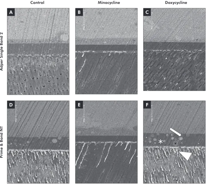
As regards SNU, signiicant differences were ob-served for both adhesives (p < 0.05). Lower % of SNU was observed for PB and SB when CH and MI was used in comparison with DO (p < 0.05) (Table 3 and Figure 1).
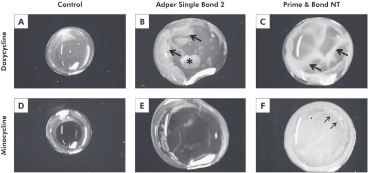
It was not possible to produce mixtures of MI and DO with acetone, as these were not soluble in acetone. MI and DO were only slightly soluble in ethanol (data not shown). Therefore SH was only tested with aqueous solutions of the aforementioned substances. Aqueous solutions of MI and DO were clear and completely transparent before mixing with
Table 2 -µTBS values and standard deviations (MPa) and fracture pattern (%) (*).
Prime & Bond NT A/M C PT Adper Single Bond 2 A/M C PT
Control 35.7 ± 4.5 A,B 24 (75.0) 0 (0) 8 (25.0) 42.1 ± 8.3 a 34 (80.9) 0 (0) 8 (19.1)
2% MI 40.2 ± 12.1 A 38 (77.6) 0 (0) 11 (22.4) 36.9 ± 16.4 a 33 (71.7) 0 (0) 13 (28.3)
2% DO 30.3 ± 8.2 B 28 (50.0) 0 (0) 28 (50.0) 40.2 ± 13.2 a 36 (75.0) 0 (0) 11 (25.0)
2% CH 38.1 ± 8.3 A 35 (87.5) 0 (0) 5 (12.5) 42.8 ± 10.4 a 32 (94.1) 0 (0) 2 (5.9)
(*) Comparisons are valid within each adhesive system. Means identified with the same upper or lowercase letters are not significantly different (Tukey’s test, p > 0.05). A/M – adhesive/mixed fracture mode; C – dentin or resin cohesive fracture mode; PT – pretest failures.
Table 3 - SNU and standard deviations (%) and statistical significance (*)
Prime & Bond NT Adper Single Bond 2
Control 27.9 ± 4.9 B,C 25.5 ± 4.9 b
2% MI 23.9 ± 3.6 A,B 16.3 ± 4.2 a
2% DO 34.3 ± 3.8 C 25.5 ± 4.3 b
2% CH 16.4 ± 5.4 A 18.1 ± 3.9 a,b
the adhesives (Figures 2A and 2D).
SB mixed with aqueous solution of DO showed a slight phase-separation (Figure 2B). No resin drop-lets, representative of phase-separation, were seen when SB was mixed with the aqueous solution of MI, and the adhesive was shown to be homogeneous and transparent (Figure 2D). PB mixed with MI and DO showed the presence of several resin droplets (Figures 2C and 2E). In contact with MI the
solu-tion turned opaque with few opaque resin droplets (Figure 2E).
Discussion
Tetracyclines and their analogues (DO and MI) are a group of MMP inhibitors. These compounds are zinc-dependent endopeptidases that play an im-portant role in the remodeling of connective tissue and are involved in embryogenesis, wound
heal-A
D
B
E
C
Control Minocycline Doxycycline
A
d
p
e
r
S
in
g
le
B
o
n
d
2
P
ri
m
e
&
B
o
n
d
N
T
F
Figure 1 - Backscattered SEM images of the resin-dentin interfaces bonded with SB (A to C) and PB (D to F). For all SB figures, the amount of silver penetration was lower and occurred practically only within the hybrid layer. Only few dentin tubules were infiltrated by silver nitrate. For PB, SNU deposition occurred almost throughout the entire thickness of the hybrid layer, mainly in
the DO group. The presence of some globules in the adhesive layer (F) probably indicates phase separation (arrow = globules;
ing, rheumatoid arthritis, and tumor invasion and metastasis.17,21 There are MMPs that break down
ibrillar collagen known as collagenases (MMP-1, MMP-8, MMP-13) and those that can affect base-ment membrane collagen (collagen IV) known as ge-latinases (MMP-2, MMP-9).
DO and MI show a basic chemical structure con-sisting of a tetracyclic naphthacene carboxamide ring system, but they differ slightly. Doxycycline presents one hydroxyl group in carbon 5 of ring B and one methyl group in the carbon 6 of ring C, whereas minocycline has one amine group in carbon 7 of ring D.17
DO and MI can inhibit collagenases and gelati-nases.23 Inhibition of MMPs can theoretically occur
at numerous levels attributable to the multiple steps involved in MMP transcription, protein synthesis, and enzyme activation through binding in the MMP active site. By binding to the active site zinc ion of the enzyme, modiied tetracyclines can alter the con-formation of the pro-enzyme molecule, thus block-ing its catalytic activity in the extracellular matrix.24
Whether or not this will bring the same beneits as those of chlorhexidine in preserving resin-dentin
bonds over time has yet to be investigated.
Although MI and DO have quite similar chemi-cal structures their results were very different in the present investigation. MI reached µTBS and SNU similar to that of CH irrespective of the adhesive sys-tem used. In Figure 2, one can observe that no phase separation was observed when MI was mixed with the SB adhesive (Figure 2C). The mixture looked like the aqueous solution of MI, with a translucent and reddish appearance. However, this was not the case when the aqueous solution of MI was mixed with the acetone-based system. Small resin droplets could be observed (Figure 2D), and although one may hypothesize that this is due to the insolubility of MI in acetone, one cannot rule out the possibil-ity that other components of the adhesive may have played a role in the lack of solvent homogeneity. It is worth mentioning that the phase separation ob-served between MI and PB systems was not enough to jeopardize the resin-dentin bonds. The explana-tion for this, however, is still unclear to the author’s understanding.
On the other hand, DO showed a very differ-ent pattern. DO cannot be used with acetone based
F
Figure 2 - Representative digital pictures of the MI and DO solutions (A and D) and the solution homogeneity after mixing
these solutions with the adhesive systems. For SB, several resin droplets (arrows) and globules (asterisk), representative of
phase-separation were observed when mixing with DO (B). No sign of phase-separation was observed when SB was mixed with
MI (E). For PB, signs of phase-separation, with many droplets, were observed in all parts of the specimens, when mixed with DO
(C, arrows) and little (F, asterisk) and opaque droplets (F, arrows) when mixed with MI. Magnification: 100×.
A
D
B C
E
Adper Single Bond 2 Control
D
o
x
y
cy
cl
in
e
M
in
o
cy
cl
in
e
adhesive systems, because lower bond strength val-ues as well as higher silver nitrate penetration were observed within the hybrid layer. This was also demonstrated by the high number of PT (Table 2), which denotes the fragility of the bonding inter-face. The lack of solubility of DO in acetone could be an explanation of why DO cannot be used as a rewetting agent for acetone-based systems. Figure 2B showed that when PB, the acetone-based system, was dropped onto the aqueous solution of DO, the material did not set properly, showing yellowish waves which probably represent phase separation domains.25
The formation of phase separation leads to en-trapment of droplets within the adhesive layer.25
These droplets themselves must reduce the µTBS of the adhesive system by acting as laws during the µTBS test.26 This is probably also responsible for
the low fatigue resistance of materials with phase separation in comparison with those in which no phase separation is seen.
For the water/ethanol based system, µTBS values similar to those of the control group were obtained with DO, which would lead one to think that this MMP inhibitor is appropriate for this type of adhe-sive. This would be true if the adhesive interface re-wetted with DO were not highly iniltrated by silver nitrate. Higher SNU within the adhesive interface indicated that the resin matrices contained several hydrophilic domains and/or hydrogel polymer for-mation.27
The amount of SNU at the SB interfaces was larger when the dentin was rewetted with DO rather than with CH and MI. This probably indicates the quality of the polymer formed within the hybrid layer when DO was applied beforehand. This may probably become more evident after some months of water storage, as this interface will be more prone to degradation through hydrolysis due to its higher initial hydrophilicity. Since hydrolytic degradation occurs only in presence of water, adhesive hydrophi-licity and water sorption are directly correlated with hydrolytic degradation.28
Further studies also should be conducted to analyze the effects of different concentrations and application times of MI aqueous solutions. In addi-tion, the association of MI with water and ethanol adhesive solutions should be investigated as well as the effects of MI application on the stability of the resin-dentin bonds over time.
Conclusions
The use of a 2% aqueous solution of chlorhexi-dine and MI did not impair resin-dentin bond strength and the quality of the hybrid layer forma-tion for both adhesives, although slight phase sepa-ration occurred for the acetone-based system. The use of 2% doxycycline should be avoided as it jeop-ardized the bond strengths and quality of the hybrid layer for both systems.
References
1. Breschi L, Mazzoni A, Ruggeri A, Cadenaro M, Di Lenarda R, De Stefano Dorigo E. Dental adhesion review: aging and sta-bility of the bonded interface. Dent Mater. 2008 Jan;24(1):90-101.
2. Spencer P, Wang Y. Adhesive phase separation at the dentin interface under wet bonding conditions. J Biomed Mater Res. 2002 Dec 5;62(3):447-56.
3. Furukawa M, Shigetani Y, Finger WJ, Hoffmann M, Kanehira M, Endo T, et al. All-in-one self-etch model ad-hesives: HEMA-free and without phase separation. J Dent. 2008 Jun;36(6):402-8.
4. Hashimoto M, Ohno H, Kaga M, Sano H, Endo K, Oguchi H. The extent to which resin can infiltrate dentin by acetone-based adhesives. J Dent Res. 2002 Jan;81(1):74-8.
5. Tjaderhäne L, Larjava H, Sorsa T, Uitto VJ, Larmas M, Salo T. The activation and function of host matrix metalloproteinases in dentin matrix breakdown in caries lesions. J Dent Res. 1998 Aug;77(8):1622-9.
6. Lee W, Aitken S, Sodek J, McCulloch CA. Evidence of a di-rect relationship between neutrophil collagenase activity and periodontal tissue destruction in vivo: role of active enzyme in human periodontitis. J Periodontal Res. 1995 Jan;30(1):23-33. 7. Wang Y, Spencer P. Hybridization efficiency of the adhe-sive-dentin interface with wet bonding. J Dent Res. 2003 Feb;82(2):350-4.
9. Martin-De-Las Heras S, Valenzuela A, Overall CM. The ma-trix metalloproteinase gelatinase A in human dentine. Arch Oral Biol. 2000 Sep;45(9):757-65.
10. Mazzoni A, Mannello F, Tay FR, Tonti GA, Papa S, Maz-zotti G, et al. Zymographic analysis and characterization of MMP-2 and -9 forms in human sound dentin. J Dent Res. 2007 May;86(5):436-40.
11. Hannas AR, Pereira JC, Granjeiro JM, Tjäderhane L. The role of matrix metalloproteinases in the oral environment. Acta Odontol Scand. 2007 Feb;65(1):1-13.
12. Carrilho MR, Carvalho RM, de Goes MF, di Hipólito V, Geraldeli S, Tay FR, et al. Chlorhexidine preserves dentin bond in vitro. J Dent. Res. 2007 Jan;86(1):90-4.
13. Stanislawczuk R, Amaral RC, Zander-Grande C, Gagler D, Reis A, Loguercio AD. Chlorhexidine-containing acid con-ditioner preserves the longevity of resin-dentin bonds. Oper Dent. 2009 Jul-Aug;34(4):481-90.
14. Hebling J, Pashley DH, Tjäderhane L, Tay FR. Chlorhexidine arrests subclinical degradation of dentin hybrid layers in vivo. J Dent Res. 2005 Aug;84(8):741-6.
15. Loguercio AD, Stanislawczuk R, Polli LG, Costa JA, Michel MD, Reis A. Influence of chlorhexidine digluconate concen-tration and application time on resin-dentin bond strength durability. Eur J Oral Sci. 2009 Oct;117(5):587-96.
16. Fardal O, Turnbull RS. A review of the literature on use of chlorhexidine in dentistry. J Am Dent Assoc. 1986 Jun;112(6):863-9.
17. Sapadin AN, Fleischmajer R. Tetracyclines: nonantibiotic properties and their clinical implications. J Am Acad Derma-tol. 2006 Feb;54(2):258-65.
18. Golub LM, Lee HM, Lehrer G, Nemiroff A, McNamara TF, Kaplan R, et al. Minocycline reduces gingival collagenolytic activity during diabetes: preliminary observations and a pro-posed new mechanism of action. J Periodontal Res. 1983 Sep;18(5):516-26.
19. Sulkala M, Wahlgren J, Larmas M, Sorsa T, Teronen O, Salo T, et al. The effects of MMP inhibitors on human salivary MMP activity and caries progression in rats. J Dent Res. 2001 Jun;80(6):1545-9.
20. Osorio R, Yamauti M, Osorio E, Ruiz-Requena ME, Pash-ley DH, Tay FR, et al. Zinc reduces collagen degradation in demineralized human dentin explants. J Dent. 2011 Feb;39(2):148-53.
21. Tallant C, Marrero A, Gomis-Rüth FX. Matrix metallopro-teinases: fold and function of their catalytic domains. Biochim Biophys Acta. 2010 Jan;1803(1):20-8.
22. Reis A, Grande RHM, Oliveira GMS, Lopes GC, Loguercio AD. A 2-year evaluation of moisture on microtensile bond strength and nanoleakage Dent Mater. 2007 Jul;23(7):862-70. 23. Golub LM, Ramamurthy NS, McNamara TF, Greenwald RA,
Rifkin BR. Tetracyclines inhibit connective tissue breakdown: new therapeutic implications for an old family of drugs. Crit Rev Oral Biol Med. 1991 2(3):297-321.
24. Corbitt CA, Lin J, Lindsey ML. Mechanisms to inhibit matrix metalloproteinase activity: Where are we in the development of clinically relevant inhibitors? Recent Pat Anticancer Drug Discov. 2007 Jun;2(2):135-42.
25. Van Landuyt KL, De Munck J, Snauwaert J, Coutinho E, Poitevin A, Yoshida Y, et al. Monomer-solvent phase sep-aration in one-step self-etch adhesives. J Dent Res. 2005 Feb;84(2):183-8.
26. Ikeda T, De Munck J, Shirai K, Hikita K, Inoue S, Sano H, et al. Effect of air-drying and solvent evaporation on the strength of HEMA-rich versus HEMA-free one-step adhesives. Dent Mater. 2008 Oct;24(10):1316-23.
27. Tay FR, Hashimoto M, Pashley DH, Peters MC, Lai SC, Yiu CK, et al. Aging affects two modes of nanoleakage expression in bonded dentin. J Dent Res. 2003 Jul;82(7):537-41. 28. Malacarne J, Carvalho RM, de Goes MF, Svizero V, Pashley


