Ultrasonography and swallowing: a critical review of the
literature
Ultrassonografia e deglutição: revisão crítica da literatura
Karoline Kussik de Almeida Leite
1, Laura Davison Mangilli
2, Fernanda Chiarion Sassi
3, Suelly Cecília Olivan
Limongi
4, Claudia Regina Furquim de Andrade
4ABSTRACT
Purpose: To identify how the deglutition function has been evaluated using ultrasound (US). Research strategy: This literature review used the PubMed database to survey international scientific publications about US and its use to evaluate deglutition. Studies were located and selected by surveying relevant articles published between January 2002 and August 2013. The survey was limited to studies on humans published in English. Selection criteria: Repeated studies (determined by overlap-ping keywords), case studies, literature reviews, letters to the editor and studies not directly related to the topic were excluded. Results: A total of 17 studies matching the inclusion criteria were identified. More than half of the studies evaluated the deglutition of healthy adults with no preference for gender. The parameters adopted for image analysis were not unanimous, and there was considerable variation among studies.
Conclusion: US proved to be a fast, non-invasive, low-cost method for evaluating objective parameters of deglutition. As a further advantage, US may be performed at bedside because the equipment is typically easy to handle and transport.
Keywords: Ultrasonography; Ultrasonics; Deglutition; Deglutition disorders; Evaluation
RESUMO
Objetivo: Identificar como a função da deglutição tem sido avaliada por meio da ultrassonografia (USG). Estratégia de pesquisa: Esta revisão da literatura levantou publicações científicas internacionais sobre a USG e seu uso na avaliação da deglutição, por meio da base de dados PubMed. Foi realizada a localização e seleção dos estudos através de levantamento de textos publicados sobre o assunto, no período de janeiro de 2002 a agosto de 2013, limitando-se a estudos em seres humanos, no idioma inglês. Critérios de seleção: Foram excluídos aqueles repeti-dos por sobreposição das palavras-chave, esturepeti-dos de caso, revisões de literatura, cartas ao editor e os não relacionados diretamente à temática.
Resultados: Foram identificados 17 estudos que corresponderam aos critérios de inclusão. Observou-se que mais da metade dos estudos ava-liou a deglutição de indivíduos adultos saudáveis, sem preferência por nenhum dos gêneros. Os parâmetros adotados para a análise das imagens não foram unânimes, havendo variação considerável entre os estudos.
Conclusão: A USG da deglutição demonstrou ser um método rápido, não invasivo, de baixo custo, que fornece parâmetros objetivos sobre a deglutição e que pode ser realizado em beira de leito, uma vez que o equipamento costuma ser de fácil manuseio e transporte.
Descritores: Ultrassonografia; Ultrassom; Deglutição; Transtornos de deglutição; Avaliação
Study conducted at the Department of Physical, Speech and Occupational Therapy, School of Medicine, Universidade de São Paulo and Clinics Hospital, School of Medicine, Universidade de São Paulo – USP – São Paulo, (SP), Brazil.
(1) Enhancement Program in Hospital Speech Therapy in Orofacial Functions, Speech Therapy Support Unit, Clinics Hospital Central Institute, School of Medi-cine, Universidade de São Paulo – USP – São Paulo, (SP), Brazil.
(2) Rehabilitation Sciences Medical Research Laboratory, Clinics Hospital, School of Medicine, Universidade de São Paulo – USP – São Paulo, (SP), Brazil. (3) Department of Physical, Speech and Occupational Therapy, School of Medicine, Universidade de São Paulo – USP – São Paulo, (SP), Brazil.
(4) Department of Physical, Speech and Occupational Therapy, School of Medicine,Universidade de São Paulo – USP – São Paulo, (SP), Brazil; Speech Pathol-ogy Service, Clinics Hospital Central Institute, School of Medicine, Universidade de São Paulo – USP – São Paulo, (SP), Brazil.
Conflict of interests: No
Authors’ contribution: KKAL: study development, literature review, collection/analysis and interpretation of data, manuscript writing; LDM: study development,
timeline development, literature review, collection/analysis and interpretation of data, manuscript writing, manuscript submission and procedures; FCS: study
development, timeline development, collection/analysis and interpretation of data, manuscript writing, manuscript submission and procedures; SCOL: co-advisor,
study development, timeline development, approval of the final version; CRFA: advisor, study development, timeline development, data analysis and interpretation,
manuscript text correction and approval of the final version.
Correspondence address: Claudia Regina Furquim de Andrade. R. Cipotânea, 51, Cidade Universitária, São Paulo (SP), Brazil, CEP: 05360-160. E-mail: [email protected]
INTRODUCTION
Ultrasound (US) involves placing a transducer on the
body part being examined to transform the echoes reflected
by the human body into signals that are electronically
de-coded to form an image
(1). US is a noninvasive examination
that provides dynamic images that focus on the soft tissues
and body structures
(2). The technique is widely applied in
clinical practice because of its low cost, safety, speed and
lack of radiation
(3).
US is a viable option for studying the oral and pharyngeal
structures involved in deglutition
(2,3). Some advantages related
to its use include the possibility of using food under regular
conditions without the presence of contrasts and/or dyes and
the portability of the device, which allows the test to be
per-formed at bedside
(2).
The structures and variables most often analyzed in
de-glutition studies are the amount and duration of hyoid bone
elevation; glottal closure (frequency, latency, response and
duration); the amplitude and speed of the vertical movement
of the tongue; the total duration of deglutition; the mobility and
function of the phonoarticulatory organs during deglutition;
the distance between the hyoid bone and the larynx during
deglutition; and the laryngeal elevation (beginning, peak and
duration)
(3,4).
The literature describes the use of US during deglutition
in both healthy and sick individuals for organ analysis or for
diagnosing dysphagia
(2,5). US can also be performed in patients
of different age groups and is considered a noninvasive, accurate
method for visualizing bolus movement in the pharyngeal phase
of deglutition during infant feeding
(6).
PURPOSE
The present study aimed to determine how US has been
used to assess deglutition based on refereed literature.
RESEARCH STRATEGY
To establish the search method, the precepts of the Cochrane
Handbook
(7)were followed.
The studies were located and selected by surveying articles
on the subject published between August 2002 and August
2013. The articles were selected from the PubMed
databa-se and were limited to studies performed on humans and
published in English. The following descriptors were used:
“Ultrasound and Deglutition”, “Ultrasound and Deglutition
Disorders”, “Ultrasound Diagnosis and Deglutition”,
“Ultrasound Diagnosis and Deglutition Disorders”,
“Ultrasonography and Deglutition”, “Ultrasonography and
Deglutition Disorders”, “Ultrasonography Diagnosis and
Deglutition”, “Ultrasonography Diagnosis and Deglutition
Disorders”.
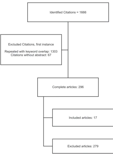
Figure 1. Search pathway for article selection Identified Citations = 1666
Complete articles: 296
Included articles: 17
Excluded articles: 279 Excluded Citations, first instance
Repeated with keyword overlap: 1303 Citations without abstract: 67
SELECTION CRITERIA
The database searches were performed independently by
the researchers to minimize the possible loss of citations and
to analyze the relevance of each retrieved citation for
selec-tion and inclusion in the study. Citaselec-tions in languages other
than English were excluded, as were those that contained
overlapping keywords. Among the full articles obtained, case
studies, literature reviews, letters to the editor and those not
directly related to the topic were excluded. Articles that were
effectively related to the research proposal were analyzed. The
researchers conducted all stages of the study independently;
when disagreements occurred, only articles with consensual
final agreement were included. By its nature, this study was
not single-blind (Figure 1).
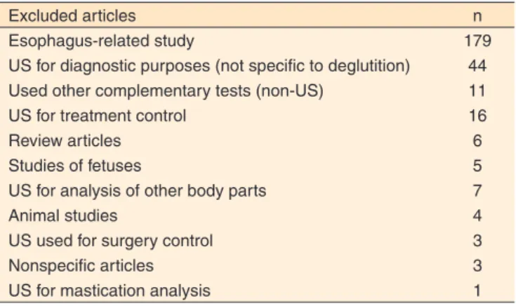
The rationale for the exclusion of 279 citations is provided
in Table 1.
DATA ANALYSIS
The 17 citations selected were critically evaluated in terms
of their objectives; number, gender and ages of the participants;
criteria and evaluation methods; results; and conclusions.
whether there was homogeneity among them.
Regarding the results and conclusions, in addition to the
general information the articles provided in these sections, we
also noted the advantages and disadvantages of the techniques
discussed in each study.
RESULTS
The results of the study are briefly described in Chart 1.
DISCUSSION
After analyzing the selected articles, we observed that the
studies that used US to assess deglutition could be divided into
three groups: studies that investigated the deglutition of healthy
individuals (52.9%)
(2,3,6,8-13); studies that compared the
deglu-tition of diseased individuals and their healthy controls
(14,15-19)(i.e., those with craniofacial abnormalities
(14)[16.7%],
stroke
(15,18,19)[50%] and neurogenic dysphagia
(16,17)[33.3%])
to that of healthy controls (35.3%)
(14,15-19); and studies that
investigated deglutition in two specific diseases
(5,20)(11.8%):
nephropathic cystinosis and amyotrophic lateral sclerosis.
Regarding the participants’ gender, most of the studies
in-cluded individuals of both genders (82.3%)
(2,3,5,8-11,13,15-20). Only
one study included only male subjects
(12)(5.9%).
The age of the participants varied considerably among the
studies. Most of the studies
(5,15-19)included adult and elderly
sub-jects (35.2%). Some of the studies
(3,9,10)divided the adults into
different age groups (17.6%), and others
(8,12)examined adults
of a particular age group (11.8%). Studies involving children
and infants were the minority
(2,6,11,13,20)(29.41%).
The parameters evaluated during the US examination
were not unanimous. In seven studies
(2,3,9,10,12,15,16)(41.1%), the
Table 1. Articles excluded because of a lack of direct relation to the topic
Excluded articles n
Esophagus-related study 179
US for diagnostic purposes (not specific to deglutition) 44
Used other complementary tests (non-US) 11
US for treatment control 16
Review articles 6
Studies of fetuses 5
US for analysis of other body parts 7
Animal studies 4
US used for surgery control 3
Nonspecific articles 3
US for mastication analysis 1
Note: US = ultrasound; n = number of citations
Chart 1. Summary of the articles included in the study
Reference Objective Method Results and Conclusions
Yabunaka et al., 2011(3)
To evaluate the movement of the hyoid bone and the chang-es arising from aging during deglutition in healthy patients using ultrasound.
- 30 adults with no complaints or history of deglutition difficulties divided into three groups: 20-39 years old (5 men and 5 women); 40-59 years old (5 men and 5 women); 60-79 years old (5 men and 5 women).
- Variable: trajectory of the hyoid bone (slowly ascending phase, rapidly ascend-ing phase, temporary pause phase and resting position phase) during deglutition of 5 ml of water.
- Mean duration of deglutition increased and peak elevation of the hyoid bone de-creased with advancing age.
- US can be a quantitative method for clinically evaluating hyoid bone movement during deglutition.
Scarborough et al., 2010(2)
To explore the parameters of maximum displacement of the hyoid bone during spon-taneous deglutition in healthy preschool children using US. To observe whether gender is associated with greater dis-placement of the hyoid bone.
- 29 subjects (16 boys and 13 girls) with a mean age of 4 years and 2 months, rang-ing from 3 years to 4 years and 10 months. - Variable: amount of hyoid bone elevation when swallowing two food consistencies (puree and liquid) at three different vol-umes (0.5, 1.5 and 2.5 ml).
- A significant gender effect was observed for all consistencies and volumes: the mean hyoid bone movement was significantly greater in females.
- The study concluded that gender can in-fluence the amount of hyoid bone elevation in children.
Galén & Jost-Brink-mann, 2010(14)
To investigate the possibility of using B-mode and M-mode US to differentiate visceral and somatic deglutition.
- 11 patients referred for orofacial myo-functional therapy (for anterior open bite or excessive overjet and visceral deglutition pattern).
- B-mode and M-mode US was applied before and after myofunctional therapy. - Variable: the amplitude and speed of the vertical movement of the tongue and total duration of deglutition of saliva (at least 6 deglutition episodes from each participant).
- 13 subjects with visceral deglutition pat-tern and normal occlusion comprised the control group.
- There was wide intraindividual variability in the variables studied, making interindi-vidual comparisons difficult.
- The M-mode images determined the amplitude and speed of the vertical move-ment of the tongue and the total duration of deglutition.
Chart 1. Summary of the articles included in the study (cont.)
Reference Objective Method Results and Conclusions
Tamburrini et al., 2010(5)
To determine the role of US in diagnosing dysphagia in pa-tients with amyotrophic lateral sclerosis (ALS).
- 9 patients underwent simultaneous static and dynamic deglutition examinations with US and videofluoroscopy (VDF), respec-tively: 5 presented with classic ALS and 4 with bulbar ALS; 8 subjects were clinically dysphagic.
- Variable: mobility/function of the orofacial myofunctional organs during deglutition, especially during the oropharyngeal phase.
- 3 volumes of water (5, 10 and 15 ml) were offered.
- Static phase: 5 patients had tongue atro-phy. Abnormal bolus position was observed in 6 patients with US and in 3 patients with VDF. Both techniques identified the inability to maintain the bolus in the oral cavity in 4 patients.
- Dynamic phase: decreased tongue move-ment was observed in 5 patients with US and in 2 patients with VDF. Disorganized movement of the tongue was observed in 3 patients with US and in 2 patients with VDF. - Multiple deglutitions were only visualized with US. The presence of stasis was not observed with US, whereas it was observed in 2 patients with VFS.
- US of the tongue is complementary to VDF, as it provides a precise description of the oral phase of deglutition.
Geddes et al., 2009(6) To develop a rough
visualiza-tion of infants during breast-feeding using US and to de-termine the accuracy of the US image of deglutition compared with respiratory plethysmogra-phy in a cohort of infants.
- 16 lactating women and their healthy infants participated in the study. The infants were between 24 and 156 days of age (mean of 57 days) and were within the normal parameters of growth for their age (mean of 4871 g).
- US was used to examine deglutition, and plethysmography was used to verify breathing.
- Variables: duration of deglutition and of deglutition apnea. To measure breathing during deglutition, breast-feeding was recorded.
- US is a noninvasive, accurate method for visualizing the movement of the milk bolus during the pharyngeal phase of deglutition. - Deglutition apnea was identified using US, and the results correlated well with the results of respiratory inductive pleth-ysmography.
- The combined use of these techniques has the potential to provide useful informa-tion in cases of breastfeeding difficulties.
Jadcherla et al., 2009(11)
To investigate the pharyn-goglottal relationship during basal and adaptive deglutition.
- 12 healthy neonates who were orally fed without structural, chromosomal, or neurological disturbances.
- Simultaneous pharyngoesophageal ma-nometry, plethysmography, electromyogra-phy (submental) and glottal US were used. - Variables: temporal changes in the kinet-ics of glottal closure (frequency, response latency and duration) during spontaneous and adaptive pharyngeal deglutition.
- Glottal adduction during basal or adaptive deglutition occurs in any respiratory phase, thus ensuring airway protection before and during deglutition.
- The duration of adduction of the pharyn-geal-glottal closure reflex suggests a state of glottal hypervigilance in preventing as-piration during deglutition or during events of high gastroesophageal reflux.
- Investigation of the pharyngoglottal rela-tionship using noninvasive methods may be more acceptable for patients and is applicable to all ages.
Huang et al., 2009(15) To evaluate the reliability of
US in stroke patients with or without dysphagia.
- 55 adults divided into 3 groups: 15 normal (control group); 20 with stroke and without dysphagia (G1); 20 with stroke and with dysphagia (G2).
- Variable: distance between the hyoid bone and larynx during deglutition. - Participants had to swallow 3 times with an interval of 1-2 minutes between each deglutition.
- The distance between the hyoid bone and the thyroid cartilage was significantly greater in normal subjects compared with stroke patients and even greater compared with patients with stroke and dysphagia. - The distance between the hyoid bone and the thyroid cartilage during deglutition was significantly smaller in normal subjects than in the stroke group.
Chart 1. Summary of the articles included in the study (cont.)
Reference Objective Method Results and Conclusions
Welge-Lüssen et al., 2009(8)
To examine whether retrona-sal olfaction combined with simultaneous gustatory stimuli affects deglutition differently than orthonasal olfaction.
- 47 healthy, non-smoking adults. - US (transducer placed on the floor of the mouth) of deglutition.
- Variable: tongue movements (speed and frequency) during deglutition and after the presentation of an olfactory stimulus and latency of deglutition. A sweet taste was presented simultaneously with an essence of an edible food (randomly presented either ortho- or retronasally) using a com-puter-controlled olfactometer.
- After retronasal stimulation, deglutition occurred significantly more rapidly and more frequently compared with deglutition after orthonasal stimulation.
- These results show that an essence of edible food presented as retronasal stimu-lation in combination with a congruent taste stimulus may influence deglutition.
Komori et al., 2008(12) To evaluate a new method of
bedside deglutition assess-ment that combined US and videoendoscopy (VED) com-pared with videofluoroscopy (VDF) alone.
- 8 healthy male volunteers aged 25-31 years with no deglutition disorders. - Simultaneous combined videoendoscopy (VED), US of deglutition and videofluoros-copy of deglutition (VDF).
- Variable: laryngeal elevation (beginning, peak and duration).
- The beginning of laryngeal elevation was identified with VDF and US.
- After the beginning, the pharynx became invisible with VED.
- The peak elevation of the larynx was iden-tified with VDF and US, and this moment was almost identical in both examinations. - The distance and duration of peak laryn-geal elevation, measured with US and with VDF, were almost equal and were positively correlated.
- This study suggests that the combined technique (US + VED) can demonstrate the deglutition function as efficiently as VDF.
Peng et al., 2007(13) To evaluate the movement of
the tongue during deglutition using a system assisted by M-mode US.
- 55 individuals (30 females and 25 males) with a mean age of 22.7 years (8 to 50 years).
- Variable: movement pattern and duration of the activity of the dorsum of the tongue. Three deglutitions of saliva from each subject were evaluated using M-mode US. The images obtained with US were video recorded and evaluated.
- The duration, amplitude and pattern of tongue movements during deglutition var-ied considerably among individuals. - M-mode US provides valid information about tongue movements without any side effects and is therefore a useful tool in the diagnosis and research of tongue functions in orthodontics and dentistry.
Sonies et al., 2005(20) To evaluate the deglutition
function of patients with cys-tinosis, with special attention to the effects of cysteamine treatment.
- 101 patients with nephropathic cystinosis diagnosis.
- Oropharyngeal US was used.
- Variables: movement of the tongue and of the hyoid bone during deglutition, duration of the oropharyngeal phase of deglutition and movements of the tongue and the hyoid bone needed to initiate and complete deglutition.
- Oral motor dysfunction of deglutition in patients with cystinosis progressively increases with age and is correlated with generalized muscular dysfunction but not with the severity of disease in general. - Long-term therapy of oral cysteamine seems to reduce the severity of oral motor dysfunction and deglutition.
- Deglutition dysfunction in patients with cystinosis presents a risk of fatal aspiration and correlates with the presence of muscle atrophy.
Kuhl et al., 2003(16) To noninvasively analyze the
vertical movement of the lar-ynx during deglutition using US techniques in patients with dysphagia and in healthy subjects.
- 18 patients (mean age: 63 ± 8 years) with dysphagia caused by neurological diseases.
- Control group was composed of 42 healthy subjects (mean age 57 ± 19 years). - Deglutition US was used.
- Variables: distance between the hyoid bone and the upper edge of the thyroid cartilage during laryngeal elevation in deglutition.
- Healthy subjects: mean distance of 220 mm (± 30) at rest; the shortest distance during deglutition of 5 or 10 ml of water was 85 mm (± 11), representing a reduction of 61% (± 3) under physiological conditions. - Neurogenic dysphagia patients: the mean relative elevation of the larynx was reduced to only 42% (± 10).
- US is a viable, noninvasive method for investigating the elevation of the larynx during deglutition.
Chart 1. Summary of the articles included in the study (cont.)
Reference Objective Method Results and Conclusions
Söder & Miller, 2002(17)
To determine the extent of intrapersonal variability in the duration of tongue movement during deglutition.
- 10 individuals diagnosed with neurogenic dysphagia.
- 10 subjects without disturbances and with no diagnosis of dysphagia (control group). - Deglutition US was used.
- Variable: total duration of movement of the tongue and the duration of the oral transport phase during deglutition. The subjects were instructed to swallow water and, after 10 seconds, to swallow saliva. The investigation continued in this manner until at least 15 dry deglutitions were recorded.
- The results indicate considerable intrap-ersonal variability in both groups. - There were no significant differences between groups.
- US was a highly suitable technique for the purpose of the study.
Chi-Fishman & Sonies, 2002(9)
To investigate the movement of the hyoid bone during the deglutition of different consis-tencies.
- 31 healthy individuals (16 male, 15 fe-male) divided into three age groups: 20-39 years, 40-59 years and 60-79 years. - Deglutition US was used.
- Variables: hyoid bone movement - dura-tion of the movement, acdura-tions, maximum amplitudes, total distances, and peak velocities - analyzed in relation to the vari-ance of viscosity, volume, age and gender. - 612 deglutitions were studied.
- The results showed that older individuals were slower to start deglutition and also presented with higher maximum vertical amplitude, higher total vertical distance and higher peak velocity compared with younger individuals.
- Males had higher values for most of the motion parameters.
- The results illustrate the importance of examining the relationships among the movement variables to better understand the tasks and strategies of motor control. - The evidence also illustrates the func-tional adaptation of the infrahyoid muscles and their compensation in healthy elderly adults.
Chi-Fishman & Sonies, 2002(10)
To examine the details of the relationships among the kine-matic variables to distinguish strategies of hyoid bone move-ment in discrete deglutition and in fast sequential deglutition.
- 30 healthy subjects divided into three age groups: 20-39 years, 40-59 years and 60-79 years.
- Variables: movement, movement duration and maximum amplitude of the hyoid bone. - Changes in the position of the hyoid bone were recorded for a total of 236 discrete deglutitions and 318 rapid sequential deglutitions.
- Rapid sequential deglutitions differ signifi-cantly from discrete deglutitions in relation to the movement of the hyoid bone. - When instructed to swallow as quickly as possible, the subjects achieved the smallest movement without increasing the peak speed. This finding suggests greater flexibility in the functional range of move-ment of the hyoid bone.
Kim & Kim, 2012(18) To analyze the movement of
the lateral wall of the pharynx using US.
- 26 individuals with stroke and dysphagia and 15 healthy individuals.
- Individuals with stroke and dysphagia were divided into two groups (A and B) based on videofluoroscopy swallow studies (VFSS). In Group A (n = 12), the subjects presented with penetration or aspiration on the VFSS, while in Group B (n = 14), there was no evidence of pene-tration or aspiration.
- Variables: movement of the lateral pha-ryngeal wall. The movement was assessed by B/M-mode US.
- Comparative analysis between the groups and the relationship between movement parameters in the pharyngeal phase was performed.
- The mean pharyngeal displacement in Groups A and B was significantly smaller compared with that of healthy individuals. - The mean duration of pharyngeal move-ment of Groups A and B was longer than that of healthy individuals.
Chart 1. Summary of the articles included in the study (cont.)
Reference Objective Method Results and Conclusions
Hsiao et al., Jan 2012(15)
To measure changes in tongue thickness of and hyoid bone displacement in patients with stroke using US examination in the submental region.
- 60 stroke patients (30 with exclusive alternative feeding and 30 with regular oral intake).
- 30 healthy subjects. Another 10 healthy subjects were included to assess the reli-ability of the examination.
- The subjects were instructed to swallow 5 ml of water. During deglutition, videoflu-oroscopy and US were performed in the mentonian region. The videofluoroscopy exam was complementary to the US for evaluating the displacement of the hyoid bone.
- The changes in tongue thickness and hyoid bone displacement were significantly smaller in stroke patients with exclusive alternative feeding compared with stroke patients with oral intake.
- No significant changes were observed be-tween the control group and stroke patients with oral intake in either the tongue thick-ness or the displacement of the hyoid bone. - US of the submental region showed good reliability and correlated well with videofluoroscopy.
lifting motion of the hyoid bone during deglutition was
asses-sed, and in four of those studies
(3,9,10,12), the duration of hyoid
movement was also considered. Four studies
(8,13,14,17)evaluated
tongue movements during deglutition (23.5%), and the others
assessed different parameters: mobility and functionality of
the phonoarticulatory organs during deglutition
(5)(5.9%);
duration of deglutition and apnea
(6)(5.9%); glottal closure and
its characteristics (frequency, response latency and duration)
(11)(5.9%); movement of the lateral wall of the pharynx
(18)(5.9%);
movement of the hyoid bone and tongue thickness
(19)(5.9%);
and tongue and hyoid bone movements during deglutition
(20)(5.9%).
For a better discussion of the studies’ methods, results
and conclusions, the articles were grouped according to the
parameters considered for the assessment, as described above.
In studies that assessed the movement of the hyoid bone -
elevation and/or duration - the overall objective was to correlate
the movement of the hyoid bone with the participant’s age,
gen-der and clinical status. A longer mean duration of hyoid bone
elevation was observed with increasing age, as was decreased
peak elevation of the hyoid bone
(3). A study conducted with
children found that gender may affect hyoid bone elevation
- the mean movement was significantly greater in females
(2).
Regarding the participant’s clinical condition, the hyoid bone
movement was significantly greater in healthy subjects
compa-red with individuals with neurogenic dysphagia
(15,16).
All of the studies that assessed the hyoid bone movement
found that US could quantitatively and reliably evaluate this
parameter.
Among the studies that assessed tongue movement during
deglutition, two subgroups could be defined. The first subgroup
aimed to determine the extent of intrapersonal variability in the
duration of tongue movements during deglutition. The results
indicated high intrapersonal variability in both healthy
individu-als and individuindividu-als with neurogenic dysphagia; however, greater
variability in the duration of the transport phase was observed in
the dysphagic group
(17). The second subgroup
(13,14)investigated
US modes using tongue movement as the parameter of interest.
High intra- and/or interpersonal variability was also found in
this parameter, and it was observed that M-mode US provided
better information about tongue movement during deglutition.
For all of the studies in this group, US provided
informa-tion about tongue movements during deglutiinforma-tion without any
side effects, and it was considered a useful tool. It should be
noted that most of the studies concluded that this parameter
exhibits high intra- and interpersonal variability, which can
often complicate data analysis, as described by Galen and
Jost-Brinkmann (2010)
(14).
The other parameters were only investigated in single
studies. Thus, the conclusions regarding the use of US and its
results apply to only one study per parameter.
One of the studies investigated the mobility and function of
the phonoarticulatory organs during deglutition. The authors
sought to determine the role of US in the diagnostic evaluation
of dysphagia in patients with amyotrophic lateral sclerosis
(ALS). The exam was conducted concurrently with deglutition
videofluoroscopy (VDF), and the study found that US provided
static and dynamic functionality data for the oropharyngeal
phase of deglutition more efficiently than VDF. Thus, the
authors concluded that US should be used as a complement
to VDF because it provides a precise description of the oral
deglutition phase
(5).
Using deglutition apnea as parameter, one study evaluated
the ability of US to identify deglutition apnea in infants
(6). The
study involved the concomitant use of US and respiratory
ple-thysmography and showed that the passage of the bolus during
pharyngeal deglutition in a nursing child can be recorded using
US, regardless of the respiration phase. Deglutition US was
correlated with deglutition apnea detected with respiratory
inductive plethysmography. Both techniques, when combined,
have the potential to provide useful information about children
who have nursing difficulties.
allowed the study of glottis movement during deglutition, and
it was concluded that investigation of the pharyngeal-glottal
relationship using non-invasive methods, such as US, may be
acceptable for and applicable to all ages
(11).
One study examined the duration of the pharyngeal phase
of deglutition based on the tongue and hyoid bone movements
needed to start and finish deglutition. For this purpose, US was
used to assess the deglutition of 101 patients with nephropathic
cystinosis. The authors found that oral motor dysfunction of
deglutition in patients with cystinosis increases progressively
with age and is correlated with generalized muscular
dysfunc-tion but not with disease severity. Based on cross-secdysfunc-tional data,
the dysfunction increased with increasing age and the number
of years without cysteamine treatment
(20).
A study conducted with healthy subjects and stroke patients
used the displacement and duration of pharyngeal movement
during deglutition as a parameter. The patients were subdivided
into two groups: the first (Group A) consisted of patients who
presented with penetration and/or aspiration of food, indicated
through VDF, and the second (Group B) consisted of patients
with no VDF findings. The mean pharyngeal displacement of
Groups A and B was significantly lower compared with that
of healthy individuals, while the mean duration of pharyngeal
movement of Groups A and B was longer than that of healthy
individuals. In conclusion, the study indicated that the use of
US to analyze pharyngeal movement may help to quantify
pha-ryngeal function and can serve as a complementary method for
the anatomical evaluation of the pharyngeal phase in patients
with stroke and dysphagia
(18).
Another study analyzed the hyoid bone movement and
tongue thickness. These parameters were compared between
healthy individuals and stroke patients with and without the
use of an alternative feeding route. The study found
signifi-cantly less hyoid bone displacement and tongue thickness in
patients fed exclusively by an alternate route compared with
stroke patients with oral ingestion. No significant differences
in either variable were observed between the control group and
the stroke patients with oral ingestion
(19).
Generally, all of the articles assessed found that deglutition
US was a fast, noninvasive, low-cost method that provides
objective information about parameters of deglutition and can
be performed at bedside because the equipment is usually easy
to handle and transport. Equally important, the authors of the
examined studies all concluded that deglutition assessment
with US has some inherent disadvantages, including the
follo-wing: the pressure exerted by the transducer on the examined
structure, transducer positioning and the lack of precise
ana-tomical markers for some of the structures being investigated.
Because of these limitations, although US was effective for
the initial diagnosis of impaired deglutition, it should be used
in conjunction with other exams that complement the
asses-sment of phonoarticulatory organ function during deglutition
to accurately diagnose dysphagia.
CONCLUSION
The heterogeneity of the studies showed that different
groups and pathologies can be assessed with US; however, the
methodological variability of the included studies hinders the
definition and generalization of the patterns found.
The present review found that US examination is effective
for assessing the components involved in the dynamics of
deglutition, especially the oral and early pharyngeal phases,
such as phonoarticulatory organ function, deglutition apnea
duration, glottal closure and its aspects, and hyoid bone
mo-vement. However, US should not replace other examinations
that assist in the assessment of the pharyngeal and esophageal
phase and in dysphagia diagnosis because US cannot identify
some dynamic components of function, such as food stasis
in the pharynx. Thus, US has been used as a complement to
VDF, plethysmography, surface electromyography (EMG),
deglutition videoendoscopy and manometry to provide more
reliable parameters of the oral phase and the beginning of the
pharyngeal phase of deglutition.
REFERENCES
1. Santos HCO, Amaral WN, Tacon KCB. A história da ultrassonografia no Brasil e no mundo. EFDeportes.com Rev Digit. 2012 [acesso em 12 jan 2014];17(167). Disponível em: http://www.efdeportes.com/efd167/a-historia-da-ultrassonografia.htm
2. Scarborough DR, Waizenhofer S, Siekemeyer L, Hughes M. Sonographically measured hyoid bone displacement during swallow in preschool children: a preliminary study. J Clin Ultrasound. 2010;38(8):430-4. http://dx.doi.org/10.1002/jcu.20733
3. Yabunaka K, Sanada H, Sanada S, Konishi H, Hashimoto T, Yatake H, et al. Sonographic assessment of hyoid bone movement during swallowing: a study of normal adults with advancing age. Radiol Phys Technol. 2011;4(1):73-7. http://dx.doi.org/10.1007/s12194-010-0107-9 4. Ardakani FE. Evaluation of swallowing patterns of the tongue using real-time B-mode sonography. J Contemp Dent Pract. 2006;7(3):67-74. 5. Tamburrini S, Solazzo A, Sagnelli A, Del Vecchio L, Reginelli A, Monsorrò M, et al. Amyotrophic lateral sclerosis: sonographic evaluation of dysphagia. Radiol Med. 2010;115(5):784-93. http://dx.doi. org/10.1007/s11547-010-0523-2
6. Geddes DT, Chadwick LM, Kent JC, Garbin CP, Hartmann PE. Ultrasound imaging of infant swallowing during breast-feeding. Dysphagia. 2010;25(3):183-91. http://dx.doi.org/10.1007/s00455-009-9241-0
7. Higgins JPT, Green S, editors Cochrane handbook for systematic reviews of interventions Version 5.1.0 [internet]; Baltimore: The Cochrane Collaboration; 2011 [acesso em 11 maio 2011]. Disponível em: http://www.cochrane.org/training/cochrane-handbook
9. Chi-Fishman G, Sonies BC. Effects of systematic bolus vviscosity and volume changes on hyoid movement kinematics. Dysphagia. 2002;17(4):278-87. http://dx.doi.org/10.1007/s00455-002-0070-7 10. Chi-Fishman G, Sonies BC. Kinematic strategies for hyoid movement in rapid sequential swallowing. J Speech Lang Hear Res. 2002;45(3);457-68. http://dx.doi.org/10.1044/1092-4388(2002/036
11. Jadcherla SR, Gupta A, Wang M, Coley BD, Fernandez S, Shaker R. Definition and implications of novel pharyngo-glottal reflex in human infants using concurrent manometry ultrasonography. J Gastroenterol. 2009;104(10):2572-82. http://dx.doi.org/10.1038/ajg.2009.411
12. Komori M, Hyodo M, Gyo K. A swallowing evaluation with simultaneous videoendoscopy, ultrasonography and videofluorography in healthy controls. ORL J Otorhinolaryngol Relat Spec. 2008;70(6):393-8. http://dx.doi.org/10.1159/000163036
13. Peng CL, Miethke RR, Pong SJ, Lin CT. Investigation of tongue movements during swallowing with M-mode ultrasonography. J Orofac Orthop. 2007;68(1):17-25. http://dx.doi.org/10.1007/s00056-007-0547-y 14. Galén S, Jost-Brinkmann PG. B-mode and M-mode ultrasonography of tongue movements during swallowing. J Orofac Orthop. 2010;71(2):125-35. http://dx.doi.org/10.1007/s00056-010-9928-8 15. Huang YL, Hsieh SF, Chang YC, Chen HC, Wang TG.
Ultrasonagraphic evaluation of hyoid-larinx approximation in dysphagic stroke pacients. Ultrasound Med Biol. 2009;35(7):1103-8. http://dx.doi. org/10.1016/j.ultrasmedbio.2009.02.006
16. Kuhl V, Eicke BM, Dieterich M, Urban PP. Sonographic analysis of laryngeal elevation during swallowing. J Neurol. 2003;250(3):333-7. http://dx.doi.org/10.1007/s00415-003-1007-2
17. Söder N, Miller N. Using ultrasound to investigate intrapersonal variability in durational aspects of tongue movement during swallowing. Dysphagia. 2002;17(4):288-97. http://dx.doi.org/10.1007/s00455-002-0071-6.
18. Kim JH, Kim MS. Lateral pharyngeal wall motion analysis using ultrasonography in stroke patients with dysphagia.Ultrasound Med Biol. 2012;38(12):2058-64. http://dx.doi.org/10.1016/j. ultrasmedbio.2012.07.028
19. Hsiao MY, Chang YC, Chen HC, Chang HY, Wang TG. Application of ultrasonography in assessing oropharyngeal dysphagia in stroke patients. Ultrasound Med Biol. 2012;38(9):1522-8. http://dx.doi. org/10.1016/j.ultrasmedbio.2012.04.017