RESUMO: “Efeito do extrato, frações e da 2,3-diidromiricetina-3-O-α-L-raminosídeo obtidos da Pradosia huberi (Ducke) Ducke em artéria mesentérica isolada de rato”. Pradosia huberi
(Ducke) Ducke (Sapotaceae), espécie Amazônica popularmente conhecida como “casca-doce” é utilizada na medicina tradicional no tratamento de gastrite. O extrato etanólico de suas cascas é rico em polifenóis que podem apresentar um grande número de atividades, incluindo efeito vasorelaxante e cardioprotetor. O objetivo deste estudo foi avaliar as propriedades farmacológicas do extrato etanólico (EPH), de frações e da 2,3-diidromiricetina-3-O-α-L-raminosídeo isolados de P. huberi, em artéria mesentérica isolada de rato. O EPH foi fracionado resultando nas seguintes frações: CHCl3, CHCl3:AcOEt (1:1), AcOEt, AcOEt:MeOH (1:1) e MeOH. Da fração MeOH foi isolada a 2,3-diidromiricetina-3-O-α-L-raminosídeo e identiicada através de espectro de RMN de 1H e 13C, além de comparações com os dados de literatura. EPH (1-100 μg/mL) promoveu relaxamento dependente de concentração no tônus vascular induzido por 10 μM de fenilefrina (CE50=17,1±2,9 µg/ mL; Emax=87,4±2,9 %, n=8). A fração MeOH também relaxou os anéis mesentéricos (CE50=31±2,0 µg/mL; Emax=54±12,5%, n=6), porém com menor eicácia quando comparado ao efeito de EPH. Tanto o efeito de EPH com de MeOH foram completamente abolidos após a remoção do endotélio vascular. A fração AcOEt:MeOH (1:1) e o lavonoide isolado induziram vasorelaxamento. O estudo demonstrou que o EPH e a fração MeOH de Pradosia huberi apresentam propriedade vasorelaxante que pode ser completamente dependente da presença do endotélio. O lavonoide isolado não é o responsável por este efeito vasorelaxante.
Unitermos: Pradosia huberi, artéria mesentérica, vasodilatação, endotélio dependente, 2,3-diidromiricetina-3-O-α-L-raminosídeo, loresta amazônica.
ABSTRACT: Pradosia huberi (Ducke) Ducke (Sapotaceae), an Amazonian species, is popularly known as “casca-doce” and used in the folk medicine for the treatment of gastritis. The ethanol extract of the bark contains mainly polyphenolic compounds, which are known to show a large number of activities, including cardioprotective and vasorelaxant effects. The aim of this study was to evaluate the pharmacological properties induced by P. huberi ethanol extract (PHEE) and fractions and 2-3-dihydromyricetin-3-O-α-L-rhamnoside derived from this extract, in isolated rat mesenteric arteries. PHEE was separated and the following fractions were obtained: CHCl3, CHCl3:AcOEt (1:1), AcOEt, AcOEt:MeOH (1:1) and MeOH. We isolated 2-3-dihydromyricetin-3-O-α-L-rhamnoside from the MeOH fraction, which was identiied by 1H and 13C NMR spectra and compared with data in the literature. PHEE (1-100 μg/mL) induced concentration-dependent relaxations of 10 μM phenylephrine-induced tone (EC50=17,1±2,9 µg/mL; Emax=87.4±2.9 %, n=8). The MeOH fraction also relaxed mesenteric rings (EC50=31±2.0 μg/mL; Emax=54±12.5%, n=6) but less effectively when compared to PHEE. Both effects were completely abolished after removal of the vascular endothelium. The AcOEt:MeOH (1:1) fraction and the isolated lavonoid were ineffective in eliciting vasorelaxation. The study demonstrates that PHEE and MeOH fraction of Pradosia huberi possess a vasorelaxant effect, which may be completely dependent upon endothelium. The isolated lavonoid is not responsible for this vasorelaxant effect.
Keywords: Pradosia huberi, mesenteric artery, vasodilatation, dependent-endothelium, 2,3-dihydromyricetin 3-O-α-L-rhamnoside, Amazon Rainforest.
20(4): 542-548, Ago./Set. 2010
A
rt
igo
Received 8 August 2008; Accepted 24 November 20089
Effects of extract, fractions and 2,3-dihydromyricetin-3-
O
-α-
L
-rhamnoside from
Pradosia huberi
(Ducke) Ducke on rat isolated
mesenteric arteries
Alessandra A. N. Medeiros,
1Fernando A. Medeiros,
1Thyago M. Queiroz,
2Josean F. Tavares, Marcelo S. Silva,
2Isac A. Medeiros
*,21Instituto de Pesquisas Cientíicas e Tecnológicas do Amapá, Av. Feliciano Coelho, Bairro do Trem, Caixa Postal 1509, 68900-260 Macapá-AP, Brasil,
INTRODUCTION
Pradosia huberi (Ducke) Ducke, Sapotaceae is a medicinal plant that is common in the Amazon Rainforest, popularly known as casca-doce, pau-doce, amapá-doce or paracauba, and used in local folk medicine in the treatment of gastric and digestive problems. This species has the nomenclatural synonym Glycoxylon huberi Ducke (Corrêa, 1986). There are few phytochemical and biological studies of P. huberi reported in the literature; however, the hydroalcoholic extract from P. huberi bark has shown antisecretory and gastroprotective activity, besides no acute toxicity (5000 mg/kg; p.o.) (Kushima et al., 2005).
Phytochemical screening of the ethanolic extract was positive for the presence of compounds such as lavonoids, terpenoids, quinones, alkaloids, tannins and saponins (Ferreira et al., 2005). Flavonoids from stem bark were identiied as 2,3-dihydromyricetin 3-O-α-L -rhamnoside, astilbin, engelitin and 2,3-dihydromyricetin (Jacquemin et al., 1985), all of which are also found in various plants. It has been reported that these lavonoids have pharmacological properties, including the following: antiinlammatory (Kanbara et al., 1994; Yun et al., 2000), anti-oxidative effects (Yang et al., 2004), inhibition of lipid peroxidation (Yun et al., 2000), block of uterine contraction in rats (Carneiro et al., 1993).
Flavonoids are plant-derived polyphenolic substances commonly found in plants and consumed in the diet. Many of these compounds possess cardiovascular protective properties (Curin & Andriantsitohaina, 2005) which can be explained by the combination of the antioxidant, antiplatelet and antiinlammatory effects along with their positive effects on restoration of endothelial function or modulation of vascular tone (Fitzpatrik et al., 1993; Woodman & Chan, 2004; Curin & Andriantsitohaina, 2005).
To date, this species has not been studied with regard to cardiovascular activity. Thus, the aim of this work was to evaluate the pharmacologic properties of
Pradosia huberi ethanolic extract (PHEE), fractions and isolated substance for vasorelaxant activity in rat superior mesenteric artery.
MATERIAL AND METHODS
Plant material
The bark of Pradosia huberi (Ducke) Ducke, Sapotaceae, was collected in the city of Porto Grande¸ Amapá State, Brazil. The species was identiied and a voucher specimen (No. 012519) was deposited in the Herbário Amapaense (HAMAB) of the Instituto de Pesquisas Cientíicas e Tecnológicas do Estado do Amapá (IEPA).
Phytochemical study
After drying at 40 oC, the plant material was pulverized (3.2 kg) and extracted with 95% EtOH under maceration at room temperature for ten days. The solvent was removed by rotary evaporation under vacuum at 45 oC, yielding 200 g of Pradosia huberi ethanolic extract (PHEE). A sample of 20 g of PHEE was separated on a silica gel column under reduced pressure with the eluents CHCl3, CHCl3:AcOEt (1:1), AcOEt, AcOEt:MeOH (1:1) and MeOH, resulting in the following yields: 0.2, 0.4, 1.2, 3.0 and 4.0 g, respectively. Samples of each fraction obtained were used for pharmacological testing. The MeOH fraction was chosen for isolation since it presented the greatest amount of constituent and showed the highest vasorelaxant activity compared to the other fraction obtained. An aliquot of 2 g of the MeOH fraction was separately submitted to Sephadex LH-20 column chromatography using MeOH as eluent, from which 28 fractions of 30 mL were collected, which after analysis by TLC were grouped according to their Rf. The fraction 14-22 was rechromatographed on Sephadex LH-20 with MeOH elution, isolating 2,3-dihydromyricetin-3-O
-α-L-rhamnoside (45 mg, Figure 1), which corresponded to 22.5% of the yield in relation to the MeOH fraction. For chemical identiication of the isolated compound, 1H and 13C NMR spectra were acquired using a Mercury Varian
spectrometer operating at 200 MHz for 1H and 50.3 MHz for 13C NMR and recorded in CD
3OD.
Animals
Male Wistar rats (250-300 g) were used for the experiments. Animals were housed under conditions of controlled temperature (21±1 oC) and lighting (light-dark cycle of 12 h), with free access to water and pelleted feed (Purina-Brazil). The study was approved by the Animal Care and Use Committees of the Federal University of Paraíba (No. 0603/07).
Drugs
The drugs used were L-phenylephrine chloride and acetylcholine chloride (both from Sigma, St. Louis, MO, USA). For the experiments, PHEE was dissolved in distilled water. All the stock solutions were prepared in distilled water and kept at 4 ºC.
Preparation of isolated rat superior mesenteric artery rings
+ 5% CO2 mixture. Rings were stabilized under a resting tension of 0.75 g for 1 h. During this time the solution was changed every 15 min to prevent the accumulation of metabolites that could otherwise lead to misinterpretations (Altura & Altura, 1970). The isometric contraction was recorded by a force transducer (Miobath-4, WPI, Sarasota, Fl, EUA) coupled to an ampliier-recorder (Miobath-4, WPI, Sarasota, Fl, EUA) and to a personal computer equipped with an analog-to-digital converter board. In some experiments, the endothelium layer was removed by gently rubbing the intimal surface of the vessels with a cotton ball. The presence of functional endothelium was assessed by the ability of acetylcholine (10 μM) to induce more than 90% relaxation of vessels pre-contracted with 10 μM phenylephrine (PHE), and the absence of relaxation in response to acetylcholine was taken as evidence that the vessel segments were functionally denuded of endothelium (Furchgott; Zawadzki, 1980). PHEE was cumulatively applied after contractile responses induced by PHE (10 μM).
Effect of PHEE, MEF, MAF and 2,3-dihydromyricetin-3-O-α-L-rhamnoside on sustained contractions induced
by phenylephrine (10 μM) in isolated preparations
from rat superior mesenteric arteries
After an equilibration period, the rings with or without functional endothelium were pre-contracted with the agonist, and once the response to the second administration of PHE (10 μM) reached a plateau, increasing cumulative concentrations of PHEE (1-100 µg/ mL), MEF(1-100 µg/mL), MAF (1-100 µg/mL) or 2,3-dihydromyricetin-3-O-α-L-rhamnoside (1-100 µg/mL)
were added to the bath. The relaxations were measured by comparing the tension developed before and after addition of PHEE, MEF, MAF or 2,3-dihydromyricetin-3-O-α-L -rhamnoside.
Data analysis
Values are expressed as means ±S.E.M. When appropriate, statistical signiicance was examined with Student's t -test or one-way ANOVA followed by Bonferroni’s post-hoc test, using Graph Pad Prism TM 4.0 software. The EC50 values were calculated by nonlinear regression of individual concentration–response curves, and p<0.05 was considered signiicant.
RESULTS
The analyses of the spectral data as well as the assignments of all carbons and hydrogens (Table 1) and comparison with literature values (Gellért et al., 1981; Jacquemin et al., 1985; Shen et al., 1993; Slimestad et al., 1994; Wu et al., 1998; Du et al., 2005) allowed the identiication of 2,3-dihydromyricetin-3-O-α-L -rhamnoside (1).
O O OH HO OH OH OH O O H OH HO H H3C
HO 2 3 4 10 5 6 7 8 9 1' 6' 5' 4' 3' 2' 1" 2" 3" 4" 5" 6"
Relaxant effect of Pradosia huberi extract,
fractions and isolated constituent on the
PHE-induced sustained contractions
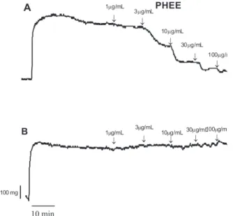
PHEE inhibited PHE-induced sustained contraction in the rat mesenteric rings in a concentration-dependent manner, in the preparations with preserved functional endothelium. EC50 of PHEE effect on contraction induced by PHE was 17.1±2.9 µg/mL, and the maximal value for the relaxant effect (Emax) was 87.4±2.9%, n=8, p<0.001***. The concentration-response curve of PHEE was completely abolished after removal of functional endothelium (Figure 1 and Figure 2A). MEF was also able to induce concentration-dependent relaxations of 10 μM PHE-induced tone with an EC50 value of 31±2.0 µg/mL. This vasodilator effect was signiicantly smaller when compared to that induced by PHEE, with a strong reduction in the Emax of 54±12.5%, n=6, as shown in Figure 2B. MEF was not able to induce relaxation in the rings without functional endothelium.
MAF did not induce a concentration-dependent vasodilator effect in the preparations, either with intact endothelium or after endothelium removal (Figure 2C). On the contrary, there was a tendency toward contraction of the rat mesenteric rings with intact endothelium at the highest concentrations of this fraction. In addition, the isolated lavonoid (2,3-dihydromyricetin-3-O-α-L -rhamnoside) was not capable of inhibiting the contractions induced by phenylephrine in the isolated preparations. On the contrary, a tendency toward contraction was observed in preparations with intact endothelium (Figure 2D).
DISCUSSION
The present study showed that PHEE exerted a vasorelaxant effect in phenylephrine-induced contractions of rat superior mesenteric rings. Removal of functional endothelium completely abolished this relaxant response to PHEE, suggesting that vasorelaxation caused by PHEE was endothelium-dependent. Furthermore, the isolated lavonoid was not responsible for this vasorelaxant effect.
The 13C NMR (APT) spectrum showed a total of twenty-one signals, including nine to no hydrogenated
carbons, eleven to methynic carbons and a methyl carbon. The spectral region between δC 168.9 and δC 95.8, characteristic of aromatic carbon signals, and the signal at δC 197.5, characteristic of carbonyl carbon signals, as well as comparison with literature data (Shen et al., 1993; Wu et al., 1998; Almeida et al., 2005; Du et al., 2005; Sinkkonen et al., 2005) and chemotaxonomy of the genus Pradosia, suggest a lavonoid skeleton such as an aglycone. Signals at δC 83.7 C and δC 76.8 are compatible with a dihydrolavonol
structure, which can be supported by the 1H NMR spectrum showing doublets at δH 4.86 and δH 4.61 (each J = 11.0 Hz), characteristic of H-2 and H-3 of the dihydrolavonol structure (Jacquemin et al., 1985; Wu et al., 1998; Du et al., 2002). The determination of the stereochemical trans-diaxial relationship between the protons at C-2 and C-3 was evident from the 11.0 Hz coupling constant (Jacquemin et al., 1985; Slimestad et al., 1994; Wu et al., 1998; Du et al., 2005). The oxymethynic carbon signals between δC 69.2-71.9, together with the methyl carbon signal at δC 17.8 (H-6", at δH 0.92, d, J = 6.2 Hz, 3H) indicated that the rhamnose sugar was attached. The 13C NMR spectrum showed a downield shift of 5.2 ppm for C-3 when compared with the data of 2,3-dihydromyricetin (Shen et al., 1993), indicating the location of the rhamnose moieties to be the C-3 (Slimestad et al., 1994; Wu et al., 1998; Du et al., 2005). The α-coniguration of rhamnose was established Figure 1. Relaxant effects of PHEE on isolated mesenteric rings
pre-contracted with phenylephrine. Panel A shows a typical recording obtained in intact endothelium rings, and panel B in removed endothelium rings.
Figure 2. Line plot showing the effects of increasing concentrations of PHEE (A), MEF (B), MAF (C) or isolated compounds of Pradosia huberi (D) on phenylephrine (10µM)-induced contraction in mesenteric rings of rats with and without the functional endothelium. Results are means ± S.E.M..
by the anomeric proton at δH 4.17 and from the 2.8 Hz
coupling constant (Slimestad et al., 1994; Wu et al., 1998). The endothelium is formed by a monolayer of cells that covers the lumen of blood vessels and serves as a secretory gland able to produce contractant as well as relaxing factors that control vascular tone (Curin & Andriantsitohaina, 2005). Under physiological conditions, there is a balance between endothelial factors released, where the effect of relaxing agents prevails. These factors include nitric oxide (NO), endothelium-derived hyperpolarizing factor (EDHF) and prostocyclin (PGI2) (Moncada & Vane, 1979; Furchgott & Zawadzki, 1980; Feletou & Vanhoutte, 1988).
Many reports show that the effect of polyphenols on the endothelium is mainly due to NO production (Andriambeloson et al., 1997; Duarte et al., 2004; Zenebe et al., 2003), increase in intracellular concentration of Ca2+ ([Ca2+]i), activation of K+ channels in the endothelium, inhibition of Ca2+-ATPases of the endoplasmic reticulum in endothelial cells (Li et al., 2000; McKenna et al., 1996), or modulation of NO levels by the action on the phosphodiesterases (PDE)
PDE-2 and PDE-4 in endothelial cells (Beretz et al., 1986a; Beretz et al., 1986b; Lugnier & Schini, 1990).
MEF also induced a concentration-dependent relaxation of the preparations pre-contracted with phenylephrine, only in intact endothelium rings (EC50=31±2.0 µg/mL; Emax=54±12.5%, n = 6). However, such effect was shown to be less potent and effective when compared to the effect produced by PHEE (EC50=17,1±2,9 µg/mL; Emax=87.4±2.9 %, n=8).
Both MAF, with the majority substances of the ethanol extract of Pradosia huberi, and 2,3-dihydromyricetin 3-O
-a-L-rhamnoside were not effective in relaxing mesenteric rings.
A particular feature of phytomedicines is their complex composition, i.e., the “phytocomplex” which includes a variety of phytochemicals with different biological activities. Some of these phytochemicals are responsible for speciic effects, while other components play an additional role. However, a wider array of effects and the healing properties are frequently guaranteed only by the phytocomplex (Pietta, 2000). We can conclude that PHEE possesses a vasorelaxant Table 1. 1H and 13C NMR spectral data of 2,3-dihydromycicetin-3-O-α-L-rhamnoside (δ (ppm), J (Hz), measured in CD
3OD.
C 2,3-dihydromyricetin-3-O-α-L-rhaminoside 1 2
δH δC δH δC δH δC
4 197.5 197.2 194.3
5 165.4 163.2 163.3
7 168.9 166.7 166.9
9 164.2 162.4 162.1
10 101.9 100.4 101.0
1’ 129.0 127.1 126.8
3’ 147.0 145.6 145.8
4’ 135.2 133.4 145.1
5’ 147.0 145.6
CH
2 4.86 (d, J=11,0 Hz) 83.7 4.90 (d, J=10.5 Hz) 83.2 5.24 (d, J=9.8 Hz) 81.5
3 4.61 (d, J=11,0 Hz) 76.8 4.38 (d, J=10.5 Hz) 71.6 4.63 (d, J=9.8 Hz) 75.6
6 5.90 (d, J=2,2 Hz) 96.3 5.89 (d, J=1.6 Hz) 95.9 5.90 (d, J=2.1 Hz) 96.0
8 5.87 (d, J=2,2 Hz) 95.8 5.85 (d, J=1.6 Hz) 94.9 5.88 (d, J=2.1 Hz) 95.0
2’ 6.50 (s) 108.0 6.40 (s) 106.9 6.88 (s) 114.7
5’ 6.74 (s) 115.3
6’ 6.50 (s) 108.0 6.40 (s) 106.9 6.74 (s) 118.7
1’’ 4.17 (d, J= 2,8 Hz) 102.7 4.07 (s) 100.0
2’’ 4.00 (dd, J=3,2; 1,4 Hz) 71.9 3.36 (br, s) 70.1
3’’ 3.41 (dd, J=9,8; 3,0 Hz) 71.9 3.42 (dd, J=9.4; 2.8 Hz) 70.4
4’’ 3.20 (dd, J=9,2; 9,2 Hz) 70.3 3.15 (dd, J=9.4; 9.4 Hz) 71.6
5’’ 2.40 (dd, J=9,4; 6,2Hz) 69.2 3.88 (qd, J=9.4; 6.2Hz) 68.9
CH3
6’’ 0.92 (d, J=6.2 Hz) 17.8 1,05 (d, J=6,2 Hz) 17.6
effect in isolated mesenteric rings and that this effect is totally dependent on the vascular endothelium. The loss of activity of the fractions and 2,3-dihydromyricetin-3-O
-α-L-rhamnoside may be due to the action of the constituents present in PHEE (phytocomplex). Since the extract consists primarily of lavonoids, these data are in line with the literature that show an endothelium-dependent vasodilator effect of lavonoids and other polyphenols (Fitzpatrick et al., 1993; Rice-Evans et al., 1996; Lemos et al., 1999).
ACKNOWLEDGEMENTS
The authors thank Instituto de Pesquisas Cientíicas e Tecnológicas do Estado do Amapá (IEPA/GEA), CNPq and Laboratório de Tecnologia Farmacêutica (LTF/ UFPB) for the inancial or technical support of this work.
REFERENCES
Almeida SCX, Lemos TLG, Silveira ER, Pessoa ODL 2005. Constituintes químicos voláteis e não-voláteis de
Cochlospermum vitifolium (Willdenow) Sprengel. Quim
Nova 28: 57-60.
Altura BM, Altura BT 1970. Differential effects of substrate depletion on drug induced contractions of rabbit aorta.
Am J Physiol 219: 1698-1705.
Andriambeloson E, Kleschyov AL, Muller B, Beretz A, Stoclet JC, Andriantsitohaina R 1997. Nitric oxide production and endothelium-dependent vasorelaxation induced by wine polyphenols in rat aorta. Br J Pharmacol 120: 1053-1058.
Beretz A, Anton R, Cazenave JP 1986a. The effects of lavonoids on cyclic nucleotide phosphodiesterases. Prog Clin Biol
Res 213: 281-296.
Beretz A, Briancon-Scheid F, Stierle A, Corre G, Anton R, Cazenave JP 1986b. Inhibition of human platelet cyclic AMP phosphodiesterase and of platelet aggregation by a hemisynthetic lavonoid, amentolavone hexaacetate.
Biochem Pharmacol 35: 257-262.
Carneiro E, Calixto J, Delle M, Franco Y, Rosendo A 1993. Isolation, chemical identiication, and pharmacological evaluation of eucryphin, astilbin, and engelitin obtained from the bark of Hymenaea martiana. Int J Pharmacog
31: 38-46.
Corrêa MP 1986. Dicionário das plantas úteis do Brasil e das exóticas
cultivadas. Rio de Janeiro: Ministério da Agricultura. IBDF. Curin Y, Andriantsitohaina R 2005. Polyphenols as potential
therapeutical agents against cardiovascular diseases.
Pharmacol Rep 57: 97-107.
Du Q, Cai W, Xia M, Ito YG 2002. Puriication of (+)-dihydromyricetin from leaves of Ampelopsis
grossedentata using high-speed countercurrent chromatography with scale-up triple columns. J
Chromatogr A 973: 217-220.
Du Q, Li L, Jerz G 2005. Puriication of astilbin and isoastilbin in the extract of Smilax glabra rhizome by high-speed
counter-corrent chromatography. J Chromatogr A 1077: 98-101. Duarte J, Andriambeloson E, Diebolt M, Andriantsitohaina R
2004. Wine polyphenols stimulate superoxide anion production to promote calcium signaling and endothelial-dependent vasodilatation. Physiol Res 53: 595-602. Félétou M, Vanhoutte PM 1988. Endothelium-dependent
hyperpolarization of canine coronary smooth muscle. Br
J Pharmacol 93: 515-524.
Ferreira ES, Medeiros FA, Medeiros AAN 2005. Estudo itoquímico da espécie Pradosia huberi (Ducke): usada na produção de itoterápicos do IEPA. Rev Cien Soc 2: 43-53.
Fitzpatrick DF, Hirschield SL, Coffey RG 1993. Endothelium-dependent vasorelaxing activity of wine and other grape products. Am J Physiol 265: H774-H778.
Furchgott RF, Zawadzki JV 1980. The obligatory role of endothelial cells in the relaxation of arterial smooth muscle by acetylcholine. Nature 288: 373-376.
Gellért M, Szendrei K, Reisch J 1981. Dihydromyricetin-3-O -rhamnoside from leaves of Catha edulis. Phytochenistry
20: 1759-1760.
Jacquemin H, Boissonnat A, Faugeras G, Tillequin F, Delaveau P 1985. Flavanoids of Glycoxylon huberi Ducke. Ann Pharm Fr 43: 521-522.
Kanbara T, Mizutani K, Tamura K, Kataoka S 1994. Extraction of astilbins from Engelhardtia chrysolepis leaves and their anti-inlammatory activities. Jpn Kokai Tokkyo Koho JP 06,256,194.
Kushima H, Hurima-Lima CA, Santos MA, Viana E, Coelho-Ferreira, Brito ARMS 2005. Gastroprotective activity of Pradosia huberi on experimentally induced gastric lesions in rodents: Role of endogenous sulphydryls and nitric oxide. J Ethnopharmacol 101: 61-67.
Lemos VS, Freitas MR, Muller B, Lino YD, Queiroga CE, Cortes SF 1999. Dioclein, a new nitric oxide- and endothelium-dependent vasodilator lavonoid. Eur J Pharmacol 386: 41-46.
Li HF, Chen SA, Wu SN 2000. Evidence for the stimulatory effect of resveratrol on Ca(2+)-activated K+ current in vascular endothelial cells. Cardiovasc Res 45: 1035-1045. Lugnier C, Schini VB 1990. Characterization of cyclic nucleotide
phosphodiesterases from cultured bovine aortic endothelial cells. Biochem Pharmacol 39: 75-84. McKenna E, Smith JS, Coll KE, Mazack EK, Mayer EJ,
Antanavage J, Wiedmann RT, Johnson RG Jr 1996. Dissociation of phospholamban regulation of cardiac sarcoplasmic reticulum Ca2+-ATPase by quercetin. J Biol
Chem 271: 24517-24525.
Moncada S, Vane JR 1979. Pharmacology and endogenous roles of prostaglandins endoperoxydes, throboxane A2 and prostacyclin. Pharmacol Rev 30: 293-331.
Pietta P 2000. Phytomedicines creating safer choices. In: Watson RR Vegetables, fruits and herbs in health promotion.
1.ed. Florida: CRC Press LLC, p.73-74.
Free Radical Biol Med 20: 933-956.
Shen C, Chang Y, Ho LK 1993. Nuclear magnetic resonance studies of 5,7-dihydrolavonoids. Phytochemistry 34: 843-845. Sinkkonen J, Liimatainen J, Koronen M, Pihlaja K 2005. Spectral
assignments and reference data: a new dihidrolavonol fron Pinus sylvestris L. Magn Reson Chem 43: 348-349. Slimestad R, Andersen OM, Francis GW 1994.
Ampelopsin-7-O-glucoside and other dihydrolavonol-7-O-glucosides from needles of Picea abies. Phytochemistry 35: 550-552.
Woodman OL, Chan ECH 2004. Vascular and anti-oxidant of lavonols and lavones. Clin Exp Pharmacol Physiol 31: 786-790.
Wu QL, Whang SP, Du LJ, Zhang SM, Yang JS, Xiao PG 1998. Cromone glycosides and lavonoids from Hipericum japonicum. Phytochemistry 49: 1417-1420.
Yang S, Zhang Y, Peng L, Ning Z, Zhang J 2004. Antioxidant properties of dihydromyricetin. Zhongguo Liangyou Xuebao 19: 82-84, apud Chemical Abstracts 129: 289-360g.
Yun BS, Lee IK, Kim JP, Chung SH, Shim GS, Yoo ID 2000. Lipid peroxidation inhibitory activity of some constituents isolated from the stem bark of Eucalyptus globulus. Arch
Pharmacal Res 23: 147-150.